Generation of Dendritic Cells from Peripheral Monocytes

describe the imagePBMCs are not just a source of many different circulating immune cell types, but also a source of potential cells that one can generate in vitro. One excellent and long-standing example of this is the generation of dendritic cells (DCs) from monocytes.  Monocyte derived DCs (mDCs) are an excellent tool for researchers to do immunological assays requiring a source of professional antigen presenting cells (APCs). While circulating B cells are capable of antigen presentation and T cell activation, they do not offer the robust response that DCs do. The generation of mDCs is a relatively simple protocol that anyone can do with just a source of PBMCs, a few important cytokines, and, of course, some media and incubator space. After this protocol, you will have obtained immature mDCs that can then be matured for use as APCs in your assay.

The first step in generating mDCs is to decide how you would like to isolate the monocyte population from your PBMCs, which serve as your precursor cells for DCs. The easiest and cheapest way is to simply plate your PBMCs on a cell culture dish and let the inherent qualities of monocytes go to work. Monocytes are unique amongst other PBMC cells in their tendency to stick to plastic. An incubation period between 1-24 hours will allow your monocytes to adhere to the dish and let you gently wash off any other PBMCs. The alternative to the adherence method for isolating monocytes is to use a magnetic antibody based technology of your choice. Several companies, such as Miltenyi Biotec, Life Technologies, and Stem Cell Technologies, offer excellent kits for this. While the adherence method is cheaper, antibody based kits give you higher monocyte recovery and purity, which may or may not matter depending on your downstream assays.

Once you have your monocytes isolated from your PBMCs, you can begin the 7 day culture to generate mDCs. Monocytes can be plated in a standard cell culture media along with two important cytokines, GM-CSF and IL-4 (50ng/mL and 100ng/mL). GM-CSF will push the monocytes down a DC differentiation pathway. IL-4 will inhibit the monocytes from differentiating into macrophages, thereby insuring they become DCs. Continue the culture for 6-8 days and be sure to refresh your cytokines every other day.

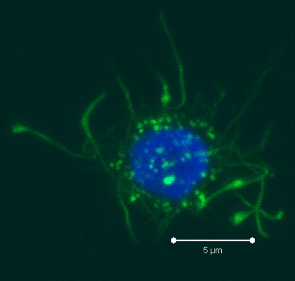
As the monocytes differentiate over the culture period, note their progress by examining them with your tissue culture room microscope. The cells should appear as fairly round and are generally 2-3 times the size of lymphocytes. It is important to note that the mDCs will not appear like the elongated cartoon DCs with long extensions you see in text books. Those DC characteristics are generally only found in tissues and not in vitro.  While you may see some cells that resemble this, those are more likely to be somewhat of a natural stromal layer, made up of cells including macrophages, that the monocyte culture develops to support cell growth. In fact, the immature mDCs will have very few if any, cytoplasmic protrusions.

DC2 resized 600Once the culture period has finished, between 6-8 days, the mDCs can be collected. The exact day is not critical, as long as you remain consistent in the day you pick for your following experiments. To collect the mDCs, gently wash the culture dishes with several streams of media by pipetting up and down. The mDCs, which are currently immature, will be somewhat floating and only loosely adherent. Because of their loose adherence, they require several rounds of gentle pipetting, but do not require cell scraping, EDTA, or trypsin treatment. Note that the culture dishes will still contain some adherent cells. Do not worry about these cells, since these are not the loosely adherent DCs we are interested in.

After completion of these steps, you should have a nice population of immature mDCs, which express CD11c, CD1c, and are CD123-. In my next post, I will cover some tips and tricks for analyzing these cells by flow cytometry. Importantly, I will also cover ways to mature the immature mDCs for use as APCs.

Colt EgelstonColt Egelston is currently a post-doctoral fellow at the Beckman Research Institute of the City of Hope, in Duarte, CA. He received his Ph.D. from Rush University in Chicago and is interested in all things immunology.


Human PBMC T cell immediate early activation markers: What are they and what do they do?

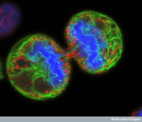
melanoma dividing cellsThere are many strategies for assessing the function of T cells from human peripheral blood mononuclear cells (PBMC).  T cells that have recently been activated through their T cell receptor (TCR) will express a series of activation markers at different time points following activation.   Activation markers include receptors such as chemokine and cytokine receptors, adhesion molecules, co-stimulatory molecules, and MHC-class II proteins.  Some of these molecules have established functions in T cell biology, while the relevance or function of others remains elusive.  Flow cytometry is the method of choice for evaluating various types of surface or intracellular markers that indicate the activation status of T cells.  However, what are these markers, what is their function in T cell biology, what T cell populations will express them, and when can they be assessed are key questions to address when deciding which markers are best for a given assay and question of interest.

In this article, the first of a short series, I will discuss two of the most commonly used immediate early activation markers for assessing the activation status of human PBMC T cells: CD69 and CD40L.

Immediate Early Activation Markers:

CD69 (AIM, Leu23, MLR3) is a signaling membrane glycoprotein involved in inducing T cell proliferation. CD69 is expressed at very low levels on resting CD4+ or CD8+ T cells in PBMC (<5-10%), and is one of the earliest assessable activation markers, being rapidly upregulated on CD4+ or CD8+ T cells within 1 hour of TCR stimulation or other T cell activators such as phorbol esters via a protein kinase C (PKC) dependant pathway.  Expression of CD69 peaks by 16-24 hours and then declines, being barely detectable 72 hours after the stimulus has been withdrawn.

The inability to upregulate CD69 following TCR activation may be associated with T cell dysfunction.  For instance, Critchley-Thorne et. al, showed that PBMC T cells from metastatic melanoma patients with lower responsiveness to interferons had reduced CD69 upregulation compared with healthy controls, and this corresponded with multiple other functional defects in T cells from these patients.  Thus CD69 expression may be a measure of T cell dysfunction in human disease.

CD40L (CD154) is a member of the TNF-receptor superfamily that functions as a co-stimulatory molecule by binding CD40 which is constitutively expressed on antigen presenting cells (APCs).  The CD40L-CD40 ligation results in the activation of multiple downstream pathways including the MAPK (JNK, p38, ERK1/2), NF-ĸB, and STAT3 transcription factors.  CD40L expression is quickly upregulated within 1-2 hours after TCR stimulation via the transcription factors NFAT and AP-1.  CD40L expression peaks near 6 hours after stimulation, and declines by 16-24hrs. CD40L expression however is biphasic, and the addition of anti-CD28 or IL-2 along with TCR stimulation leads to sustained expression for several days (Snyder et. al., 2007).

Expression of CD40L on resting PBMC CD4+ or CD8+ T cells from healthy donors is very low (<1%).  However this percentage has been shown to be significantly increased on up to 17% of CD4+ T cells and 21% of CD8+ T cells in patients with active SLE, and these differences between healthy and SLE patients were also seen following anti-CD3 stimulation of PBMCs (Desai-Mehta, et. al, 1996).  The review below by Daoussis et. al, discusses the role of CD40L expression in several other human diseases.

In summary, CD69 and CD40L are both rapidly induced following T cell activation and both exert important functions in T cell biology. Expressions of these markers have both been shown to be altered in various human diseases.  Understanding the biology of T cell activation markers will allow for the best application of these markers to specific experimental questions and assay types.


Additional Reading:

Multiparametric flow cytometric analysis of the kinetics of surface molecule expression after polyclonal activation of human peripheral blood T lymphocytes. Biselli R, Matricardi PM, D’Amelio R, Fattorossi A. Scand J Immunol. 1992 Apr;35(4):439-47.

Surface markers of lymphocyte activation and markers of cell proliferation.  Shipkova M, Wieland E.  Clin Chim Acta. 2012 Sep 8;413(17-18):1338-49.

Flow cytometric analysis of activation markers on stimulated T cells and their correlation with cell proliferation.  Caruso A, Licenziati S, Corulli M, Canaris AD, De Francesco MA, Fiorentini S, Peroni L, Fallacara F, Dima F, Balsari A, Turano A.   Cytometry. 1997 Jan 1;27(1):71-6.

T cell activation via Leu-23 (CD69).  Testi R, Phillips JH, Lanier LL. J Immunol. 1989 Aug 15;143(4):1123-8.

A whole-blood assay for qualitative and semiquantitative measurements of CD69 surface expression on CD4 and CD8 T lymphocytes using flow cytometry.  Lim LC, Fiordalisi MN, Mantell JL, Schmitz JL, Folds JD. Clin Diagn Lab Immunol. 1998 May;5(3):392-8.

Utility of flow cytometric detection of CD69 expression as a rapid method for determining poly- and oligoclonal lymphocyte activation.  P E Simms and T M Ellis.  Clin Diagn Lab Immunol. 1996 May; 3(3): 301–304.

Down-regulation of the interferon signaling pathway in T lymphocytes from patients with metastatic melanoma.  Critchley-Thorne RJ, Yan N, Nacu S, Weber J, Holmes SP, Lee PP. PLoS Med. 2007 May;4(5):e176.

Direct inhibition of CD40L expression can contribute to the clinical efficacy of daclizumab independently of its effects on cell division and Th1/Th2 cytokine production.  Snyder JT, Shen J, Azmi H, Hou J, Fowler DH, Ragheb JA. Blood. 2007 Jun 15;109(12):5399-406.

Targeting CD40L: a Promising Therapeutic Approach.  D. Daoussis, A.P. Andonopoulos, and S. C. Liossis. Clin Diagn Lab Immunol. 2004 July; 11(4): 635–641.

Hyperexpression of CD40 ligand by B and T cells in human lupus and its role in pathogenic autoantibody production. J. Clin. Investig. 97:2063-2073. Desai-Mehta, A., L. Liangjun, R. Ramsey-Goldman, and S. Datta. 1996.

Photo credit: wellcome images / Foter.com / CC BY-NC-ND

Klotho’s Potential to Reverse MS Demyelination

Klotho is a type I transmembrane protein, expressed in the brain, kidneys and reproductive organs; it is named after “Clotho”, a goddess from Greek mythology who “spins the thread of life” (the length of the threat is determinant of how long a certain individual will live). This denomination is due to the direct positive correlation between Klotho’s expression levels and life span/anti-aging phenotypes.

Klotho protein exists in two forms, membrane Klotho and secreted Klotho, and each form is associated with distinct functions. Some of Klotho’s age-suppressing functions include regulation of fibroblast growth factor-23 (FGF23) signaling, inhibition of insulin/insulin-like growth factor (IGF-1) and Wnt signaling pathways, and regulation of multiple cell-surface ion channels and growth factor receptors.

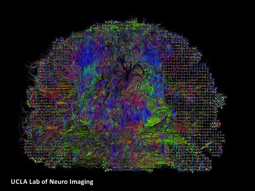
In contrast to other organ systems, the downstream effects of Klotho have not been as extensively studied within the central nervous system (CNS). This is surprising, considering that not only Klotho is present in serum and cerebrospinal fluid (CSF), but also is highly expressed in the hippocampus, choroid plexus and neurons. Disruption of the myelin sheath, either by activated pro-inflammatory cells or by protein defection within the oligodendrocytes has been previously described in aging brain, but the underlying factor stimulating the disruption was not clear. However, In 2008, Abraham’s group reported the significance of Klotho in age-related cognitive decline (ARCD), showing reduced expression of Klotho in regions of brain white matter as a function of age.

Multiple Sclerosis,White matter,Brain,Alzheimer's,cognition


In January 2013, Abraham’s group reported their new findings regarding the effects of Klotho in oligodendrocyte maturation and CNS myelination, and its relation to ARCD. They showed Klotho‘s role in inducing oligodendrocytic progenitor cells (OPCs) maturation, by enhancing the expression of myelin proteins, such as myelin-associated glycoprotein (MAG), myelin basic protein (MBP), oligodendrocyte-specific protein (OSP/Claudin11), and proteolipid protein (PLP). Based on their in vivo studies, the loss of Klotho expression is correlated to defects in myelin that result in similar progressive axonal degeneration observed in hypomyelinating and demyelinating diseases, such as multiple sclerosis (MS).

Previous studies have shown Klotho’s role in facilitating removal of reactive oxygen species (ROS) and increasing resistance to oxidative stress. Furthermore, Nagai’s team observed impaired cognitive function in Klotho-deficient mice, as well as improved cognition upon treatment with the α-tocopherol antioxidant. Thus, Abraham’s group concluded that Klotho protein may function as a neuroprotective factor in the CNS through its antioxidative stress effect.

Together, these results provide strong evidence for Klotho protein as a key player, not only in myelin maintenance, but also in supporting oligodendrocyte and OPC function in the CNS; this makes Klotho an important member of the family of proteins responsible for neuron-oligodendrocyte communication. Abraham’s group hypothesized that downregulation of Klotho may be accountable for the observed white matter myelin degeneration-mediated ARCD, hence increasing Klotho protein expression can potentially prevent damage/protect myelin in the aging brain.

Brain,memory loss,Alzheimer's,MS

Abraham’s new findings are an exciting and important initial step towards development of new neuroprotective therapeutic strategies, such as induction of endogenous remyelination, for treatment of CNS diseases characterized by oligodendrocyte cell loss, such as MS and Schizophrenia. Additionally, early defects of insulin/IGF-1 receptor signaling in Alzheimer’s disease (AD), including the deficit of glucose metabolism that anticipates cognitive decline, may be partially due to deficiencies in Klotho levels. Further investigation on the precise mechanisms involved in Klotho’s regulation within the CNS seems promising for the future of neurodegenerative disease therapy.


Further Readings:

The Antiaging Protein Klotho Enhances Oligodendrocyte Maturation and Myelination of the CNS

Gene Profile Analysis Implicates Klotho As An Important Contributor To Aging Changes In Brain White Matter Of The Rhesus Monkey

The Putative Role of The Antiageing Protein Klotho in Cardiovascular and Renal Disease