Computers meet T cells: in silico identification of mutated tumor antigens targeted by T cells

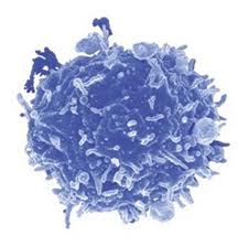
It is well accepted that T cells can recognize and kill tumors that arise in individuals but that tumor cells escape immune surveillance due to the immunosuppressive tumor microenvironment that renders these T cells dysfunctional is less understood.  Only a relatively small number of antigens that T cells recognize for tumor-killing have been identified, and the methods used to identify these antigens are quite cumbersome.  In a recent article in Nature Medicine, Robbins et al. utilize informatics methods to identify mutated tumor antigens in melanoma patients that allowed effective targeting by anti-tumor T cells.

Genome sequencing T cells

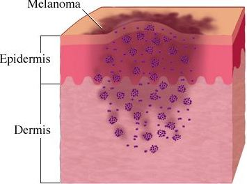
In an effort to identify clinically relevant mutated tumor cell epitopes recognized by T cells, Robbins et al. first performed whole-exome sequencing of tumor cells and matched normal cells from melanoma patients who demonstrated tumor regression following adoptive transfer of autologous tumor infiltrating lymphocytes (TILs).  Mutations in tumor cells that resulted in amino acid changes were identified and then screened using an MHC binding algorithm that predicts high affinity binding of peptide sequences to specific HLA alleles.  Candidate peptides of 9-10 amino acids in length were synthesized and pulsed with specific HLA-expressing target cell lines to load the peptides into the MHC complex.  Peptide-pulsed target cells or autologous tumor cell lines were then cultured with autologous TILs from the same donor and IFN-gamma production was assessed as a read out of T cell activation.

Three metastatic melanoma patients were assessed using this methodology.  The first patient was homozygous for HLA-A*0201, and thus mutated melanoma cell line peptides predicted to bind to the HLA-A*0201 allele were identified by the MHC-binding algorithm.  From this donor, 4 out of 55 candidate peptides elicited IFN-gamma responses from autologous T cells cultured with peptide-pulsed target cells.  Two of these mutated peptides were found to correspond to the casein kinase1α1 protein (CSNK1A1), one peptide was mapped to the growth arrest specific 7 gene (GAS7) gene, and the fourth was a fragment of the HAUS augmin-like complex, subunit 3 (HAUS3) protein.  The wild-type versions of each of these peptides bound very poorly (100-10,000 fold less) or not at all to the HLA and were not recognized by T cells.  Two other donors were assessed for predicted binding of mutated peptides to HLA-A*0101 and HLA-A*1101.  Autologous T cell responses were found to be activated in response to mutated peptides from pleckstrin homology domain containing, family M member 2 (PLEKHM2), protein phosphatase 1 regulatory subunit 3B (PPP1R3B), matrilin 2 (MATN2), and cyclin-dependent kinase 12 (CDK12) genes, but not their wild-type counterparts.  Furthermore, tumor lines were validated to express these mutated proteins.

Finally, the authors compared the reactivity of peripheral blood mononuclear cells (PBMCs) drawn before and after adoptive TIL transfer into two of these patients to determine if anti-tumor reactive T cell clones persisted in vivo.  T cells that recognized the same tumor antigens as the TILs were identified post-adoptive transfer at greater levels than prior to adoptive transfer.  Thus, T cells that recognize mutated tumor epitopes may play a clinically relevant role in mediating tumor regression.  Many questions remain, including a direct demonstration that such tumor-reactive TILs are responsible for mediating the observed tumor regression in these patients, and whether further mutation of these residues might facilitate immune escape later it the course of disease. 

Mining exomic sequencing data to identify mutated antigens recognized by adoptively transferred tumor-reactive T cells.  Robbins PF, Lu YC, El-Gamil M, Li YF, Gross C, Gartner J, Lin JC, Teer JK, Cliften P, Tycksen E, Samuels Y, Rosenberg SA. Nat Med. 2013 May 5. doi: 10.1038/nm.3161.

NetMHCpan, a method for quantitative predictions of peptide binding to any HLA-A and -B locus protein of known sequence.  Nielsen M, Lundegaard C, Blicher T, Lamberth K, Harndahl M, Justesen S, Røder G, Peters B, Sette A, Lund O, Buus S. PLoS One. 2007 Aug 29;2(8):e796.

Upcoming Oncology Conferences and Events: Sept-Nov, 2013

This listing includes upcoming Oncology-related conferences from September – November, 2013.


September

Current Trends in Urological Cancer

September 11, 2013

Wolfson Centre, The Medical School, University of Birmingham

Edgbaston, United Kingdom

Advance registration deadline: August 14, 2013

Advances in Ovarian Cancer Research: From Concept to Clinic

September 18-21, 2013

J.W. Marriott Marquis Miami

Miami, FL

Abstract submission deadline: July 8, 2013

Advance registration deadline: August 5, 2013

Cancer Vaccines

September 18-19, 2013

London, United Kingdom

Frontiers in Basic Cancer Research

September 18-22, 2013

Gaylord National Resort and Convention Center

National Harbor, MD

Abstract submission deadline: July 9, 2013

Advance registration deadline: August 6, 2013

Cancer Advance at Harvard Medical School

September 19, 2013

Harvard Medical School

Boston, MA

Clinical Genomics for Cancer Management Conference

September 23-24, 2013

Seaport Hotel

Boston, MA

Abstracts due: August 23, 2013

Advance registration deadline: August 23, 2013

17th ECCO – 38th ESMO – 32nd ESTRO European Cancer Congress

September 27th to October 1st 2013

Amsterdam, Netherlands

Advance registration deadline: Aug 6, 2013

Late Breaking Abstract Submission Deadline: Aug 7, 2013


October

UAE Cancer Congress 2013

October 3-5, 2013

InterContinental Festival City

Dubai, UAE

Abstract Submission Deadline: June 30, 2013

Early Registration Deadline: August 31, 2013

Cancer Epigenomics

October 6-8, 2013

Melia, Sitges, Spain

Abstract submission deadline June 21, 2013

Early Registration Deadline: August 2, 2013

4th International Conference on Stem Cells and Cancer (ICSCC-2013): Proliferation, Differentiation and Apoptosis

October 19-22, 2013

Mumbai, India

Abstract Submission Deadline: June 30, 2013

Early Registration Deadline: June 30, 2013

15th World Conference on Lung Cancer

October 27-30, 2013

Sydney Australia

Abstract Submission Deadline:  June 21, 2013

Early Registration Deadline: August 2, 2013


November

Bioactive Lipids in Cancer, Inflammation and Related Diseases

November 3 – 6, 2013

San Juan, Puerto Rico

Abstract Submission Deadline: August 23, 2013

Early Registration Deadline: August 16, 2013

Pediatric Cancer at the Crossroads: Translating Discovery into Improved Outcomes

November 3-6, 2013

Westin Gaslamp Quarter

San Diego, CA

Abstract submission deadline: August 28, 2013

Advance registration deadline: September 26, 2013

The Translational Impact of Model Organisms in Cancer

November 5-8, 2013

Omni San Diego

San Diego, CA

Abstract submission deadline: August 26, 2013

Early registration deadline: September 23, 2013

Translational Cancer Research for Basic Scientists

November 10-15, 2013

Omni Parker House Hotel

Boston, Massachusetts

Application deadline: May 13, 2013


Websites that list upcoming Conferences & Events in Oncology:

American Association for Cancer Research

Conference Alerts: Academic Conferences Worldwide

Genentech BioOncology

FDA APPROAVES NEW DRUGS FOR MELANOMA

Melanoma is a type of skin cancer that arises from specialized pigmented cells in our body known as melanocytes, which are responsible for the production of melanin (a pigment responsible for skin and hair color). Because most melanoma cells still make melanin, melanoma tumors are usually brown or black. It accounts for 4% of all skin cancers; however, it is responsible for the largest number of skin cancer related deaths in the world. In the U.S, according to the national cancer institute, estimated new cases and deaths from melanoma in 2013 will be 76,690 and 9,480 respectively (for details please refer to my blog titled “targeting B-RAF kinase in melanoma”).

describe the image

BRAF is a serine/threonine protein kinase that activates the mitogen activated protein kinase (MAPK) signaling pathway. Approximately 50% of melanomas harbor activating BRAF mutations among which mutations at codon 600, resulting in substitution of glutamic acid for valine (B-RAFV600E), are the most prevalent. Activated BRAF phosphorylates and activates mitogen-activated protein kinase kinase proteins (MEK1 and MEK2), which then activate downstream MAP kinases. The MAPK pathway is implicated in the regulation of proliferation and survival of tumor cells in many cancers. This suggests that both B-RAF and its downstream MEK kinase could serve as attractive targets in cancer therapeutics. In 2011, the U.S. Food and Drug Administration (FDA) approved vemurafenib for the treatment of V600E B-RAF mutated melanoma patients. As a single agent, vemurafenib resulted in some degree of tumor regression among 90% of melanoma patients early in the course of treatment. Continuing with the effort to target B-RAF and MEK, several studies tested the efficacy of other compounds to inhibit these components of the MAPK pathway. Based on international clincal trials, on May 29th, 2013, the U.S FDA approved two new drugs Tafinlar (dabrafenib) and Mekinist (trametinib) for use in advanced melanomas with B-RAF V600E mutation. Mekinist is also approved for another form of B-RAF mutilated patients, V600K, which accounts for approximately 10% of B-RAF mutated metastatic melanoma. The mutation status of the melanoma patients are detected by an FDA-approved test, such as companion diagnostic assay from bioMerieus S.A., and THxID-B-RAF.

Tafinlar (dabrafenib) is an orally bioavailable B-RAF-inhibitor which selectively binds to and inhibits the activity of mutated B-RAF (V600E). The FDA approval of dabrafenib is based on an open label multicenter phase III study where 250 were randomly assigned to receive either dabrafenib (187 patients) or dacarbazine (63 patients). Dacarbazine is an alkylating agent which is also use to treat malignant melanoma. The study observed a statistically significant increase in progression-free survival (PFS) in patients treated with dabrafenib, compared to dacarbazine. With dabrafenib, the median PFS was 5.1 months and overall response rate was 52%. The most common adverse reactions with dabrafenib were skin-related toxic effects, fever, fatigue, arthralgia, and headache.

Trametinib is an orally bioavailable inhibitor of MEK which specifically binds to and inhibits MEK 1 and 2, resulting in an inhibition of growth factor-mediated cell signaling and cellular proliferation in various cancers. The FDA approval of trametinib is based on the phase 3 open-label trials which randomly assigned 322 patients who had metastatic melanoma with a V600E or V600K BRAF mutation to receive either trametinib or dacarbazine or paclitaxel (a mitotic inhibitor used in cancer chemotherapy). The study observed a statistically significant increase in PFS in trametinib treated patients compared to other treatments. The PFS was 4.8 month for patients treated with trametinib, while with other chemotherapeutic treatments it was 1.5 months. Rash, diarrhea, and peripheral edema were the most common toxic effects noted following trametinib treatment.

GlaxoSmithKline, manufacturer of both new drugs, reported that the products would be available no later than the early part of the third quarter of 2013.

MAPK Pathway Components: Modulators of Ataxin1 Toxicity in SCA1

amyloidWith the increasing prevalence of neurodegenerative disorders in the aging population, it has become more and more important to understand the molecular pathways that regulate and advance these disorders. Due to the high level of complexity of the mammalian brain, it is very difficult to devise improved targeted treatments. The biggest limitation in neurodegenerative disease research being the lack of viable biomarkers for the elder population. Neurodegenerative disorders such as Alzheimer’s, Parkinson’s and polyglutamine diseases, share many pathogenic abnormalities such as the accumulation of misfolded proteins due to mutations rendering them resistant to degradation or over-expression of the wild type form.

In the May 2013 issue of Nature, Dr. Zoghbi and colleagues at the Baylor College of Medicine, devised a strategy to identify therapeutic entry points that influence the levels of disease-driving proteins. They applied their approach to spinocerebellar ataxia type 1 (SCA1), a disease cased by expansion of the polyglutamine tract in ataxin 1 (ATXN1), using modulation of the ATXN1 pathway as a proof-of-principle. This model was chosen for several reasons: (1) neurodegeneration in SCA1 parallels with the levels of the mutant ATXN1 protein; (2) over-expression of wild type ATXN1 results in neurodegeneration; and (3) SCA1’s pathogenic mechanisms are well characterized. In order to identify regulators of ATXN1 levels the authors developed a human medullablastoma-derived cell line containing the transgene glutamine-expanded ATXN1 fused to red fluorescent protein (mRFP-ATXN1(82Q)).  Next, to distinguish modifiers that regulate ATXN1 protein levels from those affecting transgene transcription they included an internal ribosomal entry site followed by yellow fluorescent protein downstream of ATXN1 (mRFP-ATXN1(82Q)-IRES-YFP).  Their screen focused entirely on kinases and kinase like genes based on the fact that ATXN1 phosphorylation is known to be critical for its toxicity and because kinases are pharmacologically targetable. The authors tested 1908 small interfering RNAs (siRNAs) targeting 638 genes and assessing ATXN1 levels as a readout.  Subsequently, 50 siRNAs (corresponding to 45 genes) were selected based on their ability to reduce the ratio of RFP to YFP by 2 standard deviations from the mean.

A parallel genetic screen was performed using the Drosophila SCA1 model that expresses ATXN1(82Q). This model can be identified by an external eye phenotype. Here they screened 704 alleles (337 kinase encoding, including shRNA and loss of function mutations) for those that would modify ATXN1 levels. Based on morphological and histological assessments, they identified 51 alleles (49 genes) that suppressed ATXN1 toxicity in vivo. Additionally, human cell-based screens showed 10 human modifier genes that reduced ATXN1 and it’s associated toxicity, corresponding to 8 Drosophila modifiers.  Network analysis revealed that the MAPK cascade was the most enriched in both Drosophila and human, where 6/10 genes in human belonged to the canonical MAPK pathway (ERK1, ERK2, MED2, MEK3, MEK6, and MSK1).

ATXN1(82Q) is know to impair motor performance, thus, to determine the effects of the MAPK pathway on the central nervous system, a motor performance test was carried out in Drosophila. Decreasing the MEK, ERK1/2, and MSK1 Drosophila homologues by siRNA lead to increased motor performance and lifespan. Decreasing upstream MAPK pathway homologues suppressed ATXN1(82Q) eye defects and improved motor and lifespan phenotypes.  Conversely, constitutively active RAS exacerbated ATXN1 eye degeneration.  In human cells, decreasing HRAS and FNTA lead to decreased ATXN1 protein levels, and decreasing RAS homologues reduced ATXN1 in vivo.

Previous studies by Dr. Zoghbi’s group reported ATXN1 levels were sensitive to S776 phosphorylation.  Hence, they determined that of MAPK kinases implicated here, MSK1 would be able to phosphorylate the consensus sequence associated with S776.  To prove this, they performed an in vitro kinase assay with purified MSK1 and ATXN1 and found robust ATXN1-S776 phosphorylation in both mutant and WT protein forms. Next, cerebellar fractionation assays of WT mice revealed MSK1 was enriched and had increased activity in S776 phosphorylated fractions. Alternatively, immunodepletion of MSK1 from mouse cerebellar extracts lead to decreased S776 phosphorylation.

Next, they sought to determine whether the MAPK pathway could serve as a pharmacological target for SCA1. Human cells expressing ATXN1(82Q) were treated with a PDI84352 (MEK1/2 inhibitor), GW5704 (RAF1 inhibitor), and a Ro31-8220 (MSK1 inhibitor). Pharmacological inhibition of MAPK pathway lead to decreased ATXN1(82Q). Moreover, addition of MAPK inhibitors to cerebellar slices decreased ATXN1 levels.

Lastly, to test the genetic interaction between ATXN1 and MSK1, ATXN1(154Q) knock in mice (Atxn154Q/+) were bred to Msk1+/- Msk2+/- mice. Atxn154Q/+9 week old mice display a motor phenotype that can be quantified using a rotarod test.  Breeding of Atxn154Q/+ Msk1+/- Msk2+/- mice lead to better rotarod performance. Owing to the fact that ATXN1 alterations lead to Purkinje cell degeneration, they next determined whether eliminating one copy of MSK1 could rescue the loss of Purkinje cells in another mouse model of ATXN1(82Q), B05/+.  Indeed, single copy deletion of Msk1 lead to partially suppressed Purkinje loss phenotype and double MSK1 and MSK2 single copy deletion (B05/+Msk1+/- Msk2+/-), lead to decreased levels of ATXN1.

In summary, Dr. Zoghbi’s group have devised a proof-of-principle strategy that opens many new avenues for the identification of modifiers for neurodegenerative diseases. They utilized combined cross-species genetic screens to identify novel modifiers of ATXN1, and validated in human, mouse, and Drosophila models. This study focused on an early event in pathogenesis that could possibly delay disease onset and progression for this class of neurodegenerative disorders. The RAS-MAPK-MSK1 pathway’s role identified here (phosphorylation of S776-ATXN1) provides a novel pharmacological target for SCA1 and more importantly opens new avenues for combination therapies for this disease. Neurodegenerative disease research has primarily focused on developing treatments for advanced symptoms of neurodegeneration. It would be interesting to determine what the therapeutic benefits are of targeting the RAS-MAPK-MSK1 pathway are on a more advanced form of this disease and whether there would be at least partial reversion of motor defects.


References:

Park, J., et al., RAS-MAPK-MSK1 pathway modulates ataxin 1 protein levels and toxicity in SCA1. Nature.

Emamian,E.S.etal. Serine776 of ataxin-1is critical for polyglutamine-induced disease in SCA1 transgenic mice. Neuron 38, 375–387 (2003).

Jorgensen, N. D. et al. Phosphorylation of ATXN1 at Ser776 in the cerebellum. J. Neurochem. 110, 675–686 (2009).

A bifunctional FoxP3+ regulatory T cell subset converts to pro-inflammatory helper T cells

Recently a number of studies have arisen characterizing Tregulatory cellvarious functional subsets of CD4+ FoxP3+ regulatory T cells (TREGS), as well as their plasticity and ability to differentiate into other TH subtypes.  For instance, TREGS that express RORγt were found to be the specific TREG subset that promotes pro-tumor immune functions in colorectal cancer patients.  In a recent article in Immunity, Sharma et al. identify another TREG subset: FoxP3+ TREGS that loose expression of Eos convert to a pro-inflammatory helper subtype that promotes naïve CD8+ T cells differentiation into potent effectors.

Eos is a transcription factor in the Ikaros family, and acts as an obligate co-repressor in complex with FoxP3 to inhibit expression of FoxP3-repressed genes.  In a quest to understand why TREGS in inflammatory environments were observed to become pro-inflammatory without losing FoxP3 expression, Sharma et al. examined the expression of Eos in FoxP3+ TREGS under inflammatory conditions.

Conversion of FoxP3+ TREGS into an inflammatory phenotype was demonstrated by acquired expression of IL-2, IL-17, and CD40L in the draining lymph nodes of a vaccination site compared with FoxP3+ TREGS at distant lymph nodes that did not gain this function.  In these converted inflammatory FoxP3+ TREGS, expression of Eos was rapidly lost.  IL-6 was required for downregulation of Eos, as TREGS in mice lacking IL-6 did not lose Eos expression under the same conditions.  However, IL-6 alone was insufficient for Eos downregulation, which also required interactions with MHC class II on activated dendritic cells.  Loss of Eos expression was furthermore shown to be required for acquisition of the pro-inflammatory phenotype, as TREGS with forced overexpression of Eos did not undergo this conversion.

Interestingly, not all FoxP3+ TREGS were equivalent in their propensity to lose Eos expression and become pro-inflammatory.  Thymic FoxP3+ TREGS were assessed for stability of Eos under treatment with cyclohexamide. CD38+CD69+CD103 TREGS were “Eos-labile” and specifically lost Eos expression within one hour of cyclohexamide treatment, while CD38CD69CD103+ TREGS maintained Eos expression.  Expression of other markers associated with FoxP3+ TREGS including CD25 and CTLA-4 were equivalent between these two phenotypes highlighting the inability of using these TREG markers to discriminate between these populations.  When these FoxP3+ TREGS were sorted into CD38+CD103and CD38CD103+ subsets and transferred into mice, followed by the vaccination schema, only CD38+CD103 TREGS lost Eos expression and gained CD40L and IL-2 expression. The Eos-labile TREGS do however have characteristic suppressive functions when examined in several models including protection from colitis in a Rag-deficient CD45RBHI effector cell-driven autoimmune colitis model and in vitro suppression of T cell proliferation driven by anti-CD3.

Because the Eos-labile subset was observed in the thymus as part of the natural TREG repertoire, the authors examined the signals required for development of this subset.  Again, IL-6 was required as this subset did not arise in IL-6-/- mice.  Epigenetic analysis of DNA methylation patterns comparing these FoxP3+ TREGS subsets revealed distinctive patterns of methylation yet these subsets were still much more closely related to each other as compared with FoxP3 CD4+ T cells.  Future studies will be needed to determine the nature of these epigenetic differences and which signals are controlled by IL-6.

Interestingly, the authors explored the functional contribution of the Eos-labile pro-inflammatory TREGS subset on CD8+ priming in the vaccination model.  Depletion of TREGS resulted in loss of CD8+ T cell proliferation and granzyme B expression as well as loss of CD86 upregulation on DCs, while adding back just the Eos-labile subset or IL-2 plus CD40-agonist antibodies rescued these defects.  The Eos-labile subset did not however, contribute to reactivation of memory CD4+ T cells, and thus these cells appear to play a specific role in the initial priming stages of naïve T cell activation.  Thus, despite having regulatory activity, these cells are critical in priming CD8+ T cell responses by supplying IL-2 and CD40L signals.

However, indoleamine 2,3-dioxygenase (IDO) was able to block Eos downregulation and acquisition of IL-2, IL-17, and CD40L expression.  Importantly, in a murine tumor vaccination model, blocking IDO was important for FoxP3+ inflammatory TREG induction and acquisition of anti-tumor effector CD8+ T cell responses.  The mechanism of IDO inhibition of Eos downregulation was found to be at least in part, dependent on the antagonization of the IL-6-STAT3 pathway by IDO-mediated production of kynurenine-pathway metabolites which activate the aryl hydrocarbon receptor (AhR).  Interestingly, different AhR ligands have been previously shown to differentially regulate induction of TH17 cells vs. TREGS (Quintana et al.), and kyenurine was a TREG inducing AhR ligand (Mezrich et al.).  Additionally, the contrasting effects of IL-6 and IDO will be an important factor in priming immune cell responses.

Overall, this thorough investigation identified the mechanisms that induce and inhibit this newly defined Eos-labile TREG subset that maintains FoxP3 expression and has typical suppressive TREG activity, yet is critically important in priming effector T cell immune responses.  Future studies will be needed to address how these cells balance regulatory and priming activities as well as the relationships between this subset and the many other TREG subsets described.


An inherently bifunctional subset of foxp3(+) T helper cells is controlled by the transcription factor eos.   Sharma MD, Huang L, Choi JH, Lee EJ, Wilson JM, Lemos H, Pan F, Blazar BR, Pardoll DM, Mellor AL, Shi H, Munn DH. Immunity. 2013 May 23;38(5):998-1012. doi: 10.1016/j.immuni.2013.01.013. Epub 2013 May 16.

Eos, goddess of treg cell reprogramming.  Rieder SA, Shevach EM. Immunity. 2013 May 23;38(5):849-50. doi: 10.1016/j.immuni.2013.05.001.

Control of T(reg) and T(H)17 cell differentiation by the aryl hydrocarbon receptor.  Quintana FJ, Basso AS, Iglesias AH, Korn T, Farez MF, Bettelli E, Caccamo M, Oukka M, Weiner HL. Nature. 2008 May 1;453(7191):65-71. doi: 10.1038/nature06880. Epub 2008 Mar 23.

An interaction between kynurenine and the aryl hydrocarbon receptor can generate regulatory T cells.  Mezrich JD, Fechner JH, Zhang X, Johnson BP, Burlingham WJ, Bradfield CA. J Immunol. 2010 Sep 15;185(6):3190-8. doi: 10.4049/jimmunol.0903670. Epub 2010 Aug 18.