A NEW MOLECULAR TARGET FOR BREAST CANCER THERAPY

Over expression of estrogen receptor (ER) has been implicated in over 70% of breast cancers. Thus therapy targeting ER directly or indirectly is the most important modality in the two-thirds of patients with an ER-positive early breast cancer. The mainstay of endocrine therapy targeting ER in postmenopausal women that are currently available includes selective ER modulators such as tamoxifen and raloxifene, and the ‘third-generation’ aromatase inhibitors (AIs), anastrozole, exemestane and letrozole (click here for more information: http://www.cancer.gov/cancertopics/understandingcancer/targetedtherapies/breastcancer_htmlcourse/page2).

Even though endocrine therapy is the most effective treatment for ER-positive metastatic breast cancer, its effectiveness is limited by high rates of innate (intrinsic) and acquired resistance during treatment. Only about 30% of patients with metastatic disease have objective regression of tumor with initial endocrine treatment, while another 20% have prolonged stable disease.Estrogen_Receptor_Positive_Breast_Cancer-3

Even though mutations of ER are rarely reported, other mechanisms such as ER-phosphorylation has been implicated in resistance to tamoxifen.  In addition, several clinical studies suggested potential mechanisms of resistance to endocrine therapy. Some of the mechanisms implicated include loss of ER, loss of progesterone receptor (PR), upregulation of HER-2, and response to sequential endocrine therapy.

Using a high throughput screening, a recent study by Stebbing et al.  identified a regulator of ER-α, Lemur tyrosine kinase 3 (LMTK3), and noted that LMTK3 gene amplification in both circulating free DNA and primary tumors are predictive of resistance to tamoxifen. Using an orthotopic breast cancer model with tamoxifen-resistant breast cancer cells BT474 that overexpress LMTK3, Stebbing and his colleagues noted that tamoxifen treatment along with LMTK3 knock-down resulted in significant inhibition of tumor growth compared to untreated control mice. To evaluate the clinical relevance of this observation, levels of LMTK3 were determined by immunohistochemistry in tumor samples from ER-positive breast cancer patients treated with endocrine therapy. High levels of LMTK3 were observed in non-responders compared to responders suggesting the association of LMTK3 in limiting efficacy of endocrine therapy. To identify genes and signaling pathways affected by LMTK3, a genome-wide gene expression analysis was performed using BT747 cells. One gene whose expression was found to be significantly regulated by LMTK3 was HSPB8 (heat shock 22kD protein 8). Both overexpression of HSPB8 in breast cancer and potential involvement in tamoxifen resistance have been reported by other studies. Taken together, these results suggests that LMTK3 can contribute to tamoxifen resistance.

ER targeted therapy has improved the quality of life and survival of millions of women around the world, however, resistance to therapy continues to be a major problem. Identification of the role LMTK3 in resistance would facilitate to formulate strategies to overcome this problem.

Further Reading:

http://www.cancer.gov/cancertopics/understandingcancer/targetedtherapies/breastcancer_htmlcourse/page2.

Ali S, Coombes RC. Endocrine-responsive breast cancer and strategies for combating resistance. Nat Rev Cancer. 2002;2(2):101-112.

Osborne CK, Schiff R. Mechanisms of endocrine resistance in breast cancer. Annu Rev Med. 2011;62:233-247.

Stebbing J, Filipovic A, Lit LC, et al. LMTK3 is implicated in endocrine resistance via multiple signaling pathways. Oncogene. 2013;32(28):3371-3380.


Using Mass Spectrometry for Mass T cell Epitope Discovery

Time of Flight Mass Cytometry (CyTOF) is a relatively new multiparametric technology that is far outpacing standard fluorescence-based flow cytometry in the number of parameters that can be simultaneously assessed on a single cell.  In CyTOF, rare transition element isotope-conjugated antibodies are used to label cellular antigens of interest, the magnitude of which is then quantitated by a time of flight mass cytometer, as discussed previously. Previous studies assessing 34 cell surface and intracellular proteins by this technology demonstrated the existence of high dimensional complexity in the heterogeneity of human bone marrow and CD8+ T cell populations.  In a July 2013 article in Nature Biotechonology, Newell et al., move CyTOF and the field of immunology another technological step forward by utilizing CyTOF to measure the frequencies of Rotavirus antigen-specific T cells in human peripheral blood mononuclear cells (PBMCs) and jejunal tissue with peptide-MHC tetramers.

In CyTOF, the theoretical maximum number of simultaneously assessable parameters is 100-200 depending on the instrument.  This vastly outnumbers the assessable parameters of standard fluorescence-based flow cytometry.  To date however, only approximately 40 metal ions have been utilized for antibody labeling, and the development of further metal-chelating technologies is awaited in order to utilize the maximum capacity of the CyTOF instrument.  In the current study, the authors circumvent this limitation by using a “bar-coding” methodology in which a variant combination of three out of ten metal ions are used for labeling each tetramer, allowing for up to 120 different metal combinations.

In this study, the authors sought to identify Rotavirus epitopes recognized by human CD8+ T cells in the context of the MHC class I allele, HLA-A*0201.  To date, only two Rotavirus epitopes recognized by T cells have been identified, and little is known about the phenotypic and functional diversity of antigen-specific T cells for any particular pathogen.  The technical difficulties in proper epitope prediction along with the limited number of cells attainable from human blood samples contribute to these issues.  Thus, this method represents a huge leap forward in the potential to identify significantly more antigen-specific T cell epitopes and to extensively classify these cells functionally.  Using an MHC-prediction algorithm, 77 possible Rotavirus peptides were identified that bound to HLA-A*0201.  An additional 32 positive and negative control tetramers were added for a total of 109 labeled tetramers used to stain each sample simultaneously.  This was further combined with 23-27 metal-chelated antibodies specific for cell surface and intracellular antigens to phenotypically characterize the T cells. A specialized Matlab script was used to analyze the high-dimensional data obtained following mass spectrometry of PBMC and jejunal samples.

On average, CD8 T cell populations specific for two Rotavirus-peptides plus 6-7 peptides from other viruses including influenza, EBV, and CMV, were identified on average across PBMCs from the 17 healthy donors analyzed.  These antigen-specific T cell populations were further phenotypically characterized by expression of surface and intracellular markers.   CD8 T cells specific for six Rotavirus epitopes that included the two previously identified epitopes, were recurrently detected in PBMCs from at least two individuals.  Of these, CD8 cells specific for a Rotavirus peptide from the VP3 protein were most common among healthy donor PBMCs and were phenotypically unique, being of the effector memory subtype compared with a central memory phenotype typical of the T cells specific for the other Rotavirus peptides.  VP3-specific T cells were also uniquely present in jejunal tissue obtained from obese patients that had undergone gastric bypass surgeries.  Thus, this methodology discovered at least 4 new Rotavirus peptides as well as unique characteristics of the different antigen-specific CD8 T cell populations.

In summary, this methodology of combining CyTOF technology with tetramer “bar-coding” paves the way for a vast expansion over fluorescent-based flow cytometry techniques for identifying antigen-specific T cell populations.  As vaccine strategies are an ongoing goal for treatment and prevention of infectious diseases and cancer, it is important to not only identify the peptides that can elicit T cell responses, but also functionally characterize these T cells in order to maximally promote desired immune responses.

Further  Reading:

Combinatorial tetramer staining and mass cytometry analysis facilitate T-cell epitope mapping and characterization.  Newell EW, Sigal N, Nair N, Kidd BA, Greenberg HB, Davis MM. Nat Biotechnol. 2013 Jul;31(7):623-9. doi: 10.1038/nbt.2593. Epub 2013 Jun 9.

Cracking the code of human T-cell immunity.  Harvey CJ, Wucherpfennig KW. Nat Biotechnol. 2013 Jul 9;31(7):609-10. doi: 10.1038/nbt.2626.

Cytometry by time-of-flight shows combinatorial cytokine expression and virus-specific cell niches within a continuum of CD8+ T cell phenotypes. Immunity. 2012 Jan 27;36(1):142-52. doi: 10.1016/j.immuni.2012.01.002.

Single-cell mass cytometry of differential immune and drug responses across a human hematopoietic continuum.  Bendall SC, Simonds EF, Qiu P, Amir el-AD, Krutzik PO, Finck R, Bruggner RV, Melamed R, Trejo A, Ornatsky OI, Balderas RS, Plevritis SK, Sachs K, Pe’er D, Tanner SD, Nolan GP. Science. 2011 May 6;332(6030):687-96. doi: 10.1126/science.1198704.

FIRST TARGETED THERAPEUTIC APPROACH FOR CHRONIC LYMPHOCYTIC LEUKEMIA

Chronic lymphocytic leukemia (CLL) is a slow-growing cancer in which a large number of immature lymphocytes (white blood cells) are found mostly in the blood and bone marrow. It is the most common leukemia in the Western world with incidence rates as high as ~4 per 100,000 individuals in the USA. According to the National Cancer Institute (NCI) it is estimated that in 2013, approximately 15,680 people (9,720 men and 5,960 women) will be diagnosed with CLL and 4,580 men and women will die of CLL. Even though few durable remissions were noted following treatment with chemotherapeutic agents such as chlorambucil, cyclophosphamide, and fludarabine, in the majority of cases these agents are effective for palliation but do not improve survival. An alternative treatment option using chemoimmunotherapy (combination of a chemotherapeutic agent with an anti-CD20 antibody rituximab) was found to have limited efficacy and increased toxicity. In addition, treatment options for CLL are further limited by lack of common genetic target. Nonetheless, many studies reported the association of B-cell receptor (BCR) signaling in the survival of CLL tumor-cells. A downstream component of BCR signaling, a receptor tyrosine kinase, Burton’s tyrosine kinase (BTK) was noted for activation of the Akt, ERK, NF-κB pathways associated with CLL-cell survival.

Bruton’s tyrosine kinase is essential for B-cell development and function. BTK deficiency in man or mice results in the B-cell specific immunodeficiencies X-linked agammaglobulinemia (XLA) or x-linked immune deficiency (xid), respectively. It is also implicated in the pathogenesis of B-cell cancers. Studies suggest that the levels of BTK represent a rate-limiting step in BCR signaling and thereby B-cell activation and survival. Therefore, inhibition of BTK in CLL could serve as an effective treatment strategy. In vitro studies reported that following inhibition of BTK with selective inhibitor CLL cells lose their resistance to apoptosis. Preclinical studies also demonstrated that that BTK-deficiency completely abrogated CLL development in mice.IBRUTINIBWith the accumulating evidence of the role of BCR pathway involving BTK in CLL, first targeted therapeutic approach for CLL was tested clinically with BTK inhibitors. A study published recently in The New England Journal of Medicine (July 4, 2013) by Byrd et al. reported a high frequency of durable remissions in patients with relapsed or refractory CLL with a BTK inhibitor, ibrutinib. A phase I study of ibrutinib (previously known as PCI-32765) showed mild-to-moderate toxicity and clinical antitumor activity in patients with relapsed or refractory B-cell cancers; 11 of the 16 patients in the study had CLL or small lymphocytic lymphoma. These preliminary results prompted the initiation of a phase Ib–II study of ibrutinib in CLL; this study involved two different therapeutic doses in patients with relapsed or refractory disease.

In the phase Ib-II multicenter study of ibrutinib, Byrd et al. (2013) assessed the safety, efficacy, and pharmacokinetics of this inhibitor in patients with CLL or small lymphocytic lymphoma (ClinicalTrials.gov number NCT01105247). Among 85 patients enrolled in this study, 51 received 420 mg and 34 received 840 mg ibrutinib orally once daily. In both cohorts the overall response rate was 71%. This treatment resulted in durable response were the 26-month estimated progression-free survival was 75% and the rate of overall survival was 83%. The pharmacodynamic study showed that ibrutinib was able to successfully inhibit BTK. However, disease progression was noted in 13% patients during follow-up. The most common toxicities observed during ibrutinib treatment were diarrhea, fatigue, and upper respiratory tract infection.

As compared to other single agent therapies for relapsed CLL this targeted therapy of BTK inhibition exhibited more durable responses. The durable remissions observed in this study suggest that many patients may be treated successfully with ibrutinib.

References:

1. Gribben, J.G. and S. O’Brien, Update on therapy of chronic lymphocytic leukemia. J Clin Oncol, 2011. 29(5): p. 544-50.

2.  Advani, R.H., et al., Bruton tyrosine kinase inhibitor ibrutinib (PCI-32765) has significant activity in patients with relapsed/refractory B-cell malignancies. J Clin Oncol, 2013. 31(1): p. 88-94.

3. Byrd, J.C., et al., Targeting BTK with ibrutinib in relapsed chronic lymphocytic leukemia. N Engl J Med, 2013. 369(1): p. 32-42.


Whole Blood Phospho-flow: Direct Ex Vivo Measurement of Signaling in PBMC

I previously discussed phospho-flow cytometry, a method to study intracellular protein phosphorylation events in peripheral blood mononuclear cells (PBMC) at the single cell level.  In standard phospho-flow cytometry protocols, prior to performing assays, PBMCs are first isolated from blood using density gradient centrifugation methods such as Ficoll.  However, there may be times when it is advantageous to study signaling pathways in relatively unmanipulated cells directly ex vivo.  For this, Chow et al. have established a protocol for performing phospho-flow cytometry on PBMCs directly in whole blood.

phospho_flow_cytometry

There are many advantages to isolating and cryopreserving PBMCs with the intention of later studying signaling events by methods including standard phospho-flow cytometry.  In particular, when comparisons are desired between patient groups and healthy controls, there is likely to be less confounding contributions of experimental variability to the results if all of the comparative samples are assayed together.  However, as discussed by Chow et al., pharmacodynamic monitoring as well as evaluation of constitutively activated signaling pathways in PBMCs would be best studied on cells having undergone the least manipulation.  Some signaling pathway responses may be more robust in whole blood PBMCs as well.  For example, I have found in my own assays that signaling responses to IL-6 are strongest in whole blood PBMCs compared with PBMCs following Ficoll or culture in the incubator for any amount of time.  This method can also be used to study bone marrow immune cell signaling as well as expression of intracellular molecules that are exposed by the permeabilization method chosen. In addition, looking at signaling events in murine PBMCs is difficult to do if PBMCs need to be isolated first, given the very small amount of blood that can be obtained from a mouse.  In these cases, anti-coagulated whole blood phospho-flow cytometry should be considered.

Whole blood phospho-flow cytometry is a relatively easy method.  Using 100 ul of whole blood is enough for this assay, and the stimulus (cytokine or other activating signaling molecule) is added directly to the whole blood for the preferred amount of time.  PBMCs are then fixed with formaldehyde and a Triton X-100 based buffer is added to lyse the red blood cells and permeabilize the white blood cells.  This is followed with a few washes and finally the cells can be treated with methanol to unmask phospho-epitopes, similarly to the standard phospho-flow cytometry method by Nolan and colleagues.  Chow et al. include an optional step in which the PBMCs can be stored in a freezing buffer prior to methanol treatment.  However, I have successfully stored PBMCs in 90-100% methanol at -20 or -80 ºC until staining for flow cytometry, similarly to what is done for the standard phospho-flow cytometry method by Nolan and colleagues.

As with all protocols involving treatment of cells with reagents such as methanol or Triton X-100, some epitopes may be lost and thus will not be evaluable if staining is done following these treatments.  Thus, there is an alternate method included in the protocol to stain for some antigens up front.  As a reminder however, some fluorophores are sensitive to methanol, for instance V500, and thus cannot be used to stain PBMCs prior to such treatments.  Finally, in a prior article, Chow et al. (2005), tested different methods of fixation, permeabilization and alcohol unmasking, and I have included the link to that article below as an excellent reference in the case that modulation of the protocol is required for optimal assessment of your antigens of interest.

Further Reading:

Whole blood processing for measurement of signaling proteins by flow cytometry.  Chow S, Hedley D, Shankey TV. Curr Protoc Cytom. 2008 Oct;Chapter 9:Unit 9.27.

Whole blood fixation and permeabilization protocol with red blood cell lysis for flow cytometry of intracellular phosphorylated epitopes in leukocyte subpopulations.  Chow S, Hedley D, Grom P, Magari R, Jacobberger JW, Shankey TV. Cytometry A. 2005 Sep;67(1):4-17.

Single-cell phospho-protein analysis by flow cytometry. Schulz KR, Danna EA, Krutzik PO, Nolan GP.Curr Protoc Immunol. 2012 Feb;Chapter 8:Unit 8.17.1-20.


Finding the Right Cancer Culprits Using Mutational Heterogeneity

Imagine this: you are a police officer on patrol and you receive a call that multiple 30-year-old Caucasian males were seen breaking and entering; stealing heirlooms from a nearby neighborhood. The suspects were last seen entering a convention center and, to your dismay, you arrive to find the entire convention center is an antique show containing several 30-year-old Caucasian males carrying heirlooms. What do you do to apprehend your perpetrators?  You could arrest everyone that fits the description and interrogate them. On the other hand, you could scan the crowd for clues that there is a group of people that do not belong, or also radio to the police station for more information to narrow down the crowd. Needless to say, without gaining more contextual information for prudent discernment of the situation, you may arrest the wrong men and let the criminals go free.genomes

This is where cancer genomics is today; the sophistication of sequencing techniques have allowed for datasets that can detect every genomic mutation within cancer cells. Unfortunately, mutation rates are not equal among all genes. While this may seem a non-issue, this could lead scientists to ascertain that a mutated gene is associated with cancer when, in fact, the gene that “matches the description” is more susceptible to mutation, but has no role in oncogenesis. This is exactly what occurred to researchers who found high mutation rates of olfactory genes within lung cancer1. Doubtful of the role of olfactory genes in lung tumorigenesis, these scientists ultimately concluded that the mutation of olfactory genes had no role in the transformation of the lung epithelial cells1.

In Nature, Lawrence et al. further explored this issue, showing that failure to correct for the variability of mutation rates across the genome could lead to false positives for cancer associated genes1. To illustrate the importance of incorporating heterogeneity into the methodologies of data analysis, the authors compared a datasets with similar mutation frequencies to datasets that had different average mutation frequencies and found, when failing to take into account variability of mutations, there was an increase false categorization of cancer associated genes. Furthermore, the authors demonstrate that an analysis of an increasing sample size, as seen in the “big data” datasets of  American Society of Clinical Oncology’s “CancerLinQ™”2 and the Cancer Genome Atlas3, without correcting mutation rates, may exacerbate  the amount of false positives for cancer associated genes by decreasing the threshold needed to reach statistical significance. Lawrence postulated that heterogeneity may affect the detection of appropriate cancers by failing to correct for three contextual events: heterogeneity in mutation rates amongst samples of the same cancer type (patient-specific context), heterogeneity in mutation rates based on nucleotides surrounding a sequence (sequence-specific context), and heterogeneity in mutation rates based on the time that the gene is replicated or transcribed (replication/transcription-specific context).  Using the mutated olfactory genes mentioned above, along 3083 tumor-control pairs spanning 27 different cancer types, the authors demonstrate the importance of these contextually-discerning mutation rates and construct an algorithm for further context-based analysis, called MutSigCV.

Lawrence et al. studied cancer samples of the same cancer type (3,083 tumor-normal pairs across 27 tumor types) with variable average mutation rate. The authors found that, among all pairs and tumor types, there was a 1,000-fold variance in median frequency of mutations within the sample size. In these samples, the lowest variances were amongst hematological and pediatric cancers while the highest were among tumors induced by environmental factors, such as smoking and radiation. Given the importance of having accurate knowledge of the variability of rate of mutation, this underscores the importance in treating different cancer types, as well as patients with the same cancer, with a context-specific treatment protocol.

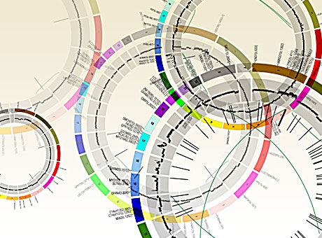
However, correcting for mutational frequencies attributed to tissue types, and mutations caused by known carcinogens and differences in cancer types, the authors still found that there was high mutational variability within certain samples of the same cancer type. Since mutation variance cannot be wholly accounted for by carcinogens, Lawrence et al. postulated that nucleotide makeup of the gene sequence may play a role in the mutation rate variability. The authors tested mutational heterogeneity in multiple tumors by assaying for 96 possible mutations (taking into account flanking bases) that were simplified into a radial chart for analysis1. Lawrence et al found that certain tumor types clustered into certain mutated sequences with the same flanking nucleotides (for instance lung cancers had a really high C to A mutations) was predominate, but still varied, within a certain cancer type.

While both variance in median mutation rates, and predominance of a specific sequence mutation, within specific cancer types was significant, the most important aspect in mutational heterogeneity seems to be in regional areas across a whole genome of cancer types, attributing to an excess of fivefold differences in median mutation rates1. Lawrence et al. credited this to two factors: the amount a gene is transcribed for the time the DNA section is replicated. The authors discovered that mutation rates are highest in genes with low rates of transcription and late DNA replication events. Comparing falsely-implicated olfactory receptor genes to known cancer associated genes, Lawrence et al. demonstrate different transcription rates and different replication times, with olfactory genes being expressed at cells with lower rates and later replication times. In contrast, cancer associated genes have higher transcription rates and earlier replication times.  In other words, while normal and cancer associated genes are both gaining mutations, the events that lead to these mutations are different. Thus, without parsing out mutational rates compared to replication and transcription, one may falsely assume that similar mutation levels must determine a cancer associated genes.

In the end, the authors surmised that “the rich variation in mutational spectrum across tumours underscores the problems with using an overly simplistic model of the average mutational process for a tumour type and failing to account for heterogeneity within a tumor type.” They state that their new analysis algorithm, MutSigCV, takes into account these context dependent nuances, allowing for cancer genomic analysis of mutations that eliminates these false positives. Using MutSigCV, Lawrence et al. was able to take a list of 450 suspected cancer associated genes in lung carcinoma and narrow the list to 11 suspected genes; genes shown to be linked to cancer1. This underscores the importance of context-specific analysis of big data in terms of cancer genomics. Without such a process, the use of whole genome sequencing for mutation rates for novel drug targets may be inadequate, sending many pharmaceutical and biotech companies toward therapeutic targets that, while look like the right suspect, are just an innocent bystanders that “fit the description”.


References:

1          Lawrence, M. S. et al. Mutational heterogeneity in cancer and the search for new cancer-associated genes. Nature, doi:10.1038/nature12213 (2013).

2          DeMartino, J. K. & Larsen, J. K. Data Needs in Oncology: “Making Sense of The Big Data Soup”. Journal of the National Comprehensive Cancer Network 11, S-1-S-12 (2013).

3          Network, C. G. A. R. Comprehensive genomic characterization of squamous cell lung cancers. Nature 489, 519-525, doi:10.1038/nature11404 (2012).


ISSCR 2013 Meeting Updates: Can Alzheimer’s Disease be modeled in a dish?

Human pluripotent stem cells (hPSCs) can differentiate into all cell types of the body, and can thereby serve as great models to examine the pathological mechanisms of various human diseases.  At the International Society for Stem Cell Research (ISSCR) 11th Annual Meeting, various stem cell experts highlighted the current human stem cell models for Alzheimer’s disease, and discussed the potential future directions of the field.

Alzheimer’s disease (AD) is the most common neurodegenerative dementia, affecting ~30 million people worldwide.  AD occurs in two main forms:  early-onset, familial AD (FAD) and late-onset, sporadic AD (SAD).  Both 40004_webare characterized by extensive neuronal loss and the aggregation of two proteins in the brain: amyloid β peptide (Aβ) and tau.  Aβ peptide is derived from the amyloid precursor protein (APP) via cleavage by two proteases, β-secretase and γ-secretase.  According to the amyloid cascade hypothesis, elevated levels of Aβ are necessary and sufficient to trigger disease 1.  Tau is synthesized in neurons and normally functions in binding to tubulin and stabilization of microtubules.  However, in AD, tau is hyper-phosphorylated, resulting in dissociation from microtubules, aggregation, and formation of neurofibrillary tangles (NFTs).  Although the pathological hallmarks of AD consist of these amyloid plaques and NFTs, how the two are related to each other and how they contribute to clinical onset and progression of AD is still under investigation.  By the time a patient manifests symptoms of a mild dementia, there is already significant neuronal loss and substantial accumulation of plaques and tangles.  One major limitation to our understanding of AD has been the lack of live, patient-specific neurons to examine disease progression.

With recent advances in reprogramming technology, scientists can now generate induced pluripotent stem cells (iPSCs), and thereby use live, patient-specific models to examine disease phenotypes in a dish.  At the ISSCR meeting, Larry Goldstein presented his lab’s recent work on using hiPSC models to study AD.  They generated iPSCs from two patients with FAD caused by a duplication of the APP gene, two patients with SAD, and two control individuals.  Next, neurons were generated from the iPSC lines by directed differentiation and fluorescence-activated cell sorting (FACS) purification 2.  Neurons from one SAD and two FAD patients demonstrated significantly higher levels of secreted Aβ and phosphorylated tau (p-tau) 3.  To determine whether there is an association between APP processing and elevated p-tau levels, they treated iPSC-derived neurons with γ-secretase and β-secretase inhibitors.  Interestingly, pharmacologic inhibition of β-secretase resulted in a significant reduction in the levels of Aβ and p-tau.  Treatment with the γ-secretase inhibitor only reduced Aβ levels, but not p-tau levels.  This suggests that products of APP processing other than Aβ might contribute to elevated p-tau levels, highlighting a potential weakness with the amyloid cascade hypothesis.

Other groups have proposed alternative hypotheses to explain AD pathogenesis.  Haruhisa Inoue presented his group’s work on using human iPSC models to examine how intracellular Aβ oligomers contribute to AD.  They generated iPSCs from one patient with FAD caused by the APP-E693Δ mutation, two patients with SAD, and three control individuals.  Corticol neurons were derived using small molecule inhibitors of bone morphogenic protein (BMP) and activin/nodal signaling as previously described 4.  Aβ oligomers accumulated in neurons derived from the FAD patient and one SAD patient, but not in the control neurons 5.  Specifically, the Aβ oligomers accumulated in the endoplasmic reticulum (ER), and triggered ER and oxidative stress in the neurons.  In addition, treatment with docosahexaenoic acid (DHA) alleviated the stress responses.  Although the drug has previously failed in some clinical trials of AD treatment, Inoue’s work suggests that DHA might be effective for a subset of patients.

In summary, Goldstein and Inoue presented convincing evidence that human iPSC models can be used to study early AD pathogenesis and patient-specific drug responses.  Although it can take decades for symptoms to manifest in patients, disease phenotypes can be observed using iPSC models.  However, the fact that only one out of two SAD patients generated a disease phenotype highlights the need of future iPSC studies to examine larger numbers of patients to account for the observed heterogeneity in AD pathogenesis.

References:

1          Hardy, J. & Selkoe, D. J. The amyloid hypothesis of Alzheimer’s disease: progress and problems on the road to therapeutics. Science 297, 353-356, doi:10.1126/science.1072994 (2002).

2          Yuan, S. H. et al. Cell-surface marker signatures for the isolation of neural stem cells, glia and neurons derived from human pluripotent stem cells. PLoS One 6, e17540, doi:10.1371/journal.pone.0017540 (2011).

3          Israel, M. A. et al. Probing sporadic and familial Alzheimer’s disease using induced pluripotent stem cells. Nature 482, 216-220, doi:10.1038/nature10821 (2012).

4          Morizane, A., Doi, D., Kikuchi, T., Nishimura, K. & Takahashi, J. Small-molecule inhibitors of bone morphogenic protein and activin/nodal signals promote highly efficient neural induction from human pluripotent stem cells. J Neurosci Res 89, 117-126, doi:10.1002/jnr.22547 (2011).

5          Kondo, T. et al. Modeling Alzheimer’s disease with iPSCs reveals stress phenotypes associated with intracellular Abeta and differential drug responsiveness. Cell Stem Cell 12, 487-496, doi:10.1016/j.stem.2013.01.009 (2013).


GEM T cells: A newly identified class of restricted α-chain TCRα/β T cells

The diversity of the T cell repertoire allows for recognition of a wide diversity of pathogens. During T cell development, T cell receptors (TCRs) undergo genetic rearrangements of their V, D, and J segments, as well as random deletions and nontemplated additions of nucleotides.  Furthermore, major histocompatibility complex (MHC) class I and II molecules are highly polymorphic.  Thus, each person has a unique and highly diverse T cell-MHC repertoire.  In addition, there are two known classes of lymphocytes with restricted diversity of their TCR α-chains, and which bind to the non/rarely polymorphic antigen-presenting molecule families CD1 and MR1.  These are the invariant natural killer T cells (iNKT cells), and the mucosa-associated invariant T cells (MAIT cells), respectively.  In the June issue of Nature Immunology, Van Rhijn et al. identify a new class of T cells with restricted TCR α-chains, termed GEM T cells, that recognize the Mycobacterium tuberculosis (Mtb) lipid glucose monomycolate presented on CD1b.

To study the human TCR repertoire recognizing CD1b, Van Rhijn et al., utilized CD1b tetramers loaded with glucose monomycolate (GMM), to isolate and clone T cells from peripheral blood mononuclear cells (PBMC) of Mtb infected donors.  Two groups of T cell clones with differing avidity for CD1b-GMM were isolated from each patient, differentiated by intermediate (CD1bint) and high (CD1bhi) CD1b tetramer staining intensities.  CD1bint T cells were diverse in their TCR α-chain sequences.  TCR α-chains of CD1bhi T cells however, all utilized the same variable and joining sequences (TRAV1-2, and TRAJ9, respectively) with few nontemplated additions, resulting in a specific complementarity-determining region 3 (CDR3) consensus sequence.  Thus, these were termed “germline-encoded, mycolyl lipid–reactive” (GEM) T cells.  These TCR α-chain sequences furthermore had to be paired with specific TCR β-chain sequences in order to recognize CD1b-GMM complexes.

Other properties of these uniquely identified GEM T cells included expression of CD4 and production of IFNγ and TNFα upon activation, two cytokines important for anti-mycobacterial responses.  GEM T cells expressed various rates of CD161, a marker widely expressed by NKT cells and MAITs, and thus GEM T cells could not be defined by expression of CD161.  In addition, sorting of TRAV1-2+ CD4+ cells from two healthy donors followed by deep sequencing of the TCR α-chain revealed identification of the GEM-specific CDR3 sequence, demonstrating that GEM T cells were present in Mtb uninfected individuals in the naïve T cell repertoire.  However, these cells become clonally expanded in Mtb infected patients, and thus likely to contribute to anti-mycobacterial immune responses.

In conclusion, GEM T cells are a newly identified third class of CD1-recognizing T cells with restricted TCR α-chain sequences.  These cells arise via VDJ recombination, and indicate that special selection mechanisms exist to generate T cells bearing this specific TCR α-chain.  Although what CD1b-self antigen complex could positively select for these cells in the thymus is unknown.  Furthermore, the role these cells play during mycobacterial infections will be an interesting avenue for future studies.

Further Reading:

A conserved human T cell population targets mycobacterial antigens presented by CD1b.  Van Rhijn I, Kasmar A, de Jong A, Gras S, Bhati M, Doorenspleet ME, de Vries N, Godfrey DI, Altman JD, de Jager W, Rossjohn J, Moody DB. Nat Immunol. 2013 Jun 2;14(7):706-13.

A ‘GEM’ of a cell.  Mitchell Kronenberg & Dirk M Zajonc. Nature Immunology 14, 694–695 (2013) doi:10.1038/ni.2644. Published online 18 June 2013.

New Findings in Cell Based Therapy for GBM

Glioblastoma multiforme (GBM) is the most common and lethalGlioblastoma type of malignant primary brain tumors that account for over 70% of all intracranial cancers.  The current course of GBM treatment consists of surgical resection of the main tumor mass, followed by administration of radiation and chemotherapy. Surgical resection of the primary tumor leads to injury to the surrounding normal tissue, while chemotherapy and radiotherapy cause toxicity to the healthy tissue in the brain.  These undesirable secondary effects of glioma treatments have a major impact on patients’ physical, cognitive and emotional functioning. Nonetheless, despite this aggressive treatment regimen and its harmful side effects, GBM remains virtually incurable, with post-diagnosis median survival persisting less than 14 months.  This dismal prognosis is due to a combination of unique anatomical features of the central nervous system (CNS), in addition to GBMs’ (glioma cells’) exceptional invasive capacity. Glioma cells infiltrate the brain’s highly dense parenchyma, migrating along the corpus callosum and creating new masses within the hemisphere contralateral to the initial tumor mass homing.

Thus, recurrence in postoperative GBM patients is in essence inevitable. Furthermore, GBMs are not only heterogeneous among individual patients, but they are also highly heterogeneous within a single tumor mass. Recent studies have shown compelling evidence of a therapeutically resistant subpopulation of malignant glioma cells that exhibit stem-cell like characteristics, such as multipotency, the ability to self-renew and invade/migrate; these tumor-initiating cells are referred to as glioma stem cells (GSCs) and are believed to be responsible for tumor initiation and recurrence in GBM patients. Furthermore, GSCs have been observed to ensconce within similar niches to neural stem cells (NSCs).

NSCs and mesenchymal stem cells (MSCs) have been shown to have an exceptional migratory ability within brain’s parenchyma and possess a notable inherent tumor tropism.  Thus, several cell-based therapy (CBT) studies and clinical models of malignant tumors have incorporated autonomous tracking of tumor cells by employing NSCs and MSCs to deliver multiple therapeutic genes to specific tumor loci. This target specific drug-delivery model has NSCs or MSCs transduced to express one pro-drug-activating enzyme, which catalyzes the conversion of a particular pro-drug into an active toxic agent, which results in the localization of the chemotherapeutics specifically at the tumor sites.  In addition to its ability to deliver effective cytotoxic damage to the tumor without causing damage to the healthy surrounding tissue, the resulting bystander effect of this system causes cell death not only to the drug-delivery-vehicle cells, but also to the surrounding glioma cells. Although a number of different enzyme/prodrug systems have been utilized in relevant studies, HSV-thymidine kinase (HSV-tk)/ deoxyguanosine analog ganciclovir (GCV) has been the most commonly tested in animal and in vitro models. HSV-tk phosphorylates GCV and produces deoxyguanosine triphosphate which is a polymerase-I inhibitor and DNA chain terminator; cell death occurs upon incorporation of the nucleotide analog within DNA chains.

In recent study published in the journal of Molecular Therapy, Blanco’s group report their findings regarding the interaction between human MSCs (hMSCs) and gliomas and the underlying mechanism for the effectiveness of hAMSC based therapy in GBMs.  In their previous work Blanco’s group showed that the administration of hAMSCs expressing HSV thymidine kinase in glioma tumors significantly promotes tumor growth, whereas induction of cytotoxicity by administration of the prodrug GCV demonstrated a significant antitumor response.

According to this new study, hMSCs differentiate to endothelial lineage (supported by the expression of CD31 marker) within tumors, and integrate in the tumor vascular system where they adopt an endothelial phenotype. Further, Blanco proposed the notion of hMSCs’ ability to home to privileged vascular structures where GSCs also reside, is the underlying characteristic that leads to the effectiveness of cytotoxic hMSCs in regulating the bystander killing of tumor cells.

Although Blanco’s study provides invaluable insights into the GSCs’ niche and its role in malignant brain tumor CBT, hMSCs’ tumor growth promotion is a quality, which makes these cells less than ideal for utilization in human clinical trials in the near future. Future studies on this mechanism using primary patient tumor cells rather than the U87 glioma cell line and human NSCs are needed to further confirm these observations.

Further reading:

Juli R. Bagó, Maria Alieva, Carolina Soler, Núria Rubio, Jerónimo Blanco. Endothelial differentiation of adipose tissue-derived mesenchymal stromal cells in glioma tumors: implications for cell based therapy. Molecular Therapy.