Upcoming Oncology Conferences and Events: Sept-Nov, 2013

This listing includes upcoming Oncology-related conferences from September – November, 2013.


September

Current Trends in Urological Cancer

September 11, 2013

Wolfson Centre, The Medical School, University of Birmingham

Edgbaston, United Kingdom

Advance registration deadline: August 14, 2013

Advances in Ovarian Cancer Research: From Concept to Clinic

September 18-21, 2013

J.W. Marriott Marquis Miami

Miami, FL

Abstract submission deadline: July 8, 2013

Advance registration deadline: August 5, 2013

Cancer Vaccines

September 18-19, 2013

London, United Kingdom

Frontiers in Basic Cancer Research

September 18-22, 2013

Gaylord National Resort and Convention Center

National Harbor, MD

Abstract submission deadline: July 9, 2013

Advance registration deadline: August 6, 2013

Cancer Advance at Harvard Medical School

September 19, 2013

Harvard Medical School

Boston, MA

Clinical Genomics for Cancer Management Conference

September 23-24, 2013

Seaport Hotel

Boston, MA

Abstracts due: August 23, 2013

Advance registration deadline: August 23, 2013

17th ECCO – 38th ESMO – 32nd ESTRO European Cancer Congress

September 27th to October 1st 2013

Amsterdam, Netherlands

Advance registration deadline: Aug 6, 2013

Late Breaking Abstract Submission Deadline: Aug 7, 2013


October

UAE Cancer Congress 2013

October 3-5, 2013

InterContinental Festival City

Dubai, UAE

Abstract Submission Deadline: June 30, 2013

Early Registration Deadline: August 31, 2013

Cancer Epigenomics

October 6-8, 2013

Melia, Sitges, Spain

Abstract submission deadline June 21, 2013

Early Registration Deadline: August 2, 2013

4th International Conference on Stem Cells and Cancer (ICSCC-2013): Proliferation, Differentiation and Apoptosis

October 19-22, 2013

Mumbai, India

Abstract Submission Deadline: June 30, 2013

Early Registration Deadline: June 30, 2013

15th World Conference on Lung Cancer

October 27-30, 2013

Sydney Australia

Abstract Submission Deadline:  June 21, 2013

Early Registration Deadline: August 2, 2013


November

Bioactive Lipids in Cancer, Inflammation and Related Diseases

November 3 – 6, 2013

San Juan, Puerto Rico

Abstract Submission Deadline: August 23, 2013

Early Registration Deadline: August 16, 2013

Pediatric Cancer at the Crossroads: Translating Discovery into Improved Outcomes

November 3-6, 2013

Westin Gaslamp Quarter

San Diego, CA

Abstract submission deadline: August 28, 2013

Advance registration deadline: September 26, 2013

The Translational Impact of Model Organisms in Cancer

November 5-8, 2013

Omni San Diego

San Diego, CA

Abstract submission deadline: August 26, 2013

Early registration deadline: September 23, 2013

Translational Cancer Research for Basic Scientists

November 10-15, 2013

Omni Parker House Hotel

Boston, Massachusetts

Application deadline: May 13, 2013


Websites that list upcoming Conferences & Events in Oncology:

American Association for Cancer Research

Conference Alerts: Academic Conferences Worldwide

Genentech BioOncology

MAPK Pathway Components: Modulators of Ataxin1 Toxicity in SCA1

amyloidWith the increasing prevalence of neurodegenerative disorders in the aging population, it has become more and more important to understand the molecular pathways that regulate and advance these disorders. Due to the high level of complexity of the mammalian brain, it is very difficult to devise improved targeted treatments. The biggest limitation in neurodegenerative disease research being the lack of viable biomarkers for the elder population. Neurodegenerative disorders such as Alzheimer’s, Parkinson’s and polyglutamine diseases, share many pathogenic abnormalities such as the accumulation of misfolded proteins due to mutations rendering them resistant to degradation or over-expression of the wild type form.

In the May 2013 issue of Nature, Dr. Zoghbi and colleagues at the Baylor College of Medicine, devised a strategy to identify therapeutic entry points that influence the levels of disease-driving proteins. They applied their approach to spinocerebellar ataxia type 1 (SCA1), a disease cased by expansion of the polyglutamine tract in ataxin 1 (ATXN1), using modulation of the ATXN1 pathway as a proof-of-principle. This model was chosen for several reasons: (1) neurodegeneration in SCA1 parallels with the levels of the mutant ATXN1 protein; (2) over-expression of wild type ATXN1 results in neurodegeneration; and (3) SCA1’s pathogenic mechanisms are well characterized. In order to identify regulators of ATXN1 levels the authors developed a human medullablastoma-derived cell line containing the transgene glutamine-expanded ATXN1 fused to red fluorescent protein (mRFP-ATXN1(82Q)).  Next, to distinguish modifiers that regulate ATXN1 protein levels from those affecting transgene transcription they included an internal ribosomal entry site followed by yellow fluorescent protein downstream of ATXN1 (mRFP-ATXN1(82Q)-IRES-YFP).  Their screen focused entirely on kinases and kinase like genes based on the fact that ATXN1 phosphorylation is known to be critical for its toxicity and because kinases are pharmacologically targetable. The authors tested 1908 small interfering RNAs (siRNAs) targeting 638 genes and assessing ATXN1 levels as a readout.  Subsequently, 50 siRNAs (corresponding to 45 genes) were selected based on their ability to reduce the ratio of RFP to YFP by 2 standard deviations from the mean.

A parallel genetic screen was performed using the Drosophila SCA1 model that expresses ATXN1(82Q). This model can be identified by an external eye phenotype. Here they screened 704 alleles (337 kinase encoding, including shRNA and loss of function mutations) for those that would modify ATXN1 levels. Based on morphological and histological assessments, they identified 51 alleles (49 genes) that suppressed ATXN1 toxicity in vivo. Additionally, human cell-based screens showed 10 human modifier genes that reduced ATXN1 and it’s associated toxicity, corresponding to 8 Drosophila modifiers.  Network analysis revealed that the MAPK cascade was the most enriched in both Drosophila and human, where 6/10 genes in human belonged to the canonical MAPK pathway (ERK1, ERK2, MED2, MEK3, MEK6, and MSK1).

ATXN1(82Q) is know to impair motor performance, thus, to determine the effects of the MAPK pathway on the central nervous system, a motor performance test was carried out in Drosophila. Decreasing the MEK, ERK1/2, and MSK1 Drosophila homologues by siRNA lead to increased motor performance and lifespan. Decreasing upstream MAPK pathway homologues suppressed ATXN1(82Q) eye defects and improved motor and lifespan phenotypes.  Conversely, constitutively active RAS exacerbated ATXN1 eye degeneration.  In human cells, decreasing HRAS and FNTA lead to decreased ATXN1 protein levels, and decreasing RAS homologues reduced ATXN1 in vivo.

Previous studies by Dr. Zoghbi’s group reported ATXN1 levels were sensitive to S776 phosphorylation.  Hence, they determined that of MAPK kinases implicated here, MSK1 would be able to phosphorylate the consensus sequence associated with S776.  To prove this, they performed an in vitro kinase assay with purified MSK1 and ATXN1 and found robust ATXN1-S776 phosphorylation in both mutant and WT protein forms. Next, cerebellar fractionation assays of WT mice revealed MSK1 was enriched and had increased activity in S776 phosphorylated fractions. Alternatively, immunodepletion of MSK1 from mouse cerebellar extracts lead to decreased S776 phosphorylation.

Next, they sought to determine whether the MAPK pathway could serve as a pharmacological target for SCA1. Human cells expressing ATXN1(82Q) were treated with a PDI84352 (MEK1/2 inhibitor), GW5704 (RAF1 inhibitor), and a Ro31-8220 (MSK1 inhibitor). Pharmacological inhibition of MAPK pathway lead to decreased ATXN1(82Q). Moreover, addition of MAPK inhibitors to cerebellar slices decreased ATXN1 levels.

Lastly, to test the genetic interaction between ATXN1 and MSK1, ATXN1(154Q) knock in mice (Atxn154Q/+) were bred to Msk1+/- Msk2+/- mice. Atxn154Q/+9 week old mice display a motor phenotype that can be quantified using a rotarod test.  Breeding of Atxn154Q/+ Msk1+/- Msk2+/- mice lead to better rotarod performance. Owing to the fact that ATXN1 alterations lead to Purkinje cell degeneration, they next determined whether eliminating one copy of MSK1 could rescue the loss of Purkinje cells in another mouse model of ATXN1(82Q), B05/+.  Indeed, single copy deletion of Msk1 lead to partially suppressed Purkinje loss phenotype and double MSK1 and MSK2 single copy deletion (B05/+Msk1+/- Msk2+/-), lead to decreased levels of ATXN1.

In summary, Dr. Zoghbi’s group have devised a proof-of-principle strategy that opens many new avenues for the identification of modifiers for neurodegenerative diseases. They utilized combined cross-species genetic screens to identify novel modifiers of ATXN1, and validated in human, mouse, and Drosophila models. This study focused on an early event in pathogenesis that could possibly delay disease onset and progression for this class of neurodegenerative disorders. The RAS-MAPK-MSK1 pathway’s role identified here (phosphorylation of S776-ATXN1) provides a novel pharmacological target for SCA1 and more importantly opens new avenues for combination therapies for this disease. Neurodegenerative disease research has primarily focused on developing treatments for advanced symptoms of neurodegeneration. It would be interesting to determine what the therapeutic benefits are of targeting the RAS-MAPK-MSK1 pathway are on a more advanced form of this disease and whether there would be at least partial reversion of motor defects.


References:

Park, J., et al., RAS-MAPK-MSK1 pathway modulates ataxin 1 protein levels and toxicity in SCA1. Nature.

Emamian,E.S.etal. Serine776 of ataxin-1is critical for polyglutamine-induced disease in SCA1 transgenic mice. Neuron 38, 375–387 (2003).

Jorgensen, N. D. et al. Phosphorylation of ATXN1 at Ser776 in the cerebellum. J. Neurochem. 110, 675–686 (2009).

Optimizing Assays to Find Rare Antigen-Specific T cells in Cryopreserved PBMCs

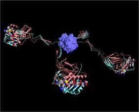
Immunomonitoring of T cell based immune responses spans a wide range of therapeutic applications such as infectious and autoimmune diseases and is particularly important for vaccine research. Regardless of the therapeutic application, immunomonitoring can be a daunting task due to the variability of methods and protocols available. There are several commonly used functional assays for the enumeration of antigen specific CD8+ T cells and there is great variability in the protocols that are used for these assays. Thus, making it increasing difficult to thoroughly interpret data obtained from multi-center clinical trials and to compare results between laboratories. In order to address some of the issues associated with immunomonitoring of clinical trials, the Association for Immunotherapy of Cancer (CIMT) formed a CIMT monitoring panel tasked to standardize protocols for assaying T cell antigen immune responses. Thirteen centers from 6 different European countries participated in this study. They were given the same samples and asked to determine the number of antigen specific T cells and assess their antigen specific function using tetramer staining and a functional assay of their choice. Common techniques used for monitoring antigen induced immune responses included ELISPOT assays, HLA-multimer staining and intracellular cytokine staining (ICS).

Pre-tested samples of peripheral blood mononuclear cells (PBMC), synthetic peptides, and PE-conjugated HLA-tetramers were distributed to each center. Using HLA-typed healthy volunteers, PBMCs were isolated by Ficoll density gradient separation. Each sample was tested for T cell reactivity against CMV and influenza. All centers received an HLA-A negative control as well as HLA-A positive samples consisting of a combination of CMV and influenza reactive PBMCs. The study comprised of 2 phases; Phase I consisted of all centers performing the assays with their commonly used protocols, and in Phase II each center received optimized protocols based on the findings from Phase I.

For Phase I’s tetramer-staining assay, the laboratories could choose to stain samples with antibodies (Ab) for CD8+ alone, CD3+CD8+, or CD4+ CD8+ and use their preferred Ab clone, fluorescent dye, and Ab concentration. For the functional assays synthetic peptides were provided and each group could choose either the INF-γ ELISPOT assay, FACS-based intracellular INF-γ staining or both with their antigen concentration of choice ranging from 1-10 g/ml. To reduce variability in FACS analysis, sample plots were provided as well as gate settings and quadrants. Tetramer-staining data reported included; number of viable cells post-thawing, cytometer model, number of lymphocytes and/or CD8+ cells analyzed. Data was presented as percent of tetramer-positive cells among CD8+, CD3+CD8+, or CD4+ lymphocytes depending on what antibody cocktail was chosen. For the functional assays each center reported the type of ELISPOT plates used, reagents and conditions used, and number cells tested.

Tetramer results from the Phase I study showed the number CD8+ cells analyzed significantly affected the sensitivity of tetramer staining. Antigen-specific T cell reactivity when less than 30,000 CD8+ T cells were counted resulted in only 70% responsiveness detected. In contrast, when more than 30,000 CD8+ cells were counted, an 89% response was observed. Although, when antigen-specific T cells were present at high frequencies the number of counted cells did not matter. Interestingly, Ab clone variability, Ab concentration, or cytometer type did not result in any significant differences. Thus, the main factors affecting antigen-specific T cell reactivity by tetramer staining is the number of CD8+ cells used. For Phase II it was then recommended at least 1 x10⁶ PBMCs are used for this assay.

The majority of groups chose the INF-γ ELISPOTas their functional assay. Results showed a large amount of heterogeneity between the centers. Some centers included a resting phase after thawing the cells, of 2-20 hours, resulting in 73% positive reactivity (number of spot forming cells per seeded PBMC). In contrast, not allowing a resting phase resulted in only detecting 30% of the positive cells. Additionally, intra-center replicate reproducibility was significantly affected by the number of replicates used, where duplicates often failed the Student t test and triplicates were sufficient to reach statistical significance. Addition of allogenic-APCs for binging and presentation of the synthetic peptides was found to have a negative effect on detection response (28% of all responses vs. 58%). When looking at the number of cells seeded per well, those with more than 4 x10⁵ PBMC detected 71% positive samples and those with less than 4 x10⁵ only detected 43%. Granted, when antigen specific T cells were available at high frequencies the number of counted cells did not affect the response rates. Consequently, Phase II’s minimum requirements for the INF-γ ELISPOT protocol included: (1) triplicates should be performed for each test antigen (2) avoid using allogenic-APCs (3) include a resting phase (4) use over 4 x 10⁵ PBMCs per well.

Another interesting finding from this study was that lab experience in performing these assays had no effect on the performance of the assays compared to labs that had just adopted the techniques. Further highlighting the importance of developing standardized protocols for immunomonitoring assays. This study did not however, address specific detection limits for the ELISPOT assays, the variability between ELISPOT plate readers, nor serum source effects on background and specificity. In addition, it was not reported whether live/dead cell stains where included in the tetramer assays and how combinations of these may have had an effect on the sensitivity of the assay.

Overall, this study identified several factors that should be generally implemented when performing tetramer staining and INF-γ ELISPOT assays with cryopreserved PBMC samples. Furthermore, these protocol modifications are particularly important when assaying antigen-specific T cell populations present at low frequencies.

Reference:

The CIMT-monitoring panel: a two-step approach to harmonize the enumeration of antigen-specific CD8+ T lymphocytes by structural and functional assays. Britten CM, Gouttefangeas C, Welters MJ, Pawelec G, Koch S, Ottensmeier C, Mander A, Walter S, Paschen A, Müller-Berghaus J, Haas I, Mackensen A, Køllgaard T, thor Straten P, Schmitt M, Giannopoulos K, Maier R, Veelken H, Bertinetti C, Konur A, Huber C, Stevanović S, Wölfel T, van der Burg SH. Cancer Immunol Immunother. 2008 Mar;57(3):289-302. Epub 2007 Aug 25.
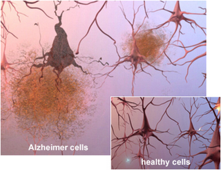
Deep Brain Stimulation Shows Increased Cerebral Metabolism in Alzheimer’s Patients

Alzheimer’s disease (AD) is the most common form of dementia typically presenting itself after the age of 60, due to protein misfolding in the brain.  According to the National Institute on Aging, currently it is estimated that 5.1 million Americans suffer from AD. Dimentia, is defined as the loss of cognitive function including thinking, remembering, reasoning abilities, and behavioral abilities.  The severity of the disease ranges from pre-dementia (early onset) characterized by small affects on a persons functioning all the way to advanced where a person requires complete dependence on others for daily living.

AD was originally identified by Dr. Alois Alzheimer in 1906 after a brain biopsy of a woman showed abnormal clumps in her brain, now known as Amyloid plaques.  Amyloid plaques originate from amyloid-β (Aβ) deposits which are insoluble fibrous protein aggregates arising from misfolded proteins and polypeptides.

The dominant hypothesis for AD neuropathology is that Aβ plaque formations initiate a cascade that is followed by loss of neurons and synapses in the cerebral cortex and central subcortical regions as well as abnormal levels of neurotransmitters acetylcholine (decreased levels, leading to decreased cognition) and glutamate (increased levels, leading to neuronal over-activation and cell death).  The combined effects being progressive cell death in select regions of the brain. Currently available treatments for AD include drugs that target acetylcholine, glutamate and the amyloid cascade pathway.  These treatments are only efficacious at treating some of the symptoms they don’t stop the underlying decline and death of neurons. Additionally, it is now believed that Aβ plaques are necessary but not sufficient for the cognitive and neurodegenerative effects seen in AD patients. Therefore, the lack of efficacy in currently available treatments and the much needed understanding of the secondary effects due Aβ plaques, shows there is a dire need for safer and more efficacious therapies which can delay and reverse the effects of AD as well as modulate neuronal function in the affected neural circuits.

In a recent TEDx Talk, Dr. Andres M. Lozano from the Toronto Western Research Institute (TWRI) talks about a novel technology used for treating neuronal disorders, deep brain tissue stimulation (DBS). Dr. Lozano’s team have studied the effects of DBS on a variety of neurodegenerative disorders including AD and Parkinson’s disease showing exciting breakthroughs. DBS is surgical procedure where a brain pacemaker is implanted in an affected region of the brain.  The pacemaker can then be controlled externally and directed to send electrical impulses to a specific location.

The human brain consumes about 25% of the total body glucose levels.  In AD patients, it is well known that glucose uptake/metabolism is significantly impaired. Using positron emission tomography (PET) scans with the radiotracer [18F]-2-deoxy-2-fluoro-D-glucose (measures regional cerebral glucose metabolism) Dr. Lozano’s team found that DBS resulted in increased glucose metabolism in AD patients after 1 yr of treatment (http://clinicaltrials.gov/show/NCT00658125). More importantly, the increased metabolism correlated with better outcomes in global cognition, memory, and quality of life.  Based on preclinical studies, it is believed that DBS functions by inducing the generation of new neurons through electrical impulses.  Although, the effects of DBS on Aβ breakdown remain unclear and are under investigation in preclinical models.

It is important to note that not all patients displayed the same level of benefit from DBS.  In addition, all patients in this study had relatively mild AD and higher basal levels of cognitive function and glucose compared to more advanced patients. Therefore, it is possible that the effects of BDS in advanced AD patients will not be as beneficial, although this remains to be determined. It will be interesting to see what the long term effects of DBS will be and whether BDS will be sufficient to keep AD progression at bay.

Further Reading:

Increased Cerebral Metabolism After 1 Year of Deep Brain Stimulation in Alzheimer Disease. Gwenn S. Smith, Adrian W. Laxton, David F. Tang-Wai, Mary Pat McAndrews, Andreea Oliviana Diaconescu, Clifford I. Workman, Andres M. Lozano. Arch Neurol. 2012;69(9):1141-1148. doi:10.1001/archneurol.2012.590.

Memory rescue and enhanced neurogenesis following electrical stimulation of the anterior thalamus in rats treated with corticosterone. Clement Hamani, Scellig S. Stone, Ariel Garten, Andres M. Lozano, Gordon Winocur. Experimental Neurology.Nov;232(1):100-104. doi: 10.1016/j.expneurol.2011.08.023.

Cancer Biomarkers: How they are used for personalized medicine

As promising cancer treatments emerge the need for improved detection and characterization methods are still evident.  Identification of novel biomarkers is a promising area of cancer research and development but because of the high complexity and heterogeneity of tumors much remains to be learned.

What is a cancer biomarker? A biomarker is a biological molecule that can be found in the blood, bodily fluids or tissue of interest (i.e. tumor) that can give information about the molecular characteristics of a tumor.

Specimens for biomarker discovery

Potential biomarker biological molecules

  • DNA (copy number, methylation states, mutations)
  • RNA (mRNA, microRNA)
  • Protein (phosphorylation, post-translational modifications)
  • Metabolic products

Tools for cancer biomarker identification

Biomarkers can be used as tools for diagnosis (detect the presence of cancer), prognosis, tracking cancer progression, and assessing treatment efficacy.

In cancer, a biomarker is often a protein that is mutated or is expressed at higher levels in the cancerous cells compared to the normal tissue.  There are various proteins whose mutated status is shared by multiple types of cancers these include inactivating mutations of tumor suppressor proteins such as the cell cycle regulators p53, PTEN, and retinoblastoma protein (RB) and activating mutations of proto-oncogenes such as Ras and Myc.  Another prominent cancer biomarker is the cell proliferation marker, Ki67 that can be used not only as a prognostic indicator but also to assess the efficacy of a treatment where reduction in Ki67 expression indicates reduced cellular proliferation.

Types of biomarkers and their uses

types_of_biomarkers_uses.jpg

  1. Prognostic biomarker: Knowing the key molecular changes in a patient’s cancer allows a doctor to determine whether the patient is likely to have a poor outcome and thus more aggressive treatment is necessary.
  2. Predictive biomarker: Understanding the molecular characteristics about a patients cancer can lead to tailoring of drug treatments with a higher likelihood of efficacy.  For example, patients with certain kinase domain mutations on EGFR would possibly not respond to EGFR targeted treatments such as erlotinib. Additionally this gives the added benefit reducing a patients exposure to possible toxic side effects from a drug they may not have benefited from.
  3. Pharmacodynamic biomarker: Using biomarkers drug dosing could be tailored to each patient. Dosing a drug that has a specific molecular target can be decided based on its ability to decrease the activity of its biological target. For example, if a patient shows high activity for a particular kinase a targeted drug could be dosed up for that patients specific needs.

biomarker_drug_selection

With the advent of genomic and proteomic technology and improved data mining algorithms, it is now easier and faster to identify biomarkers.  Unfortunately, a major limitation in biomarker research and discovery is the need for biopsy samples and their limited availability for research. Another issue is that most biopsy samples available are taken from a patient during the initial diagnosis. Less available are samples from patients post treatment initiation or with advanced disease where the molecular characteristics of their cancer may have very likely changed.

Luckily, there is great interest in developing and improving current technologies to utilize blood specimens for protein, metabolic products, CTCs and circulating DNA as alternative non-invasive sources to identify and screen for cancer biomarkers.

Further reading:

Mining the plasma proteome for cancer biomarkers

The cancer biomarker problem

Taming the dragon: genomic biomarkers to individualize the treatment of cancer

Proteomics for Cancer Biomarker Discovery


Clinical Trials: What’s New in Oncology?

Novel Immunotherapy Shows Promise for Various Types of Cancers

Since the approval of Provenge and Yervoy immunotherapies, development of anti-cancer immune therapies has gained a lot of momentum. Bristol-Myers Squibb’s and Ono Pharma’s antibodies targeted toward PD-1 and PD-L1 molecules are showing great promise for the treatment of non-small cell lung cancer, melanoma, kidney cancer and ovarian cancer. PD-1 is a molecule found on T-cells, when PD-1’s ligand PD-L1 binds to it T-cells loose activity or die. These targeted antibodies block the binding of ligand to receptor and in doing so maintain anti-tumor T-cell activity.   

Investigational Drug Trastuzamab Emtansine Delays Progression of Advanced HER2-positive Breast Cancer

Trastuzamab emtansine  (T-DM1) is a combination drug containing the trastzamag (Herceptin) antibody attached to chemotherapeutic agent DM1. DM1 is toxic when delivered alone into the bloodstream, combining it with an antibody which has specificity for a given antigen limits its potential wide range toxicity to only cells positive for Her2. Women treated with T-DM1 benefited from a 3 month progression free survival compared to patients treated with lapatinib (Her2/neu and EGFR inhibitor) and chemotherapeutic, capecitabine (DNA synthesis inhibitor) combination treatment.

The FDA is expected to decide on the approval of T-DM1 on Feb. 26, 2013.

Improved Therapy for Rare Form of Brain Cancer
Brain tumor resized 600
Clinical trials conducted on patients with anaplastic oligodendrogliomas, a rare form of brain cancer (anaplastic oligodendrogliomas account for less than 10% of brain cancers) were found to live much longer if treated with a combination of chemotherapy and radiation therapy rather than radiation alone. These findings came after a long term (10 yr) follow up in patients whose tumors had mutations or deletions in both chromosomes 1 and 19 which account for about half of all cases.  Patients who lacked those mutations did not show any benefit to the combination treatment.  This work highlights the importance of genetic screening of a patients cancer and tailoring of cancer treatments.  

Promising New Treatment for Drug-Resistant Leukemia

Chronic Myeloid Leukemia (CML) patients who have failed all therapeutic options now have a new drug option, ponatinib. This drug is efficacious at inhibiting various mutations of the BCR-ABL fusion protein known to cause CML. First and second-generation BCR-ABL inhibitors imatinib, desatinib and nilotinib are effective in the treatment of CML but eventually acquired resistance develops towards these treatments or in some cases there is no response. Resistance to these therapies is largely attributed to mutations on BCR-ABL. Ponatinib is said to overcome these limitations based on its intelligent design that renders it capable of blocking BRC-ABL’s various mutations.

The FDA approved ponatinib on Dec. 14, 2012 for the treatment of CML and Philadelphia chromosome-positive acute lymphoblastic leukemia (ALL). 

Characterization of Myeloid Suppressor Cells in the Tumor Microenvironment

Myeloid suppressor cells (MSCs) such as of macrophages and myeloid derived suppressor cells (MDSCs) are thought to be key players in cancer promotion and resistance to therapy.  MSCs originate from the myeloid lineage in the bone marrow and circulate the bloodstream. They are recruited from the peripheral blood to tissues or tumor sites by cytokines such as colony stimulating factor-1 (CSF-1) where they can differentiate into macrophages or MDSCs. Their normal role is to protect a host from possible autoimmune reactions and to subdue over active immune responses.

Macrophages can exist in various activation states depending on the cues they receive from their environment. Classically activated or M1 macrophages are said to be anti-tumor and pro-inflammatory.  Signaling from cytokines such as GM-CSF, TNF, IFN-γ, or microbial stimuli such as LPS promote an M1 response. M1 macrophages are said to be cytotoxic, and secrete reactive oxygen species (ROS), and thus can damage tissue. Alternatively activated or M2 macrophages are said to be pro-tumorigenic and anti-inflammatory. Cytokines such as IL4, IL-13 and IL-10 are said to promote an M2 activation state, although the designation of IL-10 as an M2 skewing cytokines remains controversial in the field. M2 macrophages downregulate T-cell activity, secrete high levels of growth factors, angiogenenic factors and matrix remodeling enzymes. Thus, macrophages can either promote an anti-tumor inflammatory response or suppress it depending on the cytokines (signals) and they encounter.

The different activation states of macrophages can be characterized by variations in cell surface and intracellular marker expression levels.  Both M1 and M2 macrophages express myeloid markers CD11b and CD33, monocyte marker CD14 as well as macrophage marker, glycoprotein CD68. M1 macrophages express high levels of pro-inflammatory cytokines IL-12, IL-23, and low levels of the anti-inflammatory cytokine IL-10 while the reverse is true for M2 macrophages. The mannose receptor CD206 and scavenger receptor CD163 are expressed at elevated levels in M2 macrophages.  CD68, CD163 and CD206 markers are used largely for immunohistological characterization of macrophages although flow cytometry analysis with these marker is also possible.

Retrospective studies have been carried out on breast, melanoma, pancreatic, and non-small cell lung cancer specimens to name a few, assessing the phenotype of tumor associated macrophages based CD68, CD163, and CD206 expression levels.  In general, a negative or poor prognosis was associated with higher levels of M2 versus M1 macrophages.
It is important to note that variations of the M2 phenotype exist.  Additionally, these phenotypes are quite plastic and it is therefore possible for macrophages to switch between activation states.

MDSCs are a heterogeneous population of immature myeloid cells that share many functions with tumor associated macrophages. Although their most noted function is a strong ability to suppress T-cell proliferation and activity. MDSCs consist of polymorphonuclear (granulocytic) and monocytic cells (PMN and MO-MDSC). PMN-MDSCs do not express HLA-DR while MO-MDSC express low levels of HLA-DR and thus both are poor antigen presenting cells. CD14 and VEGFR1 are markers that can be used to differentiate between PMN and MO-MDSCs populations.  PMN-MDSC phenotype is VEGFR1+CD14 and MO-MDSCs are VEGFR1CD14+.

Based on the pro-tumorigenic properties of MSCs they present novel targets for anti-cancer therapies.  Pre-clinical studies in mouse models suggest blockade of MSC recruitment to tumors in combination with chemotherapies and anti-angiogenic treatments have beneficial effects in delaying the onset cancer cells resistance to therapy.

Further reading:
Transcriptional Profiling of the Human Monocyte-to-Macrophage Differentiation and Polarization: New Molecules and Patterns of Gene Expression

Myeloid Cells in the Tumor Microenvironment: Modulation of Tumor Angiogenesis and Tumor Inflammation

Tumour-associated macrophages are a distinct M2 polarised population promoting tumour progression: Potential targets of anti-cancer therapy

Spring Quarter 2013: Oncology Conferences and Events

Noncoding RNAs in Development and Cancer
January 20 – 25, 2013
Vancouver, British Columbia, Canada
IT’S NOT TOO LATE TO SUBMIT YOUR ABSTRACT. Email your formatted abstract to abstracts@keystonesymposia.org or call +1 970-262-1230/+1 800-253-0685.
Abstracts received can no longer be considered for oral short talks; poster display opportunities remain open. There will be an additional USD 50 administrative fee (USD 100 total) for abstracts received after the initial abstract deadline.

Cancer Immunology and Immunotherapy
January 27 – February 1, 2013
Vancouver, British Columbia, Canada
IT’S NOT TOO LATE TO SUBMIT YOUR ABSTRACT. Email your formatted abstract to abstracts@keystonesymposia.org or call +1 970-262-1230/+1 800-253-0685.
Abstracts received can no longer be considered for oral short talks; poster display opportunities remain open.
There will be an additional USD 50 administrative fee (USD 100 total) for abstracts received after the initial abstract deadline.

Gastrointestinal Cancers Symposium
Housing and Early Registration Deadline: December 19, 2012
Moscone West Building
San Francisco, California

Immune Responses in Tumor Microenvironment Workshop
Jan 30, 2013
San Diego, CA

Lung Development, Cancer and Disease
February 5 – 10, 2013
Taos, New Mexico, USA
IT’S NOT TOO LATE TO SUBMIT YOUR ABSTRACT. Email your formatted abstract to abstracts@keystonesymposia.org or call +1 970-262-1230/+1 800-253-0685.
Abstracts received can no longer be considered for oral short talks; poster display opportunities remain open.
There will be an additional USD 50 administrative fee (USD 100 total) for abstracts received after the initial abstract deadline.

Tumor Metabolism
February 24 – March 1, 2013
Keystone, Colorado, USA
IT’S NOT TOO LATE TO SUBMIT YOUR ABSTRACT. Email your formatted abstract to abstracts@keystonesymposia.org or call +1 970-262-1230/+1 800-253-0685.
Abstracts received can no longer be considered for oral short talks; poster display opportunities remain open.
There will be an additional USD 50 administrative fee (USD 100 total) for abstracts received after the initial abstract deadline.

4th Cancer Targets & Therapeutics Conference
25 Feb 2013 → 26 Feb 2013
Las Vegas, United States

State-of-the-Art Molecular Imaging in Cancer Biology and Therapy
An AACR-SNMMI Joint Conference
February 27-March 2, 2013
San Diego, CA
Advance registration deadline: Sunday, January 13

Epigenetic Marks and Cancer Drugs
March 20 – 25, 2013
Santa Fe, New Mexico, USA

AACR Annual Meeting 2013
Program Committee Chairperson: José Baselga
April 6-10, 2013 • Washington, DC
Abstract submission deadline: Thursday, November 15
Early registration deadline: Friday, December 21

Avon Walk for Breast Cancer
Houston
April 20-21, 2013

Making Strides Against Breast Cancer
Albuquerque NM
Sunday, April 28, 2013
7:30 AM to 10:00 AM

Making Strides Against Breast Cancer
Portland, OR
Saturday, May 11, 2013
9:00 AM to 1:00 PM

The Hippo Tumor Suppressor Network: From Organ Size Control to Stem Cells and Cancer 
May 19 – 23, 2013
Monterey, California, USA

Accelerating Anticancer Agent Development and Validation Workshop
May 8-10, 2013
Bethesda, MD

Synthetic Lethal Approaches to Cancer Vulnerabilities
May 17-20, 2013
Bellevue, WA

How immune cells can promote cancer progression

Did you know that the immune system can actually help promote cancer?

The tumor microenviroment is a complex milieu containing stromal cells (such as immune cells and fibroblasts), signaling molecules such as cytokines, and extracellular matrix. There is growing evidence that immune cells in the tumor microenvironment can be tricked by tumor cells to help the cancer grow by promoting angiogenesis (new blood vessel formation), suppressing the anti-tumor immune response, and promoting growth by secretion of growth factors. Immune cells are also thought to aid in the metastatic process as well as confer resistance to various chemotherapies.  It is therefore extremely important to further understand the interplay between cancer cells and cells in the tumor microenvironment.

Immune cells present in the tumor microenviroment include effectors of adaptive immunity (immunity guided by specific identification of pathogens) such as T-cells, dendritic cells, and to a lesser extent, B-cells. Also present are cells of the innate immune system (non-specific identification of pathogens) such as macrophages and other myeloid derived cells, leukocytes, and rarely natural killer cells.

Cytotoxic T-cells, can kill tumor cells by secreting cytotoxic substances such as perforin, granzymes, and granulysin.  Their activity can be regulated by various cytokines or signals from helper T-cells or other cells in the tumor microenviroment.

The myeloid lineage in tumors, generally termed myeloid suppressor cells (MSC), are considered key in the aberrant growth promotion of tumor cells and suppression of the anti-tumor immune response.  They are considered the major inflammatory cells of many solid tumors, including breast and prostate. MSCs in tumors include, tumor associated macrophages (TAM), polymorphonuclear and monocytic myeloid derived suppressor cells (PMN and MO-MDSC)Similar to T-cells, MSC activity can also be modulated by signaling factors from the microenvironment and can be induced to become more anti-tumor and pro-inflammatory.

MSCs share similar functions and their role in cancer promotion is said to be several fold.  First, they can suppress the adaptive immune response and thus function as regulators of anti-tumor T-cell activity. Second, they can induce angiogenesis through secretion of vascular endothelial growth factors (VEGFs) and matrix remodeling enzymes.  Additionally, they can also promote growth and proliferation by secreting growth factors such as epidermal growth factor (EGF), fibroblast growth factors (FGFs) among others.

In non-pathological conditions, myeloid derived cells play a large role in wound repair also by promoting angiogenesis, growth and proliferation. Therefore, it is easy to deduce that during chemotherapy or any type of anti-tumor treatment a dying tumor cell may appear as a wound that needs repair or healing.

Further research to better understand the interplay of tumor cells and the microenvironment as well as how to better fine-tune the tumor microenvironment against cancer is imperative for the development of better therapeutic agents.

DNA sequencing from peripheral blood test detects cancer

Aberrant alteration of chromosomal DNA drives the development and progression of cancer. There are a variety of alterations that promote tumorigenesis including aneuploidy, chromosomal translocation, gene amplification, and point mutations.

The ability to identify these abnormalities in cancer patients is central to disease diagnosis, staging, and treatment. The current methods that are used clinically to identify chromosomal changes rely on molecular analyses of tissue from tumor biopsies. While biopsy samples provide a wealth of information about the molecular abnormalities in tumors, they often require invasive procedures which may be prone to sampling error. The ability to detect chromosomal changes that cause cancer in peripheral blood samples may allow earlier and more accurate diagnosis.

In a recent study in Science Translational Medicine, researchers at Johns Hopkins University show that it is possible to get detailed information about the molecular characteristics of a tumor’s chromosomal DNA from peripheral blood samples. The authors exploit the fact that dead or dying tumor cells frequently dump their contents into the bloodstream. A major component of these intracellular contents is the chromosomal DNA that contains the deleterious alterations that drive tumor growth. The authors isolated this circulating cell-free DNA (CFDNA) from both cancer patients (colon and breast cancer, specifically) and healthy volunteers and used whole genome sequencing (WGS) to assess for chromosomal abnormalities. The authors saw chromosomal abnormalities such as, chromosomal copy number changes and genomic rearrangements, in the CFDNA specifically from cancer patients and not from healthy volunteers. Interestingly, the chromosomal abnormalities that the authors detected corresponded to common mutations seen in these types of cancers. Previous studies have shown that it is possible to observe oncogenic changes in chromosomal DNA from the peripheral blood of cancer patients. However, these methods required prior knowledge of what chromosomal changes might be present—that is, the investigators could only find the specific mutations that they were looking for. The current study demonstrates that it is possible to measure chromosomal changes in tumors using blood samples without advanced knowledge of the mutations that caused the cancer. This opens up the possibility of being able to fully characterize the unique molecular defects in a patient’s tumor and allowing for individual tailoring of therapy. The authors also compare chromosome arm alterations from colorectal cancer cell lines and xenografts to the blood from the colon cancer patients.  They found that both showed ≥5 chromosomal alterations compared to healthy volunteers (less than 2.4 alterations).

Although this technology is promising, substantial obstacles must be overcome before WGS on peripheral blood becomes a widely-used clinical technique. First, the sensitivity of WGS depends on the amount of mutant CFDNA obtained for sequencing. Chromosomal abnormalities that are present in small amounts may be missed (i.e. small tumors). Of note, the patients analyzed in this study all had advanced disease. Further investigations into whether this technique can identify chromosomal abnormalities during early stage disease or in instances of diagnostic uncertainty are warranted. Second, it is not clear to what extent the chromosomal abnormalities detected in peripheral blood represent the molecular defects in actual tumors. Are there additional mutations contained in tumor tissues that do not show up in the blood? Further study is necessary comparing peripheral blood sequencing analyses to those performed on biopsy samples obtained from the same patient. This will be especially important for applications which seek to use the information garnered from WGS of peripheral blood to guide treatment decisions. Finally, the sequencing techniques used in this study are expensive and preclude routine clinical use at this time. Although, based on the current trend of rapidly deceasing costs associated with next-generation DNA sequencing technologies it is plausible that clinical testing of this sort will become affordable in the near future.