Proof-of-principle study of the first-ever autologous iPSC-derived cell transplant in non-human primates

Shinya Yamanaka was awarded the Noble Prize for Medicine last year for his work on cellular reprogramming and creating induced pluripotent stem cells (iPSC).  Shinya Yamanaka found four transcription factors (Oct-3/4, Sox2, c-Myc, and Klf4) that determine pluripotency and he was able to reprogram differentiated adult cells into pluripotent cells that can then be re-differentiated into fully specialized tissue.  His findings raised great expectations, especially in the field of cellular therapy and regenerative medicine.  However, the path to the therapeutic use of iPSC is long and not without complications.  It was believed that iPSCs would avoid any immunogenic response because these cells can be developed from a patient’s own somatic cells. However, Zhao et al. challenged this notion when they discovered that iPSCs derived from C57BL/6 (B6) mice by the standard retroviral approach formed teratomas once transplanted into syngenic host mice and induced a rapid T cell dependent immune response1.

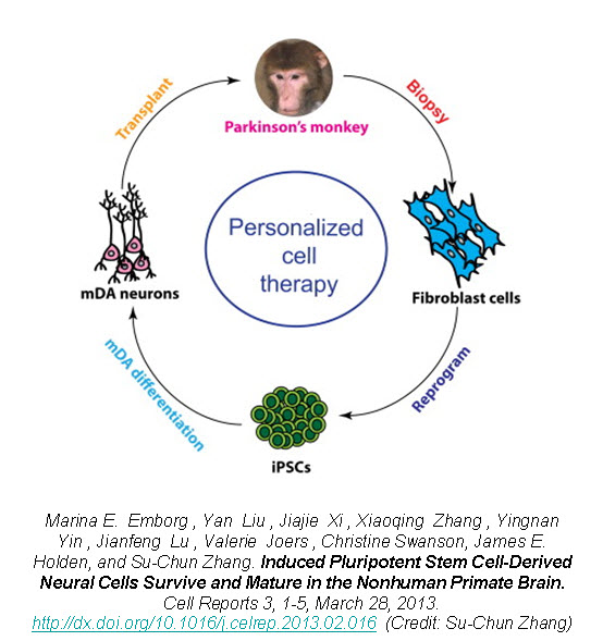
Recently, a group of researchers led by Dr. Su-Chun Zhang at the Waisman Center on the University of Wisconsin-Madison campus have shown that utilizing iPSCs as an autologous cell therapy is feasible and without any immunological reaction or rejection.  Dr. Zhang was the first to derive neural cells from embryonic stem cells (ESCs), as well as from iPSCs, and now has shown the first proof-of-principle that autologous iPSC-derived cells can engraft and survive in the primate brain.

In a new study published in Cell Reports, Zhang’s group reports on the successful generation of iPSCs from fibroblasts obtained from 8-10 year old rhesus monkeys (Macaca mulatta) using retroviruses containing the four Yamanaka factors, subsequent neuronal differentiation and cell transplantation back into the donor monkeys2.  The rhesus iPSCs were differentiated into neuroepithelia with the characteristic neural tube-like rosettes and expression of neuroectoderm transcription factors Pax6 and Sox1.  The neuronal rosettes were then expanded and further differentiated into neurons so that by the time for cell transplantation (day 42), 37% of the cells were bIII-tubulin+ neurons, 16% were S100b+ immature astrocytes, and 47% were Nestin+ progenitors.

primate ipsc cell pic
To examine the feasibility of transplanting autologous iPSC-derived neural progenitors in Parkinson’s disease, Zhang created parkinsonism in the rhesus monkeys by unilateral intracarotid artery injection of a neurotoxin 1-methyl-4-phenyl-1,2,3,6,-tetrahydropyridine (MPTP).  A year to 18 months after the MPTP infusion, all the monkeys developed a stable hemiparkinsonian condition distinguished by the characteristic tremors, bradykinesia, imbalance and impairment in motor skills.  At day 30 of cell culture the iPSC-derived neural progenitors were labeled with a GFP (green fluorescent protein) lentivirus and cells were transplanted at day 42 into the striatum and substantia nigra of the same monkey from which the iPSC were derived.

The monkey did not receive any immune suppression and engraftment of the GFP-labeled iPSC-derived neural progenitors was assessed at 6 months post-transplantation using stereological analysis on serial coronal sections.  Distinct grafts were present in the injected regions where 63% of the cells were microtubule-associated protein 2 (MAP2)+ neurons, 22% were glial fibrillary acidic protein (GFAP)+ astrocytes, and 10% were myelin basic protein (MBP)+ oligodendrocytes.  The GFP+ neurons showed long fibers extending into the surrounding host tissue and some neurons were present outside of the graft region expressing markers of mature neuronal differentiation.  Additionally, there was an absence of markers for pluripotent stem cells (OCT4, NANOG, SOX17, and Brachyury) and no positive staining for Ki67 (labels mitotic cells) indicating that the grafted progenitors had terminally differentiated.

Zhao et al. showed that the teratomas formed from transplanted iPSCs had an immunological rejection by syngenic mice1.  However, Zhang demonstrates that the autologous iPSC-derived neural cell transplants in the primate brain do not undergo immunological rejections suggested by the lack of CD3 and CD8 (lymphocyte markers) staining.  There was minimal response of endogenous glia (astrocytes and microglia) since staining for human leukocyte antigen D-related (HLA-DR; microglia and macrophage marker) was seen throughout the brain including the grafted regions.

Despite the positive engraftment and differentiation of the iPSC-derived neural progenitors, they did not see any behavioral improvement in the parkinsonian monkeys.  A potential explanation is that the GFP+ neurons were mostly g-Aminobutyric acid (GABA+) and few were positive for tyrosine hydroxylase (TH+).  TH catalyzes the formation of L-DOPA, the rate-limiting step in the biosynthesis of dopamine.  Deficiency in TH has been implicated in giving rise to parkinsonian characteristics3.  Also, the number of transplanted cells may not have been enough to replace the dopamine-making cells in the primate brain.

However, this study provides hope for cell therapy using autologous iPSC-derived cells.  The iPSC-derived neural progenitors survived and differentiated into mature neurons, astrocytes, and oligodendrocytes in the primate brain with no evidence of immune rejection or teratoma formation.  The transplanted cells structurally integrated into the host brain and with characteristic features of neurons indicating by extending long processes and features of oligodendrocytes indicated by staining for myelin basic protein, suggestive of myelination.  This proof-of-principle study of the first-ever transplant of iPSC-derived cells back into the same non-human primate presents hope for personalized regenerative medicine and the neurodegenerative patient population.

Further reading:

1. Zhao, Tongbiao; Zhang, Zhen-Ning; Rong, Zhili; and Xu, Yang.  Immunogenicity of induced pluripotent stem cells.  Nature. 474, 212–215, 9 June 2011

2. Emborg ME, Liu Y, Xi J, Chang X, Yin Y, Lu J, Joers V, Swanson C, Holden JE, and Zhang Su.  Induced pluripotent stem cell-derived neural stem cells survive and mature in the nonhuman primate brain.  Cell Reports 3, 1-5, March 28, 2013.

3. Goodwill KE, Sabatier C, Marks C, Raag R, Fitzpatrick PF, Stevens RC. Crystal Structure of tyrosine hydroxylase at 2.3a and its implications for inherited neurodegenerative diseases. Nature Structural Biology 4 (7): 578–585, 1997

Receptor Tyrosine Kinase “Hijacking” in Glioblastoma

Francis Collins, the director of the National Institutes of Health’s Human Genome Research Institute, commented in a “Brave New Pharmacy” (Time Magazine, June 2001) that a new era of drug discovery was upon us where “if you understand the genetic basis of a disease, then you can predict what protein it produces and set about developing a drug to block it.”  One such success is the development of Trastuzumab, an antibody against the extracellular domain of HER-2 (Human Epidermal Growth Factor Receptor 2 also known as ErbB-2) which was found to be over-expressed in 15-30% of breast cancers.  However, targeting other ErbBs that are found in cancer has not been successful.

ErbB1 (also know as Epidermal Growth Factor Receptor or EGFR) has also been found to be over-expressed in a variety of tumors.  EGFR is a 170,000 dalton transmembrane glycoprotein with intrinsic tyrosine kinase activity and family members include EGFR, ErbB2 (HER-2), ErbB3 and ErbB4.  The predominant ligand for EGFR is epidermal growth factor (EGF), a 53-amino acid polypeptide, as well as the EGF family members transforming growth factor a (TGF-a), amphiregulin, heparin-binding EGF, β-cellulin, neuregulin and epiregulin.  These proteins share a high binding affinity for EGFR and, upon binding to the receptor, induce EGFR dimerization, internalization and auto-phosphorylation which triggers signaling events involved in proliferation, migration, survival, and angiogenesis.  Since EGFR signaling induces numerous mitogenic effects, EGFR over-expression and/ or gain-of-function mutations (EGFRvIII) can promote oncogenic transformation.

EGFR inhibitors have been developed to treat cancers that are caused by EGFR up-regulation such as breast, colorectal, head and neck, non-small cell lung carcinoma, pancreatic renal cell, squamous cell and thyroid cancer.  EGFR inhibitors are either protein-tyrosine-kinase (PTK) inhibitors that bind to the tyrosine kinase domain or monoclonal antibodies that bind to the extracellular component of EGFR, preventing actual substrates from binding to the receptors and therefore preventing activation of EGFR.  These drugs include Iressa (Gefitinib), Tarceva (Erlotinib), Erbitux (Cetuximab), Tykerb (Lapatinib), Vectibix (Panitumumab), and Caprelsa (Vandetanib).

However, there are numerous genetic mechanisms of resistance to anti-EGFR therapy including acquisition and/ or selection for secondary EGFR mutations, additional mutations in effectors resulting in constitutive activation of signaling pathways downstream of EGFR and co-occurrence of other amplified or mutated RTKs that bypass the EGFR pathway.  EGFR mutations, which have been found in gliomas, non-small cell lung cancer, breast and ovarian cancer, have diminished response to EGFR therapy most likely due to conformational changes that affect intracellular domains involved in ATP binding sites.  These mutations may also overwhelm the contribution of other signaling pathways for cell survival, thus allowing the cancer cells to increase their dependence on the EGFR signaling pathway for survival.

In the March issue of Cancer Discovery, a team of researchers identified a unique mechanism by which glioblastomEGFR switch pic
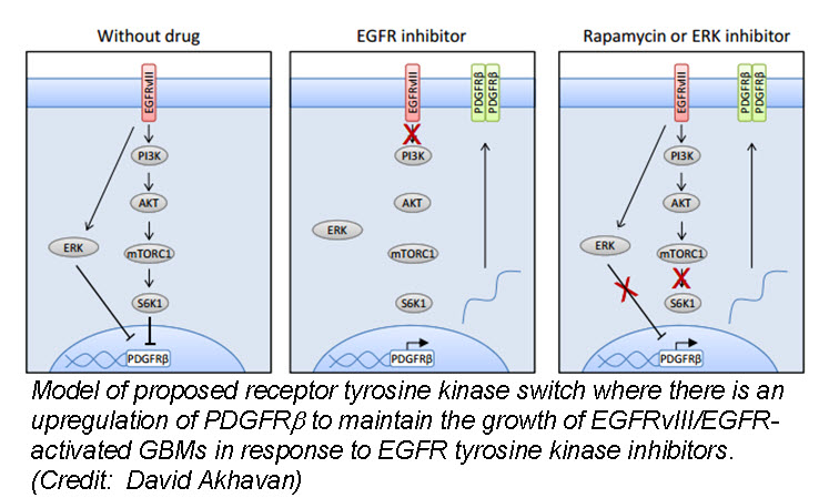
a (GBM) cells develop resistance to anti-EGFR therapy.  They demonstrate for the first time that an EGFR-dependent cancer can escape targeted therapy by developing dependence on another non-amplified, non-mutated RTK.  Specifically, they show that GBMs with EGFR mutations evade EGFR tyrosine kinase inhibitors (TKI) by transcriptionally de-repressing platelet-derived growth factor receptor β (PDGFRβ).  Cell lines, patient-derived tumor cultures, and xenotransplants showed that the persistently active EGFR mutation (EGFRvIII) suppressed PDGFRβ expression via mTORC1 and ERK-dependent mechanisms but that EGFR TKI treatment de-repressed PDGFRβ allowing the tumors to become “addicted” to a non-amplified, non-mutated RTK for continued growth and resistance to targeted treatment.

Tumor tissue from GBM patients in a phase II clinical trial for an EGFR TKI (Lapatinib) revealed a reciprocal relationship between the activation of PDGFRβ and EGFRvIII.  Tissue analysis from one patient before and after therapy revealed that Lapatinib treatment significantly reduced EGFR activation, but with a concomitant increase in PDGFRβ expression, supporting their in vitro and in vivo data that pharmacologic inhibition of EGFR results in RTK switching to PDGFRβ signaling.

We have targets and we have drugs, but RTK inhibitors have resulted in unfulfilled promises.  Acquired drug resistance has presented a significant challenge for personalized cancer therapy.  Despite being able to identify druggable RTK mutations in patients as well as second site mutations, non-genetic adaptive resistance mechanisms are able to “rewire” their circuitry through pathway crosstalk and release of inhibitory feedback loops.  To further develop kinase cancer drugs, scientists need to combine RTK inhibitors with other agents (chemotherapy, radiation, other small molecules etc.) as well as target multiple tumor-promoting signaling pathways, either with drug combinations or with a single multi-targeted compound.


Further reading:

Akhavan D, Pourzia AL, Nourian AA, Williams KJ, Nathanson D, Babic I, Villa GR, Tanaka K, Nael A, Yang H, Dang J, Vinters HV, Yong WH, Flagg M, Tamanoi F, Sasayama T, James CD, Kornblum HI, Cloughesy TF, Cavenee WK, Bensinger SJ, Mischel PS.  De-repression of PDGFRβ transcription promotes acquired resistance to EGFR tyrosine kinase inhibitors in glioblastoma patients.  Cancer Discovery. 2013 Mar 27. [Epub ahead of print]

Deric L. Wheeler, Emily F. Dunn, and Paul M. Harari.  Understanding resistance to EGFR inhibitors—impact on future treatment strategies.  Nature Reviews Clinical Oncology. 2010 September; 7(9): 493–507.

James Perry, Masahiko Okamoto, Michael Guiou, Katsuyuki Shirai, Allison Errett, and Arnab Chakravarti.  Novel Therapies in Glioblastoma.  Neurology Research International.  Volume 2012 (2012), Article ID 428565, 14 pages

Mechanical Properties of 3D Cell Culture Affect Stem Cell Phenotype

The field of regenerative medicine holds great promise as we gain greater understanding of how stem cells differentiate into the many cell types found in our bodies.  However, the clinical applications of these stem cells have been hampered by the challenges in replicating the in vitro capabilities of these cells once transplanted into the body.  This is partially due to the fact that most stem cell work is based on culturing cells in a flat two-dimensional (2D) format.

Cell culture in 2D has been routinely used in laboratories for over 40 years.  Cells are grown on flat dishes made of coated or uncoated polystyrene plastic or glass that are very stiff and arguably primitive.  These cells attach and spread on the surface to form unnatural cell attachments to other cells and to deposited proteins that are denatured on this synthetic surface.  Thus, the culturing of cells in 2D does not accurately reproduce the extracellular matrix (ECM) found in native tissue resulting in an alteration of many complex biological responses.  The native microenvironment provides mechanical signals, soluble factors, communication between neighboring cells and communication between the cell and its matrix.  This spatial and temporal organization affects normal cell fate including division, proliferation, migration, differentiation and apoptosis.

To overcome these challenges scientists have developed several three-dimensional (3D) culture methods such as cell spheroids, micro-carrier cultures, scaffolds, or tissue-engineered models.  Cell spheroids, self-assembled spherical clusters of cell colonies, are simple, reproducible and similar to physiological tissues compared to other methods involving ECM scaffolds and hydrogel systems (water-swollen polymer networks).  They are created from single culture or co-culture techniques such as hanging drop, rotating culture, non-adhesive surfaces or concave plate methods.  A similar method is the development of epithelial tissues to form polarized sheets.  However, as the size and complexity of the 3D model increases, so does the requirement for a scaffold which will ideally produce features naturally found within the ECM required for native cell function.

In this 3D cell culture environment, cells synthesize and secrete a flexible and pliable extracellular matrix in their native configuration.  Gap junctions are increased in 3D culture allowing cells to communicate with each other via exchange of ions, small molecules and electrical currents.  Surface adhesion molecules and receptors critical for cell function are also maximized.  The various effects of 3D culture versus 2D culture on differentiation, drug metabolism, expression, cell function, morphology, proliferation, viability, response to stimuli and in vivo relevance are vast and ever expanding (JUST THE FACTS: Specific effects of 3D vs. 2D cell culture).

In a recent study published April 11, 2013 online in Advanced Functional Materials, researchers at Case Western University describe how micropatterning technology influences stem cell fate decisions.  Micropatterning technology is the use of a technique to influence the network pattern of microgels aka intelligent hydrogel systems.  While this technology has recently become an important tool for spatially controlling stem cell microenvironment, very little is known about the effect of the size of the micropatterned regions, which influences hydrogel stiffness and transport properties, on stem cell behavior.  The researchers developed a 3D micropatterned hydrogel system that was either single-crosslinked or dual-crosslinked and evaluated human adipose-derived stem cell (hASC) behavior.  The cells grew into clusters in the singly-crosslinked regions where the size of the hASC clusters depended on the micropattern size (increased cell-cell interactions may have promoted cell proliferation), while hASCs encapsulated in the dual-crosslinked regions remained mostly isolated and had lower proliferation rates.  Interestingly, osteogenic (bone) and chondrogenic (cartilage) differentiation of the hASCs increased as the micropattern size increased but there was no effect on adipogenic (fat cells) differentiation.  The researchers believe that controlling local biomaterial properties may allow them to guide the formation of complex tissues.

Another study published March 24, 2013 in Nature Materials describes how mechanotransduction (how cells take information about its physical environment and t3D Traction imageranslate that into chemical signals) can influence stem cell fate.  Researchers from University of Pennsylvania showed that cell fate is regulated by cell-generated tension that is enabled through cell-mediated degradation of the covalently crosslinked matrix.  When cultured on “softer” 2D covalently crosslinked gels (RGD-modified methacrylated hyaluronic acid hydrogels), mesenchymal stem cells (MSCs) differentiated into adipocytes when cultured in bipotential adipogenic/ osteogenic media.  In contrast, MSCs cultured on “harder” 2D alginate gels differentiated into chondrocytes.  This phenomenon was not present in 3D hydrogels and was attributed to the inability of cells to degrade the covalent cross-linked bonds resulting in MSCs differentiating into adipocytes.  Introduction of proteolytically cleavable crosslinks and utilizing 3D traction force microscopy, revealed that MSC differentiation into bone cells was dependent on the cells to better anchor themselves into the environment and degradation signals.

These two studies provide insight into how the microenvironment can affect the fate of stem cells.  Understanding how the microenvironment influences stem cell behavior is important for tissue engineering approaches.  Cell-based assays have the potential to provide reliable data for regenerative medicine but scientists need to bridge the in vitro and in vivo gap by growing cells within a microenvironment that establishes physiological cell-cell and cell-substrate interactions that regulate proliferation and differentiation.  Hence, 3D models will provide more reliable and meaningful therapeutic results compared to 2D tests.


Further reading:

Haycock JW.  3D cell culture: a review of current approaches and techniques.  Methods Mol Biol. 2011; 695:1-15

Oju Jeon, Eben Alsberg.  Regulation of Stem Cell Fate in a Three-Dimensional Micropatterned Dual-Crosslinked Hydrogel System.  Advanced Functional Materials.  Article first published online: 11 April 2013

Wei Song, Naoki Kawazoe, and Guoping Chen.  Dependence of Spreading and Differentiation of Mesenchymal Stem Cells on Micropatterned Surface Area.  Journal of Nanomaterials.  Volume 2011 (2011), 9 pages

Sudhir Khetan, Murat Guvendiren, Wesley R. Legant, Daniel M. Cohen, Christopher S. Chen and Jason A. Burdick.  Degradation-mediated cellular traction directs stem cell fate in covalently crosslinked three-dimensional hydrogels. Nature Materials. Published online 24 March 2013


How Cardiosphere-derived Cells Regenerate the Injured Heart

According to the Centers for Disease Control, approximately one million heart attacks (Myocardial Infarction, or MI) occur per year in the U.S. [1].  Loss of functioning heart muscle due to an MI can result in congestive heart failure which is the leading cause of death and disability inAmerica.  It is estimated that about half of those with congestive heart failure die within five years of diagnosis [1].  New therapies are desperately needed to treat and prevent the clinical complications that follow a heart attack. Unfortunately, the heart muscles (cardiomyocytes) do not ordinarily regenerate once damage has occurred by MI and thus many researchers are looking into stem cell therapies for heart failure and disease.

The widely accepted view that the heart was not capable of regeneration was established in 1925 by Karsner et al. who demonstrated that the heart grew larger due to cardiac hypertrophy (increase in cell size) as opposed to hyperplasia (increase in cell number) [2].  However, in 2009, Bergmann et al. demonstrated that the human heart was capable of self-renewal by measuring the age of cardiomyocytes in individuals exposed to carbon-14 generated by nuclear bomb tests during the Cold War [3].  The work of Hsieh et al. showed that adult heart regeneration was due to an actual subset of progenitor cells as opposed to increased proliferation of resident cardiomyocytes [4].  This group used an α-myosin heavy chain Cre-Lox transgenic mouse model (Mer-CreMer-ZEG mouse), where 100% of the cardiomyocytes are b-galactosidase (b-gal)  positive but activation of Cre recombinase by tamoxifen results in 80% EGFP (Enhanced Green Fluorescent Protein) positive cardiomyocytes and 20% b-gal positive cardiomyocytes.  After myocardial infarction there was a 15% increase in b-gal positive cardiomyocytes indicating that cardiomyocytes were being generated from stem/ progenitor cells, which were never exposed to Cre recombinase. Thus, while there is insufficient capacity of mammalian adult heart tissue to undergo self-repair, injured myocardium can increase cardiomyocyte generation from non-myocyte precursors.

While protocols aimed to mobilize endogenous stem/ progenitor cells to sites of injury or disease would be greatly beneficial to cardiac patients, transplantation of autologous or allogeneic cells is also a promising alternative.  Cell therapies may result in phenotypic replacement of heart cells, reduce scar formation, increase survival and function of endogenous cardiac cells by increasing in vivo bioavailability of growth factors, and exert immunomodulation by releasing soluble molecules and express immune-relevant receptors (chemokine receptors and CAMs (cellular adhesion markers)).  The first clinical trial using a stem cell therapy for cardiac disease was performed over a decade ago using autologous bone marrow cells (BMCs) in patients after acute MI which reported improved heart function and myocardial perfusion [5].  Since then, numerous adult stem cell therapies targeting cardiac disease have been launched with the majority of therapies involving BMCs [6].

describe the imageRecently, a phase I clinical trial based on administration of autologous cardiosphere-derived cells (CDCs) to assess safety in patients with left ventricular dysfunction after MI was completed by Dr. Eduardo Marban atCedars-SinaiMedicalCenter[7].  The formation of cardiospheres was first described by Messina’s group who showed that human and mouse heart explants generated a layer of fibroblast-like cells over which small, phase-bright cells migrated, and once these phase-bright cells are transferred to non-adherent plates, they generate three-dimensional spherical structures [3].  Marban’s group modified Messina’s protocol by placing the cardiospheres in adherent plates where the cells begin to grow in monolayer, hence cardiosphere-derived cells, allowing for easier and faster expansion.  Promising preclinical data which showed a reduction in infarct size and improved cardiac function after transplantation of CDCs in a porcine animal model prompted the phase I clinical trial [8].

This trial, known as CADUCEUS (CArdiosphere-Derived aUtologous stem CElls to reverse ventricUlar dySfunction), enrolled patients 2-4 weeks after MI (with left ventricular ejection of 25-45%) who received either standard of care (no intervention, usual medical management after MI), low cell dose (12.5 million) or high cell dose (25 million) of autologous CDCs (ClinicalTrials.gov NCT00893360).  Percutaneous endomyocardial biopsies were used to obtain the heart tissue necessary for generation of the predetermined dose of autologous CDCs (usually within 36 days of sampling) and cells were delivered through an angioplasty catheter in the infarct-related artery. Results from the trial showed that the CDCs were relatively safe: no patients died, developed cardiac tumors or experienced a major adverse cardiac event (MACE).  Preliminary efficacy suggested that patients treated with CDCs had reductions in scar mass and increases in viable heart mass as measured by magnetic resonance imaging (MRI), as well as increases in regional contractility and systolic wall thickening at 12 months compared to controls [7].  However, the trial did not confirm whether there was true increased myocardial mass, whether the CDCs themselves could possibly distort the myocardial architecture and/or how the CDCs potentially could regenerate the injured heart.

Recently Marban’s group has shed light on the potential mechanism of how the CDC therapy reduces scarring and regenerates healthy tissue after an MI, published in EMBO Molecular Medicine [9].  They applied lineage tracing systems using the Mer-CreMer-ZEG mouse with additional labeling tools to investigate the cellular origins of regeneration in an adult mouse after surgical induction of MI followed by intramyocardial injection of mouse CDCs.  Impressive results revealed that after MI new cardiomyocytes arise from both pre-existing cardiomyocytes and undifferentiated progenitors and that transplanted CDCs further up-regulate host cardiomyocyte proliferation and recruitment of endogenous progenitors to the infarct site.  The increase in cardiomyocyte proliferation and progenitor recruitment was accompanied by structural and functional changes in the infracted heart, specifically decreased scar size, increased infracted wall thickness and myocardium (cardiomyocyte hypertrophy was excluded), and increased cardiac function.  Interestingly, these effects occurred despite robust CDC engraftment indicating that CDCs do not contribute to phenotypic replacement but promote heart regeneration by indirect means.  These promising findings indicate that stem cell therapies may stimulate dormant surviving cells after injury and boost natural repair mechanisms.

However, there still remain many challenges for developing cell therapies for cardiac disease.  First, the technology is still immature.  With autologous approaches, patient-to-patient variability can arise such as purity and potency of the cells.  Harvesting the source material, obtaining a sufficient quantity of cells, delivery of the end product and the optimal site of delivery will need to be established.  In the future, Marban’s group plans on using allogeneic CDCs obtained from donor organs, which allows for an off-the-shelf-therapy, to treat patients following MI.  A thorough cell biological characterization of the CDCs will be required to understand the molecular identity and mechanism(s) of action of these cells.  Second, the underlying mechanism(s) of cardiac repair and/ or regeneration after MI remain elusive.  Additionally, treating chronic or congestive heart failure with the aim of generating new contracting heart muscle is more complex.  Third, it is difficult to make conclusive statements about the results obtained from many of the clinical trials since they have small patient size and therefore limited statistical power.  It is unclear how the findings will generalize to a larger population of patients.  Fourth, most of the clinical trials use surrogate endpoints which are measures of an effect of a certain treatment expected to predict clinical benefit (or harm, or lack of benefit or harm) such as measuring cardiac function.  Even though surrogate endpoints may correlate with a real clinical endpoint, they do not have a guaranteed relationship.  Therefore, clinically meaningful endpoints such as improvement in overall survival and prevention of future heart failure are needed.  In the CADUCEUS trial there was only enhanced regional structure/ function but no significant effect on global functional endpoints such as ejection fraction.  A phase II double-blinded placebo-controlled clinical trial will need to be performed to assess true efficacy.  It will be several more years before we have a clear understanding of the true potential of cell therapy in cardiac disease.  In the meantime, studies to understand the cellular sources and underlying cellular mechanisms involved in cardiac regeneration are still needed.


1.         CDC. Heart Disease Facts.  updated October 16, 2012; Available from: http://www.cdc.gov/heartdisease/facts.htm.

2.         Karsner, H.T., O. Saphir, and T.W. Todd, The State of the Cardiac Muscle in Hypertrophy and Atrophy. Am J Pathol, 1925. 1(4): p. 351-372 1.

3.         Carvalho, A.B., B.K. Fleischmann, and A.C. Campos de Carvalho, Cardiac Stem Cells, in Resident Stem Cells and Regenerative Therapy, R.C.d.S. Goldenberg, Editor. 2012, Academic Press: Waltham, MA. p. 141-155.

4.         Hsieh, P.C., et al., Evidence from a genetic fate-mapping study that stem cells refresh adult mammalian cardiomyocytes after injury. Nat Med, 2007. 13(8): p. 970-4.

5.         Strauer, B.E., et al., Repair of infarcted myocardium by autologous intracoronary mononuclear bone marrow cell transplantation in humans. Circulation, 2002. 106(15): p. 1913-8.

6.         Sheridan, C., Cardiac stem cell therapies inch toward clinical litmus test. Nat Biotechnol, 2013. 31(1): p. 5-6.

7.         Makkar, R.R., et al., Intracoronary cardiosphere-derived cells for heart regeneration after myocardial infarction (CADUCEUS): a prospective, randomised phase 1 trial. Lancet, 2012. 379(9819): p. 895-904.

8.         Johnston, P.V., et al., Engraftment, differentiation, and functional benefits of autologous cardiosphere-derived cells in porcine ischemic cardiomyopathy. Circulation, 2009. 120(12): p. 1075-83, 7 p following 1083.

9.         Malliaras, K., et al., Cardiomyocyte proliferation and progenitor cell recruitment underlie therapeutic regeneration after myocardial infarction in the adult mouse heart. EMBO Mol Med, 2013. 5(2): p. 191-209.