New Findings in Cell Based Therapy for GBM

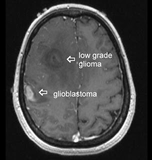
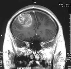
Glioblastoma multiforme (GBM) is the most common and lethalGlioblastoma type of malignant primary brain tumors that account for over 70% of all intracranial cancers.  The current course of GBM treatment consists of surgical resection of the main tumor mass, followed by administration of radiation and chemotherapy. Surgical resection of the primary tumor leads to injury to the surrounding normal tissue, while chemotherapy and radiotherapy cause toxicity to the healthy tissue in the brain.  These undesirable secondary effects of glioma treatments have a major impact on patients’ physical, cognitive and emotional functioning. Nonetheless, despite this aggressive treatment regimen and its harmful side effects, GBM remains virtually incurable, with post-diagnosis median survival persisting less than 14 months.  This dismal prognosis is due to a combination of unique anatomical features of the central nervous system (CNS), in addition to GBMs’ (glioma cells’) exceptional invasive capacity. Glioma cells infiltrate the brain’s highly dense parenchyma, migrating along the corpus callosum and creating new masses within the hemisphere contralateral to the initial tumor mass homing.

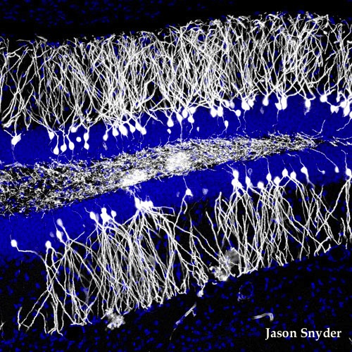
Thus, recurrence in postoperative GBM patients is in essence inevitable. Furthermore, GBMs are not only heterogeneous among individual patients, but they are also highly heterogeneous within a single tumor mass. Recent studies have shown compelling evidence of a therapeutically resistant subpopulation of malignant glioma cells that exhibit stem-cell like characteristics, such as multipotency, the ability to self-renew and invade/migrate; these tumor-initiating cells are referred to as glioma stem cells (GSCs) and are believed to be responsible for tumor initiation and recurrence in GBM patients. Furthermore, GSCs have been observed to ensconce within similar niches to neural stem cells (NSCs).

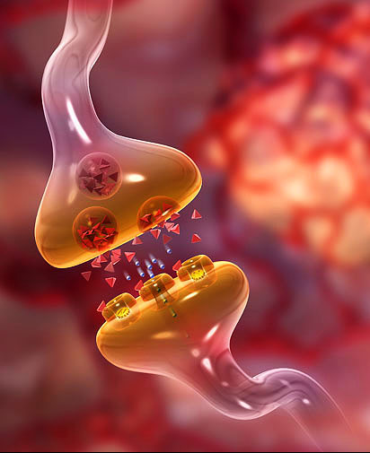
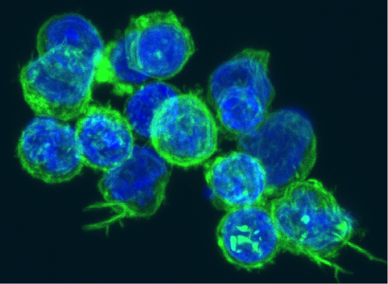
NSCs and mesenchymal stem cells (MSCs) have been shown to have an exceptional migratory ability within brain’s parenchyma and possess a notable inherent tumor tropism.  Thus, several cell-based therapy (CBT) studies and clinical models of malignant tumors have incorporated autonomous tracking of tumor cells by employing NSCs and MSCs to deliver multiple therapeutic genes to specific tumor loci. This target specific drug-delivery model has NSCs or MSCs transduced to express one pro-drug-activating enzyme, which catalyzes the conversion of a particular pro-drug into an active toxic agent, which results in the localization of the chemotherapeutics specifically at the tumor sites.  In addition to its ability to deliver effective cytotoxic damage to the tumor without causing damage to the healthy surrounding tissue, the resulting bystander effect of this system causes cell death not only to the drug-delivery-vehicle cells, but also to the surrounding glioma cells. Although a number of different enzyme/prodrug systems have been utilized in relevant studies, HSV-thymidine kinase (HSV-tk)/ deoxyguanosine analog ganciclovir (GCV) has been the most commonly tested in animal and in vitro models. HSV-tk phosphorylates GCV and produces deoxyguanosine triphosphate which is a polymerase-I inhibitor and DNA chain terminator; cell death occurs upon incorporation of the nucleotide analog within DNA chains.

In recent study published in the journal of Molecular Therapy, Blanco’s group report their findings regarding the interaction between human MSCs (hMSCs) and gliomas and the underlying mechanism for the effectiveness of hAMSC based therapy in GBMs.  In their previous work Blanco’s group showed that the administration of hAMSCs expressing HSV thymidine kinase in glioma tumors significantly promotes tumor growth, whereas induction of cytotoxicity by administration of the prodrug GCV demonstrated a significant antitumor response.

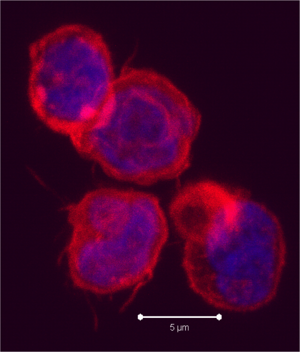
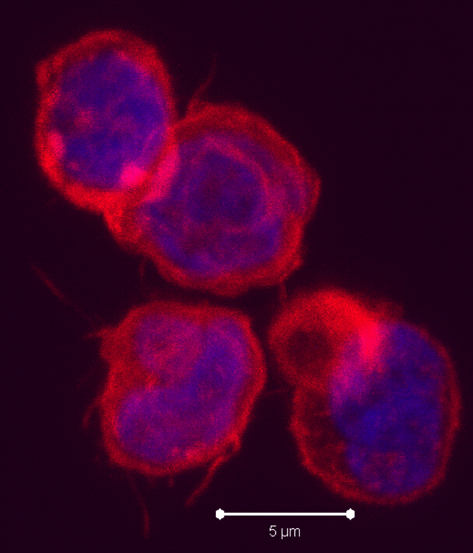
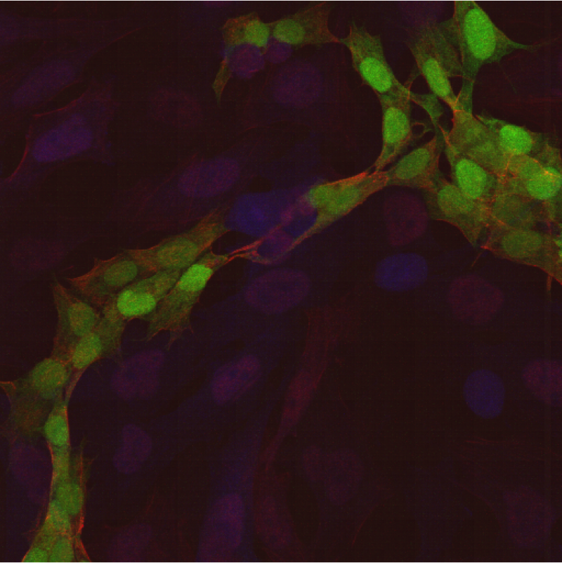
According to this new study, hMSCs differentiate to endothelial lineage (supported by the expression of CD31 marker) within tumors, and integrate in the tumor vascular system where they adopt an endothelial phenotype. Further, Blanco proposed the notion of hMSCs’ ability to home to privileged vascular structures where GSCs also reside, is the underlying characteristic that leads to the effectiveness of cytotoxic hMSCs in regulating the bystander killing of tumor cells.

Although Blanco’s study provides invaluable insights into the GSCs’ niche and its role in malignant brain tumor CBT, hMSCs’ tumor growth promotion is a quality, which makes these cells less than ideal for utilization in human clinical trials in the near future. Future studies on this mechanism using primary patient tumor cells rather than the U87 glioma cell line and human NSCs are needed to further confirm these observations.

Further reading:

Juli R. Bagó, Maria Alieva, Carolina Soler, Núria Rubio, Jerónimo Blanco. Endothelial differentiation of adipose tissue-derived mesenchymal stromal cells in glioma tumors: implications for cell based therapy. Molecular Therapy.

Phase 1 Trial: Tolerance To MS Autoantigens Using Peptide-Coupled PBMCs

Multiple sclerosis (MS) is a degenerative inflammatory disease of the brain and the spinal cord, with its onset of symptoms occurring between the ages of 20 and 40. MS is categorized into two major forms: the most common form which accounts for 85%–90% of MS cases is relapsing-remitting MS (RRMS) whose victims usually develop secondary progressive MS (SPMS) over time. The second category, termed primary progressive MS (PPMS), accounts for approximately 10%–15% of MS cases that present with disability from the onset of the disease, progressing steadily with very little to no remissions in symptoms. It is not clear which factors are responsible for differentiating these different courses. In fact, up to date, there is little known about the underlying factors responsible for the complex heterogeneity, such as variation in immune abnormalities, observed among MS patients.Multiple sclerosis T cells

Although the etiology of MS remains unclear, it is predominantly considered to be driven by CD4+ T-cells autoreactivity to self-antigens expressed in the central nervous system (CNS), particularly to the myelin antigens. Three myelin sheath proteins that have been recognized as key autoantigens in MS include myelin basic protein (MBP), myelin oligodendrocyte protein (MOG), and proteolipid protein (PLP). Previous studies suggest epitope spreading may occur during the immune response to these three antigens in relapsing-remitting MS models. This notion is further supported by the existence of different target myelin epitopes in MS patients, which may be indicative of changes in the specificity of T-cell pathogenic response over time. These observations suggest the involvement of epitope spreading in MS, while providing a viable cause for the unfavorable efficacy reported from the several MS clinical trials that utilized a single antigen/peptidic-epitope in their therapeutic approach. In other words, previous clinical trials targeted pathogenic T-cells that are reactive against a single target antigen/epitope, which do not take into account the change of specificity in the pathogenic response overtime.

Antigen-coupled cell tolerance is a therapeutic approach aimed at antigen-specific T-cell tolerance via coupling target peptide(s) to carrier agents. In a recent study published in Science Translational Medicine, Lutterotti’s group report promising outcomes from their first-in-man MS clinical trial, demonstrating antigen-specific tolerance by autologous myelin peptide–coupled cells that utilizes a single infusion of autologous peripheral blood mononuclear cells (PBMCs) as the carrier cells. Seven myelin peptides which are believed to be key targets of autoreactive CD4+ T-cells in MS peptides (MOG1–20, MOG35–55, MBP13–32, MBP83–99, MBP111–129, MBP146–170, and PLP139–154), were chemically bound to the surface of patient-isolated PBMCs in the presence of the chemical cross-linker 1-ethyl-3-(3-dimethylaminopropyl)-carbodiimide (EDC), followed by reinfusion back to the patient. Here, Lutterotti’s group validated the safety and feasibility of this antigen-coupled cell tolerization therapeutic approach in nine MS patients. Furthermore, they reported promising tolerability resulting from their approach, since patients’ immune autoreactivity to myelin peptides were reduced by 50 to 75 percent.  While all nine patients in this study displayed T-cell reactivity to at least one of the seven targeted myelin-peptides, seven were RRMS patients and two were SPMS patients.


describe the imageThese results support the epitope-spreading hypothesis, which indicates that MS patients make antibodies against one or a few myelin proteins, but as the disease progresses, the autoimmune response spreads to other myelin sheath epitopes. In their recent publication, Lutterotti et al. provide sufficient evidence necessitating emphasis not only on the specific target antigens, but also on the facility to inhibit epitope spreading, preferably prior to diversification of the CD4+ T-helper cell autoreactivity.


Lutterotti’s study is a significant step towards finding an effective strategy not only for treatment of MS, but also other T-cell-driven autoimmune disorders. Nonetheless, this therapeutic method must be tested on a much larger sample and geographically distinct population to demonstrate whether the efficacy reported here would be consistent in most if not all MS subtypes. The phase II of this clinical trial is planned to take place in the near future, during which more will be known about the long-term safety and efficacy of this technique. Regardless, the data presented by this study, at the very least, have set the requirement for future antigen-specific therapies to include the ability to target not only the previously activated autoreactive T-cells, but also the naïve autoreactive T-cells specific for several myelin epitopes.

Further Reading:

Antigen-Specific Tolerance by Autologous Myelin Peptide–Coupled Cells: A Phase 1 Trial in Multiple Sclerosis

Potential of BDNF in Treating Neurodegenera­tive Disorders

Neurodegenerative diseases are characterized by usually fatal and progressive nervous system dysfunction caused by the death of neurons in the brain and spinal cord. In terms of human suffering and economic cost, neurodegenerative disorders carry an immense disease burden. However, despite extensive clinical research, especially in developing disease-modifying therapeutics, there is no effective medicine that halts or even slows any neurodegenerative disease. Currently in the United States, over 5 million Americans suffer from Alzheimer’s disease (AD), 1 million from Parkinson’s (PD), 400,000 from multiple sclerosis (MS), 30,000 from amyotrophic lateral sclerosis (ALS), and 30,000 from Huntington’s disease (HD). Thus, modification of current therapeutic research strategies and a more aggressive approach is a goal of increasing urgency.

clinical trials,alzheimer's,parkinson's,huntington's,clinical research,phase III

Thus far, the majority of clinical research for treatment of neurodegenera­tive diseases has utilized disease-modifying therapeutics, which either prevent or target elimination of the pathogenetic causes or neurotoxins resulting from the disease. The basis for this approach revolves around several characteristics implicated in neurodegenerative diseases, such as accumulation of neurotoxic substances, autophagy and inflamma­tion, as well as aggregation of misfolded proteins in neurodegenerative disorders, such as amyloid-β (Aβ) aggregates in AD or the mutant Huntington protein in HD, which take place prior to neuronal death. However, data obtained from several Phase III clinical trials indicate low efficacy of these treatments, specially in advanced stages of most neurodegenera­tive disorders; this is mainly due to the poor understanding of the underlying mechanisms of these disorders, hence the lack of knowledge of whether the targeted disease-characteristics are the cause or a symptom of the disease. Furthermore, the inability in early and accurate diagnosis of most neurodegenerative disorders impedes the early evaluation of therapeutic efficacy of new therapeutics.

Although the underlying cellular processes contributing to HD, PD and AD differ, one common denominator in all these neurodegenerative diseases is the presence of inadequate neuronal communication, induced by the loss of synapses. Neuronal communication is carried out via synaptic transmission at neuronal synapses. A change in the properties of synaptic transmission due to brain’s ability to dynamically reorganize itself by forming new neuronal synapses is referred to as synaptic plasticity; compensation for injury as well as adjustment of neural activity in response to new stimuli or changes in their environment are among the most critical known functions of synaptic plasticity. Thus, degeneration of synapses leads to the loss of synaptic plasticity, preventing neuronal stimulation and eventual cell death.

Alzheimer's Disease,Parkinson's,Huntington's,BDNF

According to a recent Review article published by scientists from GlaxoSmithKline in the advanced online edition of the Nature Reviews Neuroscience, Lu and colleagues present a compelling notion in treatment of neurodegenerative disorders; they propose a synaptic repair strategy targeting pathophysiology, which directly underlies the clinical syndromes. Unlike neuronal loss, synapse loss is reversible and synaptic dysfunction has the ability to be repaired, which allows the potential of neuronal repair prior to neuronal death. Furthermore, synaptic repair approach can be utilized for any neurological disease, regardless of the type or origin of the toxic byproduct. Lu’s group has proposed utilization of the synaptogenic molecule brain-derived neurotrophic factor (BDNF) as a potential synaptic repair therapeutic agent.

BDNF, an abundant neurotrophin expressed throughout the central nervous system, binds to NTRK2/TRKB and has been identified as one of the key neural signals regulating neuronal survival, neurogenesis and the only neurotrophin factor associated with synaptic plasticity in humans. Furthermore, in addition to its neuroprotective attribute, previous studies have demonstrated the key role of BDNF in cognitive functions, and synaptic deficit repair despite the presence of accumulated toxic proteins.

In this review, Lu’s team suggest potential routes of activating the BDNF pathway, as well as the importance of developing of a more reliable technique for quantification of synaptic changes in clinical settings, as essential tools in building effective disease-modifying therapies for neurodegenerative disorders. Although the notion of synaptic repair is an attractive one, the utilization of this strategy is still not feasible for the late stages of the disease, in which the irreversible neuronal death has already occurred. Furthermore, since most neurodegenerative diseases are diagnosed after the onset of neuronal death, emphasis on late stage treatment must remain a priority in neurodegenerative clinical research.


Further Reading:

BDNF-based synaptic repair as a disease-modifying strategy for neurodegenerative diseases

New in MS Research: Interplay Between IFN-beta, B-cells and Monocytes

Multiple sclerosis (MS) is a chronic autoimmune inflammatory disease of the central nervous system (CNS), characterized by the presence of scar tissues (plaques) localized within the brain’s white matter and spinal cord. These plaques are results of myelin-degeneration (demyelination) and axonal death. Although MS has been classically considered to be a T-cell-mediated disease, the high efficacy of B cell-depleting therapies have demonstrated the critical role of B-lymphocytes and the humoral immune response in MS pathogenesis, albeit the underlying mechanisms remain unclear. In approximately 90% of MS patients, there is increased levels of intrathecally synthesized IgG in the MS-plaques as well as Cerebral Spinal Fluid (CSF), which manifests B-cell clonal expansions within the CNS.

B-cell-lineage cells differentiate into antibody-secreting plasma cells that are the source of persistent IgG, in the presence of key factors such as interleukin-6 (IL-6), B-cell-activating factor of the TNF family (BAFF) and a proliferation-inducing ligand (APRIL). IL-6 promotes terminal differentiation of B cells to plasma cells and is essential for the survival and Ig secretion. In conjunction with APRIL, BAFF regulates, B-cell survival, differentiation and is essential for initiation of T-cell independent B-cell responses. 

B cell resized 600

Type I IFNs (IFN-α, IFN-β, IFN-κ, and IFN-ω) are cytokines expressed by many cell types in response to viral or microbial infections, which bind to- and trigger specific Toll-like receptors (TLRs) that induce a large number of genes modulating and linking the innate and the adaptive immune responses.

Despite the development of other new treatments, IFN-β has been the first-line disease-modifying drug treatment for patients with relapsing-remitting multiple sclerosis (RRMS). Thus, understanding the molecular mechanisms of the anti-inflammatory effect of IFN-β in RRMS may provide insight into MS pathogenesis.  

TLRs are a family of non-catalytic pattern recognition receptors that recognize and bind to specific molecular patterns of pathogen-derived and endogenous damage-associated components. In addition to their key role in mediating innate immunity, TLRs have also been shown to play an important part in the activation of the adaptive immune system by inducing proinflammatory cytokines such as TNF-α, IL-1, IL-6, IL-12, and IFN.

Several studies have shown that of the 11 TLRs identified in humans, endosomal TLRs 7, 8 and 9 which recognize pathogen-derived and synthetic nucleic acids, also recognize endogenous immune complexes containing self-nucleic acids in certain autoimmune disorders such as MS. Interestingly, B-cells express both TLR7 and TLR9. TLR7 recognizes guanosine- and uridine-rich single-stranded RNAs (ssRNAs), whereas TLR9 recognizes hypomethylated CpG-rich double-stranded DNAs.  Upon activation by their specific ligands, these TLRs induce B cell proliferation and differentiation into Ig-secreting cells.

describe the image

In a recent study published in the European journal of Immunology, Coccia’s group has demonstrated the essential interactions between monocytes and B cells for the release of effective humoral immune response that elicits TLR7-mediated -induced B-cell differentiation into Ig secreting cells. Furthermore, they have shown a clear deficiency in this cross-talk interaction in MS patients; the peripheral blood mononuclear cell (PBMC) of MS patients exhibit substantially lowered TLR7-induced Ig production (compared to Healthy donors). However, results obtained after one-month long IFN-β therapy showed that lower humoral immune response in MS subjects was replenished, through IFN-β–induced secretion of TLR7- triggering cytokines, which mediated the selective increase in IgM and IgG to levels comparable to Healthy donors’. This data revealed that the IFN-β enhancement of TLR7-induced B-cell responses in MS patients occurs in at least two steps: 1) Regulation of TLR7 gene expression, and 2) Secretion of B-cell differentiation factors, in particular IL-6 and BAFF.

Finally, the last and perhaps the most significant finding of Coccia’s new study, is reporting, for the first time, the presence of a defect in TLR7 gene expression and signaling in monocytes of MS patients. Lack of TLR7-driven IgM and IgG production, absence of IL-6 and a significant reduction in BAFF expression in samples of MS patient-IFN-β treated PBMCs that were depleted of monocytes, evince IFN-β therapeutic mechanism by fine-tuning monocyte functions, through stimulation of TLR7 which subsequently effects B cell differentiation.

The discovery of the tight regulation of both TLR expression and TLR-induced responses in maintenance of immune environment’s homeostasis, as well as IFN-β-mediated- TLR7 function recovery are indicative of the critical changes in PBMC microenvironment induced by IFN-β therapy; within this microenvironment, leukocyte subsets establish critical immune regulatory interactions which determine the fate of the host’s immune tolerance processes.

Coccia’s new study has revealed new insights, which are not only crucial for the better understanding of the MS immunopathology, but also significant for development of new MS therapeutic strategies which target TLR expression and/or TLR-induced responses.


Further Reading:

IFN-β therapy modulates B-cell and monocyte crosstalk via TLR7 in multiple sclerosis patients.

Promising advances in Recurrent GBM treatment

Glioblastoma multiforme (GBM) are malignant brain tumors, classified by the World Health Organization (WHO), as grade IV tumors of neuroepithelial tissue and are the most common and deadly intracranial tumors, accounting for more than 70% of all brain tumors.  The current course of GBM treatment entails surgical resection followed by administration of radiation and chemotherapy.  However, despite this aggressive regimen and their devastating side effects on the patient, there are several obstacles that hinder their effectiveness; surgical resection of the primary tumor leads to injury to the surrounding normal tissue, while chemotherapy and radiotherapy cause toxicity to the healthy tissue in the brain. Furthermore, some anatomical feature unique to the central nervous system (CNS) such as the blood brain barrier (BBB) and the densely packed structure of brain’s parenchyma inhibits effective drug delivery throughout the brain.

GBM,glioma,FDA,brain tumors

Despite improvements in surgical techniques and post-operational delivery of chemotherapeutics and radiation, the prognosis for GBM patients remains dismal, making these lethal tumors virtually incurable. One of the most prominent intrinsic behaviors of GBM is its invasiveness, such that single glioma cells travel a distance from the tumor mass and invade adjacent brain tissue.  Due to the inherent capability for malignant glioma cells to rapidly proliferate and metastasize away from the primary tumor mass, surgical resection of the tumor is almost always followed by tumor recurrence (rGBM) with foci located as close as 1 centimeter from the resection cavity or as far as the opposite hemisphere. Although the history of glioma treatment dates back to the 19th century, the median survival of patients remains less than 14 months post-diagnosis.

Approximately two-third of rGBM patients are incapable of enduring additional surgical resections. Thus, for over 2 decades, Laser interstitial thermal therapy (LITT) has been used for extirpation of several malignant tumors, especially for patients with rGBM. LITT refers to utilization of low-powered thermal energy to locally cytoreduce the tumor tissue through transdermal thermocoagulation. Nonetheless, a number of technical barriers of the LITT device have impeded its widespread efficacy; two of these major barriers include lack of real-time monitoring of the tissue during treatment, as well as the control of the LITT energy (wavelength) output to correspond with the real time status of the brain tissue (healthy vs. tumor).

MRI,GBM,rGBM,brain tumors

In a recent study published in the Journal of Neurosurgery, Andrew Sloan’s group (Monteris® Medical) reported their results from the First-In-Man (FIM) Phase I clinical trial of their NeuroBlate™ Thermal Therapy System (also know as AutoLITT), which consists laser treatment of recurrent/progressive brain tumors in combination with intraoperative magnetic resonance imaging (MRI) technology. This system overcomes some of the major obstacles to LITT; without the use of radiation, it employs high-resolution MRI images of the brain in real time, which allows surgeons to visualize the progress of tumor ablation at all times, thus directing and controlling the laser deposition to increase treatment efficacy and minimizing harm to the surrounding healthy tissue.

The FDA has designated NeuroBlate™ Thermal Therapy System as safe, since no severe clinical toxicity or procedure-related neurological deficits were caused by this system. This Safety Trial was conducted on 10 patients (median age 55) with recurrent or progressive GBM in whom standard radiotherapy with or without chemotherapy had failed. Based on this study, not only NeuroBlate treatment did not cause any adverse side effects in the subjects, but it also resulted in higher response and survival times than expected.

The technological advancements of NeuroBlate™ Thermal Therapy System which allow a minimally-invasive, and possibly safer method of performing resections, not only for GBM, but perhaps also utilized for the future surgical resection of other cancers. Nonetheless, further investigation is needed to optimize NeuroBlate™ Thermal Therapy System in increasing the mean percentage of treated tumor at the intent-to-treat dose, and determine the long-term efficacy of this system compared with traditional surgical resection.

Further Reading:

Results of the NeuroBlate System first-in-humans Phase I clinical trial for recurrent glioblastoma

Does Salt Contribute to Autoimmunity?

Several recent findings have identified variants in predominantly immune-response genes as underlying contributors in autoimmune diseases’ development. Nonetheless, along with other autoimmune disorders, there has been a significant increase particularly in incidences of multiple sclerosis (MS) and type-1 diabetes. These observations imply that in addition to the genetic causality, specific changes in environmental factors are significant contributors to the recent increases in autoimmune disorders.

Maintenance of immune system’s homeostasis is dependent on highly specified regulation of pro-inflammatory and regulatory CD4+ helper T-cell populations. TH17 cells are pathogenic pro-inflammatory T-cells that produce a proinflammatory cytokine, Interleukin 17A (IL-17) and while they play a major role in immune defense against extracellular pathogens, they also play a major role in induction of autoimmune disorders, such as MS, psoriasis, rheumatoid arthritis, and type-1 diabetes, for which T-cell modulation is a common treatment regime.

CD4 T-cells,TH17,MS,autoimmune

Three previously identified factors, Interleukin 6 (IL-6), Transforming growth factor β (TGFβ) and Interleukin-23 subunit alpha (IL-23) are key players in the induction of naïve CD4+ T-cells to differentiate into TH17 cells. However, the complete mechanism and factors responsible for stimulating naïve CD4+ T-cells’ differentiation into TH17 cells is not well understood. Understanding of these underlying factors is crucial for developing therapeutic strategies to control TH17 cell differentiation.

Three complementary, collaborative studies published in Nature this week report compelling insights on the regulatory mechanism of TH17 cell differentiation and subsequent inflammatory response, while revealing evidence of interplay of genetics and a new environmental factor involved in autoimmune disease susceptibility.

By utilizing a unique approach, consisting of combination of transcriptional profiling, novel computational methods and nanowire-based short interfering RNA (siRNA) delivery to construct, Regev’s group was able to and identify 12 novel regulators and decipher the complex transcriptional network of TH17 differentiation; they found that TH17 differentiation is regulated by two intra-connected, but antagonistic networks, such that one module promotes TH17 differentiation and proliferation while suppressing the development of other T-cells, whereas the antagonist module suppresses TH17 cells.

By upregulating the expression of IL-23 receptor (IL-23R) on TH17 cells, IL-23 sustains the TH17 inflammatory response and induces its pathogenic effector functions. Through transcriptional profiling, Kuchroo’s group found that a serine/threonine kinase,  Serine/threonine-protein kinase 1 (SGK1), an essential node downstream of IL-23 signaling, is crucial for regulating IL-23R expression and stabilizing the TH17 cell pathogenic effector function by inhibiting the molecule that represses IL-23R expression.

Previous studies have shown that SGK1 regulates Na+ transport and NaCl (salt) homeostasis in cells. Interestingly, Kuchroo’s study shows the positive correlation between increased concentration in salt and SGK1 expression, followed by upregulation of IL-23R and enhanced TH17 differentiation in vitro as well as in vivo.

Consistent with Kuchroo’s finding, Hafler’s group reported that excess NaCl uptake can affect the innate immune system, and provided compelling data suggesting the possibility of the direct positive correlation between increased salt intake and incidence of autoimmune disease.

Previous studies have suggested the involvement of environmental factors, such as viruses, smoking, lack of sunlight and Vitamin D in different autoimmune disorders.  Salt, as the new potential environmental factor in autoimmune diseases is certainly an intriguing notion; not only due to the relevant molecular mechanisms reported in the three new studies, but also due to the fact that in most developed countries, the consumption of processed foods (such as fast foods), which contain over 100 times more NaCl compared to non-processed meals, has increased significantly over the past three decades. Nonetheless, future epidemiological studies in humans are needed to further investigate the possible correlation between salt consumption and autoimmunity incidence.


Further Readings:

Induction of Pathogenic TH17 Cells by Inducible Salt-sensing Kinase SGK1

Dynamic Regulatory Network Controlling TH17 Cell Differentiation

Sodium Chloride Drives Autoimmune Disease by The Induction Of Pathogenic TH17 Cells

New Clues for Chemotherapy-Induced Cognitive Impairment

Cancer is the formation of a malignant neoplasm, initiated by a cell that escaped apoptosis upon which mutations during DNA replication were not repaired; this cell, no longer regulated, continuously proliferates while each progeny will carry the previous mutation(s), while generating new ones, eventually resulting in a population of unregulated cells (malignant tumors) that will metastasize and take over the host’s body.  Thus, the conventional cancer treatment consists of surgical resection of the tumor mass, followed by administration of agents which kill any diving (mitotic) cells in the body (chemotherapy and radiation therapy). Consequently, these agents will not only target proliferating cancer cells, but they also destroy healthy proliferating cells, hence the side effects associated with chemotherapy, such as hair loss, nausea and cognitive deficits. This article emphasizes on chemotherapy’s role in disrupting processing speed, working memory and attention in humans.

Adult neurogenesis is the post-natal process of generating functional neurons (and glial cells) from adult neural precursors/progenitor cells (NPCs) throughout life. The two regions in the adult brain in which neurogenesis occurs are the subventricular zone (SVZ) of the lateral ventricle and the subgranular zone (SGZ) of the dentate gyrus (DG) in the hippocampus. Newly generated cells in the SGZ can differentiate into functional neurons and integrate into the adult hippocampus’s DG as granule cells. Granule cells are involved in memory formation and many aspects of learning, with the exception of long-term memory storage.

Neurogenesis,chemotherapy,cancer,memory loss

Over 50% of cancer patients undergoing chemotherapy report significant cognitive impairment and declines in their overall cognitive processing, collectively referred to as “chemo-brain”. Thus, chemotherapy-induced loss of newly generated neurons in the hippocampus and impeding adult neurogenesis as the cause of such cognitive decline is a compelling notion. Furthermore, one of the highly “cognitive” oscillations in the human brain is the theta rhythm, which is mainly generated in the hippocampus and is also associated with processes of learning and memory. This rhythmic slow activity is also the most efficient synchronized electroencephalographic (EEG) activity that can be recorded from the brain.  It has been suggested that since synchronized oscillatory activity implements communication between functionally related structures during the process of learning, a chemotherapy-induced disruption in theta activity may obstruct inter-regional communication and result in learning deficits.

In a recent study published in the European Journal of Neuroscience, Shors’s group reported that prolonged systemic chemotherapy disrupts both the structural and functional integrity of the hippocampus, resulting in highly specific learning impairments. Their results show that chemotherapeutic agents instigate the learning deficits described in ‘chemo-brain’ via decreases in hippocampal adult neurogenesis and theta activity. Interestingly however, they are not responsible for the disruption of the hippocampus-independent memory for previously (pre-treatment) learned associations. In this study, the effects of chemotherapy on hippocampal adult neurogenesis, theta activity and learning were investigated through evaluating associative learning in adult male Sprague–Dawley rats by recording the hippocampal local-field potentials after several weeks of cyclic administration of the chemotherapeutic agent temozolomide (TMZ).The results revealed that TMZ’s effects on learning and theta activity were specific to a task in which an association had to be formed between temporally related but separate events, while no affects were observed in the expression of an already acquired trace memory.

Cancer,Chemotherapy,Hippocampus,neurogenesis

TMZ is a small lipophilic monofunctional DNA alkylating agent, commonly used to treat metastatic malignant melanomas as well as tumors of the central nervous system (CNS), such as Glioblastoma Multiforme (GBM). Shors’s group also showed evidence of TMZ’s selective affect on neurogenesis, and not glia generation. They proposed the reason for this observation to be the possible differences in DNA repair mechanisms between neural precursors and glia. This notion is further supported by previous reports indicating that unlike TMZ, chemotherapeutic agents that do not readily cross the blood brain barrier (BBB), lower hippocampal neurogenesis and give rise to abnormal dendritic morphology. Additionally, it has been shown that cells surviving radiation therapy tend to differentiate into glial cells rather than neurons.

While most cancer patient undergoing chemotherapy experience short-term memory loss and difficulty performing complex tasks, about 15% of patients experience long-lasting cognitive problems due to long-term chemotherapy treatment. Identifying the underlying cause of some of these cognitive deficits is a major step towards finding of alternative agents or modifying current ones to eliminate or alleviate these issues.

Further Readings: 

Chemotherapy Disrupts Learning, Neurogenesis and Theta Activity in the Adult Brain.

Cognitive Side Effects of Cancer Therapy Demonstrate a Functional Role for Adult Neurogenesis.

Changing the Rate and Hippocampal Dependence of Trace Eyeblink Conditioning: Slow Learning Enhances Survival of New Neurons.

Autoimmunity in Active Relapsing-Remitting Multiple Sclerosis

Multiple sclerosis (MS) is a chronically progressive, neuroinflammatory autoimmune disease of the central nervous system (CNS), mediated in part by CD4+ T-cells, which have escaped regulation and recognize myelin protein peptides.

CD4+ CD25+ regulatory T-cells (Tregs) are a subpopulation of suppressor T-cells that play a major role in maintenance of peripheral immune tolerance by active suppression of potential auto-aggressive T-cells. In contrast to patients with secondary progressive MS (SPMS) who have normal Treg function, patients with relapsing-remitting MS (RRMS) have functionally impaired Tregs; this lack of regulatory suppression leads to the infiltration of pathogenic CD4 T-cells  into the CNS and the subsequent neuroinflammation.

Although patients with RRMS have a lower Treg number and function, previous studies have shown no correlation between therapeutic response and increased Treg number. However, based on data obtained from several autoimmune animal models, it has been speculated that the resistance of pathogenic CD4+ effector T cells (Teffs) to suppression by Tregs may be responsible for the failed tolerance in autoimmunity. Moreover, Teff resistance has been reported in some human autoimmune diseases such as type 1 diabetes mellitus (T1D), rheumatoid arthritis (RA), and psoriasis. Teff resistance is stimulated by several factors, including tumor necrosis factor–α (TNF-α), interleukin-4 (IL-4), IL-12, IL-6, IL-7, IL-15, IL-21 and the maturation state of CD4 T-cells.

In a recent study published in Nature, Schneider’s group demonstrated the presence of Teff resistance in individuals with aggressive RRMS and the role of interleukin-6 (IL-6) in promoting Teff resistance to Tregs.

MS,CD4 T-cells,effector,regulatory

Previous studies have shown the implication of IL-6 in MS pathology; IL-6 has been shown to inhibit apoptosis in T-cells, it is required for the differentiation of T-helper 17 (TH17) cells, and local exposure to IL-6 can result in the development of Teffs resistant to suppression. During establishment of an immune response-derived inflammation, IL-6 levels elevate rapidly and bind to the IL-6 receptor α (IL-6Rα) on the CD4 T-cell’s surface. Next Glycoprotein 130 (gp130) is recruited to this IL-6-IL-6R complex, which results in activation and phosphorylation of the signal transducer and activator of transcription 3 (STAT3). Otherwise, IL-6 cytokine can bind soluble IL-6Rα (sIL-6Rα) in the serum and induce the phosphorylation of STAT3 by forming a complex that signals through membrane-bound gp130. In addition to the genetic correlation between variants in the STAT3 locus and MS susceptibility, a significant increase in phosphorylated STAT3 (pSTAT3) as well as IL-6Rα expression on CD4+ T cells, have been reported in RRMS patients.

In Schneider’s recent study, the role of Teff resistance in RRMS’s failed tolerance was investigated by comparing Teffs from RRMS patients and healthy individuals, via Treg suppression assays; the obtained results show that Teff resistance is present only in the Teffs of RRMS patients with active disease (two or more clinical exacerbations or presence of one or more gadolinium-enhancing lesions on MRI within 2 years of sampling) and not those with inactive/mild disease.

Furthermore, by performing suppression assays in the presence of the STAT3 inhibitor (blocking STAT3 phosphorylation), they observed enhanced suppression, indicating a positive correlation between the degree of Teff resistance and increased pSTAT3 in response to IL-6; their data imply that an increase in IL-6Rα expression on CD4+ T-cells and enhanced IL-6mediated phosphorylation of STAT3 are major contributors to the impaired suppression observed among their RRMS subjects. They hypothesized that in active RRMS patients, the increased pSTAT3 and resistance of the pathogenic CD4 T-cells to regulation mediated by Tregs, is due to the elevated IL-6 production by microglia, astrocytes, endothelial cells, neurons, oligodendrocytes, or infiltrating T-cells in the CNS.

T-cells,autoimmune,Multiple Sclerosis,MS

Schneider’s new findings suggest utilization of IL-6Rα expression and IL-6 mediated pSTAT3 as new therapeutic markers for determining disease activity as well as evaluating responsiveness to immunomodulatory therapies, such as tocilizumab (an IL-6Ra antagonist) in RRMS. Another significant aspects of this study is the unconventional technical approaches utilized in assessing the impact of IL-6 on suppression within an antigen-presenting cells (APCs)-free system, as well as ensuring consistency of activation and source of Tregs via a bead-based stimulation assay and in vitro–generated Tregs respectively. These unique techniques are key to the conclusions drawn from this study and useful for future MS research.


Further Reading:

In Active Relapsing-Remitting Multiple Sclerosis, Effector T Cell Resistance to Adaptive Tregs Involves IL-6-Mediated Signaling.   

In vitro Treg Suppression Assays.

The Promise of Immortalized Neural Stem Cells in CNS Cell-Based Therapies

Cell replacement therapy (CRT) and cell-based therapy (CBT) have provided promising therapeutic strategies for treatment of several human neurological diseases such as Parkinson’s disease (PD), Huntington’s disease (HD), multiple sclerosis (MS), amyotrophic lateral sclerosis (ALS), Alzheimer’s disease (AD) and malignant gliomas (GBM).  The four most-studied cell types considered viable candidates for development of CRT and CBT for these neurological diseases consist of embryonic stem cells (ESCs), induced pluripotent stem cells (iPSCs), mesenchymal stem cells (MSCs) and neural stem cells (NSCs). Although generation of different types of neurons and glial cells in vitro have been demonstrated by all these pluripotent cells, there are significant obstacles to the clinical utilization of stem cell-derived neurons or glial cells in CBT: First concern, aroused by previous studies, involves the long- term survival and phenotypic stability of stem cell-derived neurons or glial cells in vivo following transplantation. Second limitation is the high risk of any highly purified populations of neuronal cell type derived from ESCs, iPSCs, MSCs or NSCs, containing other neuronal/glial cell types, which may cause unfavorable interactions among grafted cells and/or with host central nervous system (CNS). Finally, the subpopulation (regardless of how small) of ESCs, iPSCs, MSCs or NSCs that did not completely differentiate, introduce a significant risk of tumorigenesis within the host CNS following transplantation. Furthermore, there are practical caveats, such as sustainable clinically approved, industrial quantity of these cells, which remain to be addressed.

In a recent review article published in the Journal of Neuropathology Seung U. Kim’s group have proposed utilization of immortalized human NSC lines as the cell-source for CBT in neurological diseases, as the best suited candidate. Kim’s group have previously generated clonally derived several immortalized human NSC lines, one of which has been particularly well characterized and currently used as a glioma therapy agent in phase II clinical trials. This particular line, named HB1.F3, was originally obtained from a fetal human telencephalon at 15 weeks gestation and immortalized by an amphotropic replication-incompetent retroviral vector, pLCN.v-myc, which encodes the v-myc oncogene.  This method of immortalization is not only safe, but also overcomes the issue of spontaneous differentiation, resulting in a non- tumorigenic, homogeneous NSC line.

Stem Cells,Cell-based therapies,PD,AD,ALS

HB1.F3’s exhibit normal human karyotype of 46XX, they are self-renewing and multipotent, capable of differentiating into neurons, astrocytes and oligodendrocytes, both in vivo and in vitro.  They express genes that encode for neurotrophic factors, such as for nerve growth factor (NGF), brain-derived neurotrophic factor (BDNF), neurotrophin-3 (NT-3), glial-derived neurotrophic factor (GDNF), ciliary neurotrophic factor (CNTF), hepatocyte growth factor (HGF), insulin-like growth factor (IGF)-1, basic fibroblast growth factor (bFGF), and vascular endothelial growth factor (VEGF), which can potentially make them a therapeutic agent rendering neuroprotection for neurons affected by injury or disease.

Kim’s group has reported functional improvement in a rat model of PD following HB1.F3 transplantation into the striatum. In yet another study, they show functional recovery in HD rat model, upon intravascular (iv) administration of HB1.F3s; their data suggests that the improvements observed here is due to the neuroprotection provided by HB1.F3s’ secretion of BDNF, since this factor has been previously shown to block neuronal injury under pathological conditions in animal models of HD. Another interesting outcome to this study is the integration of HB1.F3s in the striatum and homing to the site of neuronal injury, following their iv administration, indicative of their ability to freely cross the BBB. 

In AD patients, low levels of acetylcholine (ACh) is one contributing cause of cognitive impairment. The lack of sufficient ACh is due to the decreased activity of choline acetyltransferase (ChAT) that synthesizes ACh. Kim’s group transduced HB1.F3s, over-expressing the ChAT gene (F3.ChAT) and transplanted these NSCs into the brain of AD animal models. Their results show the functional recovery of presynaptic cholinergic system and fully restored learning and memory. Moreover, they generated motor neurons from HB1.F3s- encoding Olig2 basic helix loop helix (bHLH) transcription factor gene with sonic hedgehog (Shh) protein (F3.Olig2-Shh)- and transplanted them into L5 of the spinal cord of ALS animal model. This resulted in significantly delayed onset of the disease and prolonged average survival.

Neural Stem Cells,Huntington's,Parkinson's,Alzheimers

Utilization of HB1.F3s in human clinical trials was one of the first FDA permitted clinical trials in the United States, to use genetically modified human stem cells in maligant brain tumor CBT. Furthermore, the findings reported here do indicate that immortalized human NSCs are an effective source of cells for genetic manipulation and gene transfer into the CNS, for treatment of several neurological disorders. However, autologous iPSC-derived CNS cells seem to be a more promising strategy for CRT. This is mainly due to the risks associated with introducing immortalized cells, which may not survive long term post-transplantation. Nonetheless, all the mentioned stem cell sources have interesting characteristics that make each type suitable for treating different disorders.


Further Reading:

Neural Stem Cell-Based Treatment for Neurodegenerative Diseases

Contact and Encirclement of Glioma Cells in Vitro is an Intrinsic Behavior of a Clonal Human Neural Stem Cell Line.

Recent Findings in Multiple Sclerosis Treatment

Multiple sclerosis (MS) is believed to be a neurodegenerative autoimmune disorder, in which the body’s immune system attacks its own healthy tissue, specifically the myelin sheath surrounding the axons of the central nervous system (CNS). The word sclerosis refers to the scar tissues or the plaques within brain’s white matter, observed through the Magnetic Resonance Imaging (MRI) of MS patients’ brains. These plaques are results of myelin-degeneration (demyelination) and axonal death. The progression of MS symptoms is directly proportional to the failure of remylination by oligodentrocytes, leading to neurodegeneration; this demyelination disrupts the proper conduction of action potentials from CNS to different target organs and will eventually results in permanent disability caused by chronically demyelinated plaques. Some of the common symptoms include changes in sensation, muscle weakness, abnormal muscle spasms, or difficulty moving and maintaining balance, problems in speech or swallowing, as well as visual problems.

Multiple Sclerosis,MS,MRI,Inflammatory

Therefore, in addition to inhibiting the autoimmunity, preventing permanent neurodegeneration as well as functional recovery of oligodentrocytes, are current therapeutic targets in MS clinical research. The focus of this article is to discuss the several recent studies that have reported promising results to this end.

Acid-Sensing Ion Channels (ASICs) are neuronal voltage-insensitive cationic channels activated by extracellular hydrogen ions (H+), and mediate entering and excessive accumulation of sodium (Na+) and calcium ions (Ca2+) inside the neuron’s cytoplasm. This intra-axonal accrual of Na+ and Ca2+ ions causes cellular injury and subsequent neurodegeneration in the CNS. Over-expression of ASIC1 has been observed in acute MS lesions (oligodendrocytes and axons with an injury co-express ASIC1 in chronic MS lesions) and believed to play a role in the development of irreversible tissue damage.

Moreover, amiloride, a potassium sparing diuretic (causes excretion of large amounts of potassium from the body), blocks ASICs by acting as “channel-blocker” and has been used for hypertension and congestive heart failure management. In a recent translational clinical study, effects of ASIC1-inhibition was tested in 14 patients with primary progressive MS, by comparing the rates of brain atrophy and tissue damage before and during amiloride treatment for 3 years. The results of this preliminary study show a significant decrease in the rate of whole-brain atrophy during the amiloride treatment period, which indicates reduced neurodegeneration (cell damage) through ASIC blocking. Although further studies with larger populations are needed to confirm the robustness of these observations, this is a safe, inexpensive promising potential neuroprotective MS treatment that may be utilized in conjunction with anti-inflammatory agents.

MS,Multiple sclerosis,Myelin,autoimmune,axon

As mentioned previously, failure of oligodendrocytes to remyelinate leads to the severe clinical impairments associated with MS, which makes myelin- regeneration a significant therapeutic goal. Even though oligodendrocyte precursor cells are present, they fail to mature and myelinate in MS brain. Some of the key factors stimulating migration, maturation and survival of myelinating oligodendrocytes are components of extracellular matrix (ECM).

The ECM component in areas with MS lesions have significant differences when compared with the healthy adult brain tissue: two of these ECM abnormalities are the increased expression of Laminin, as well as upregulation of Fibronectin molecule that is absent in the normal brain’s white matter. Therefore, it has been speculated that fibronectin expression in the injury environment may inhibit oligodendrocyte maturation, contributing to remyelination failure in MS plaques.

A recent study suggests that the MS inflammation in the CNS causes astrocytes to accumulate fibronectin, which impair remyelination within the chronic lesions. These findings offer new strategic clinical approaches to promote remyelination through inhibiting fibronectin aggregation and its clearance from the inflammatory sites within the parenchyma.

Interestingly, another group have recently demonstrated a strong remyelinating effect of testosterone mediated by its receptor. They propose promotion of remyelination in males with MS, through utilizing synthetic drug which specifically bine the brain androgen receptors, employing testosterone as remyelinating agents.

Although there is no approved method to efficiently treat MS up to date, there are increasing reports on not only the disease’s underlying mechanisms, but also promising clinical strategies that are rapidly moving to human clinical trials. To learn about the role of the Blood Brain Barrier and other 2013 findings in MS, visit http://info.sanguinebio.com/neurology.