NEW IMAGING TECHNIQUE DISTIGUISHES TUMORS FROM NORMAL TISSUE

Even though the main objective of tumor surgery is to remove tumors as much as possible without disturbing the adjacent normal tissues, the task is very challenging in the operating room as neoplastic tissue is hard to distinguish from the adjacent healthy tissue. Thus, the portion of tumor still remained in the body after surgery causes recurrence, treatment failure, and poor outcome.

Surgery is an important treatment modality for brain tumors. Therefore, distinguishing normal tissue from tumor is extremely important for brain tumor surgery owing to the risk of damaging functional brain structures. Removal of healthy tissue can cause neurologic problems, but leaving tumor tissue behind can allow the cancer to grow and spread again. This is a major problem with glioblastoma multiforme (GBM), the most common form of malignant brain tumor in adults. Glioblastoma tumors grow quickly and are difficult to treat. The tumors infiltrate normal brain tissue and can’t be easily singled out. Therefore, tools designed to safely maximize the removal of tumor tissue are warranted. So far, experimental attempts to tell the difference between tumors and normal tissue during surgery have had limited success until recently, a new study by Ji et al. (2013) reported successful separation of  tumor-infiltrated brain tissue from surrounding healthy tissue in mice using stimulated Raman scattering (SRS) microscopy. Ji and colleagues provided evidence that SRS microscopy can be used to delineate tumor tissue in a human GBM xenograft mouse model, both ex vivo and in vivo, and in human brain tumor surgical specimens.

brain.scan_

Raman spectroscopy is a technique to study the interactions (vibrational, rotational, and other low-frequency modes) between matter and radiation in a system. It is named after the Indian noble laureate Dr. C.V. Raman (1930) who described the effect of light impinges upon a molecule and its interactions with the electron cloud and the bonds of that molecule. Chemical bonds in molecules have their own sets of vibration frequencies, and produce unique patterns of scattered light called Raman spectra. These spectra can be used as fingerprints to identify and differentiate different molecules in a complex environment. Developed on the concept of Raman spectroscopy, SRS is now emerging as an imaging technique to image biological tissues based on the intrinsic vibrational spectroscopy of their molecular components such as lipids, proteins, and DNA. Being free from the drawbacks of the dye-based methods, this label-free imaging technique exhibits high chemical selectivity enabling its use in complex biological applications including brain imaging.

To this end, Ji and colleagues used SRS microscopy to the problem of distinguishing protein-rich glioblastomas from more lipid-rich surrounding tissue and showed that it can be used to detect glioma ex vivo in human GBM xenograft mice, with results that correlated with the interpretation of hematoxylin and eosin (H&E)–stained slides by a surgical pathologist. Most importantly, this study demonstrated that SRS microscopy can detect extensive tumor infiltration in regions that appear grossly normal under standard bright-field conditions. This study suggests that SRS holds promise for improving the accuracy and effectiveness of cancer surgery. However, several challenges remain to be overcome including making a handheld surgical device with motion correction to acquire images from within a surgical cavity.

Reference:

Ji M, Orringer DA, Freudiger CW, Ramkissoon S, Liu X, Lau D, et al. Rapid, label-free detection of brain tumors with stimulated Raman scattering microscopy. Sci Transl Med. 2013;5:201ra119.


Tumor Immunotherapy: The mechanism of action of anti-CTLA-4 antibodies requires FcgR-dependant TREG depletion

Ipilimumab is an anti-CTLA-4 antibody used for treatment of metastatic melanoma, one of only two cancer immunotherapeutic drugs approved to date by the FDA.  CTLA-4 is a negative regulatory molecule expressed by activated T cells as well as by negative regulatory T cells (TREGs).  CTLA-4 is related to the T cell co-stimulatory receptor CD28, and acts to suppress T cell function by competing with CD28 for binding to CD80 and CD86 on antigen presenting cells and recruiting inhibitory molecules into the TCR signaling synapse.  Thus, the mechanism of action of Ipilimumab has been presumed to involve releasing anti-tumor effector T cells from CTLA-4-inhibition and/or limiting TREG activity in the tumor and therefore resulting in an increase in the ratio of effector T cells/ TREGs within the tumor.   However, two recent articles demonstrate that Ipilimumab has an additional mechanism of action: FcgR-dependant depletion of intra-tumoral TREGs.

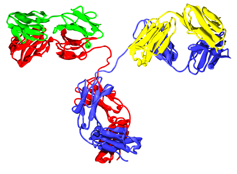
Fcg receptors are a multi-family class of immunoglobulin (IgG)-binding receptors that initiate either activating or inhibitory signals when engaged.  Activating receptors contain cytoplasmic immunoreceptor tyrosine-based activation motifs (ITAM) and activate the FcgR-expressing cell to mediate functions including antibody-dependant cell mediated cytotoxicity (ADCC) and phagocytosis of the antibody-labeled target cell.  FcgRIIB is the single inhibitory Fcg receptor in mice and humans and contains a cytoplasmic immunoreceptor tyrosine-based inhibitory motif (ITIM) which instead downregulates cellular responses.  There are four classes of IgG molecules in both humans and mice, and each bind to different Fcg receptors with varying affinity.  Thus differential affinities of IgG subclasses to functionally different Fcg receptors are thought to mediate the variation in clinical effectiveness of different antibodies targeting the same antigen.

Ipilimumab functions to increase the ratio of effector T cells to TREGS in the tumor microenvironment and has been shown to require binding to both types of T cells for maximal anti-tumor effectiveness.  However, how Ipilimumab differentially modulates these cell types remains to be understood.  A recent study published in The Journal of Experimental Medicine by Simpson et. al sought to clarify the mechanism by which Ipilimumab functions to alter the ratio of effector T cells/ TREGs in a murine tumor model.   Interestingly, the effects of Ipilimumab were found to be tissue-dependant.  In tumors which had high levels of infiltrating CD11b+ macrophages expressing the ADCC-activating FcgRIV, TREGS were selectively depleted in an FcgR-dependant manner, while effector T cells were instead expanded.  In lymph nodes lacking significant levels of these macrophages, frequencies of both effector T cells and TREGS were increased.  Tumor-associated TREGS expressedhigher levels of CTLA-4 than their effector T cell counterparts, or than TREGS present in the lymph node, indicating that higher CTLA-4 expression levels mediate ADCC via macrophages in the tumor.  Furthermore, the presence of FcgRs and hence TREG depletion was required for Ipilimumab’s effects.   Thus is appears that the mechanism of action of Ipilimumab on the effector T cell compartment is two-fold: directly targeting effector T cells to release inhibition via blocking CTLA4 activity, as well as by ADCC-mediated depletion of TREGS.

A second article in the same issue of The Journal of Experimental Medicine by Bulliard et al also explored the role of FcgR engagement on the effects of Ipilimumab as well as an agonistic antibody (DTA-1) targeting the T cell activating receptor GITR (TNFR glucocorticoid-induced TNFR-related protein), which is also expressed on both activated T cells and TREGs.  This study concluded that a major mechanism of action for both antibodies involved engagement of activating FcgRs leading to ADCC-mediated TREG depletion from the tumor.  Even though GITR-activation in effector T cells promotes activities including cytokine production and proliferation, the agonistic properties of this antibody alone were not effective in the absence of activating FcgR engagement.  Thus, even for functionally different (antagonistic versus agonistic) immunotherapeutic antibodies targeting these same T cell populations, FcgR-mediated ADCC of TREGs appears to be a critical mechanism for anti-tumor effects.

These studies highlight several important principles for the field of tumor immunotherapeutics.  Antibody targeting can elicit multiple effects, dependant on expression levels of the target, the isotype of the antibody, and the FcgR-expressing cell types present in the tissue.  Thus knowing the nature of the immune populations present in various types of tumors that are able to mediate ADCC, the FcgRs expressed by these cells, and the expression levels of the target molecule on immune populations that would be ideally targeted for elimination versus activation/inhibition will be critical areas for the forwarding of this field.  Furthermore, as FcgRs are polymorphous in humans, and affect IgG binding affinities, taking these genetic variations into account will be critical in the future of personalized medicine.

Further reading:

Fc-dependent depletion of tumor-infiltrating regulatory T cells co-defines the efficacy of anti-CTLA-4 therapy against melanoma.  Simpson TR, Li F, Montalvo-Ortiz W, Sepulveda MA, Bergerhoff K, Arce F, Roddie C, Henry JY, Yagita H, Wolchok JD, Peggs KS, Ravetch JV, Allison JP, Quezada SA. J Exp Med. 2013 Jul 29.

Activating Fc γ receptors contribute to the antitumor activities of immunoregulatory receptor-targeting antibodies.  Bulliard Y, Jolicoeur R, Windman M, Rue SM, Ettenberg S, Knee DA, Wilson NS, Dranoff G, Brogdon JL. J Exp Med. 2013 Jul 29.

Fcgamma receptors as regulators of immune responses.  Nimmerjahn F, Ravetch JV. Nat Rev Immunol. 2008 Jan;8(1):34-47.

BLOCKADE OF CTLA-4 AND PD-1 ENHANCED TUMOR REGRESSION IN MELANOMA

One of the primary roles of the immune system is the specific identification and elimination of tumor cells on the basis of their expression of tumor-specific antigens or molecules induced by cellular stress. This process is referred to as tumor immune surveillance. In this process the immune system recognizes malignant and/or pre-malignant cells and removes them. However, tumor cells do escape from tumor immune surveillance, and therefore, therapies targeted to enhance antitumor immunity is currently in development.

Blockade of immune checkpoints  is the most promising approach to activate therapeutic antitumour immunity. Immune checkpoints refer to a group of inhibitory pathways connected into the immune system that are important for maintaining self-tolerance. In peripheral tissues immune surveillance also modulates the duration and amplitude of physiological immune responses in order to minimize collateral tissue damage. Studies have suggested that tumor cells adopt many immune-checkpoint pathways as a major mechanism of immune resistance. Immune checkpoint receptors cytotoxic T-lymphocyte-associated antigen 4 (CTLA-4, also known as CD152) and programmed death 1 (PD-1) receptor appear to play important roles in antitumor immunity and have been most actively studied in the context of clinical cancer immunotherapy.

monoclonal3CTLA-4 is expressed on T cells and down modulates the amplitude of T cell activation. Several preclinical studies demonstrated significant antitumor responses following blockade of CTL4-A with limited immune toxicities. This led to the development of two fully humanized  CTLA-4 antibodies ipilimumab and tremelimumab. In clinical trials, ipilimumab demonstrated survival benefits for patients with metastatic melanoma, and was approved by the US Food and Drug Administration (FDA) for the treatment of advanced melanoma in 2010.

On the other hand, PD-1limits T cell effector functions within tissues. Tumor  cells block antitumor immune responses in the tumor microenvironment by upregulating ligands (PDL1 and PDL2) for PD1. Several studies detected increased PD1 expression by tumor infiltrating lymphocytes and the increased expression of PD1 ligands in melanoma, ovarian, lung, renal-cell cancers and in lymphomas. This provided an important rationale to target PD1 in order to enhance antitumor immunity. The fully human antibody nivolumab was found to produce durable objective responses in patients with melanoma, renal-cell cancer, and non-small-cell lung cancer.

Even though individual blocking of CTLA-4 and PD-1 have shown substantial clinical antitumor activity, studies suggest that blocking a single inhibitory receptor only leads to up-regulation of the unblocked pathway. Therefore, in order  to enhance antitumor immunity within the tumor microenvironment it appears to require simultaneous blockade of multiple negative co-stimulatory receptors. In preclinical studies, concurrent inhibition of CTLA-4 and PD-1 resulted in more pronounced antitumor activity than blockade of either pathway alone. On the basis of these observations, a phase I study was conducted to investigate the safety and efficacy of combined inhibition of CTLA-4 and PD-1in advanced melanoma patients and published recently in The New England Journal of Medicine (July 11, 2013). In their study, Wolchok and collagues (2013) treated 53 patients concurrently, and 33 patients sequentially with nivolumab and ipilimumab. Rapid responses were observed in concurrent-regimen cohorts as compared with sequential-regimen cohorts. The objective response rate in the concurrent-regimen cohorts was 40% along with 53% patients exhibited tumor regression of 80% or more. The objective response rate in the sequenced-regimen cohorts was 20% and 13% patients had tumor regression of 80% or more. In both groups, treatment related adverse events were managed with the use of immunosuppressants.

Collectively this study suggested that combined blockade of CTLA-4 and PD-1 would be more effective to enhance antitumor immunity compared to single inhibition of either CTLA-4 or PD-1.

References:

1.  Swann, J.B. and M.J. Smyth, Immune surveillance of tumors. J Clin Invest, 2007. 117(5): p. 1137-46.

2.   Pardoll, D.M., The blockade of immune checkpoints in cancer immunotherapy. Nat Rev Cancer, 2012. 12(4): p. 252-64.

3.   Topalian, S.L., et al., Safety, activity, and immune correlates of anti-PD-1 antibody in cancer. N Engl J Med, 2012. 366(26): p. 2443-54.

4.   Wolchok, J.D., et al., Nivolumab plus ipilimumab in advanced melanoma. N Engl J Med, 2013. 369(2): p. 122-33.

Techniques in Cytology – Cytospin: Cytospin Cell Resuspension Solution

In this blog series opening article, I discussed how cytospins could be used to separate cancer cells from non-cancer cells1. This cytology method and subsequent staining of the resultant cells are a key component of diagnosis and screening of diseases such as cancer. The cytospin technique can be used on any single cell suspensions of any source such as peripheral blood mononuclear cells (PBMCs), effusions, cerebral spinal fluid (CSF), bronchial lavages, fine-needle aspirates, culture cells, etc.

In cytospins, single cell suspensions are spun onto a microscope slide by use of a cytocentrifuge. A cytocentrifuge spins cells at an angle, at low speeds, and accelerates and decelerates gradually. The fluid from the suspension is absorbed onto filter paper while the centrifuge is spinning. This allows the cells to adhere to the slide in a monolayer format. The cell settling rate is determined by the rotational speed of the centrifuge and the size and density of the cells.  Large or dense numbers of cells settle quickly. Small or sparse numbers of cells settle slowly.

One question often asked is what is the optimum resuspension solution for cells that will undergo the cytospin process? The type of resuspension solution used is dependent on the cells.  A good rule of thumb is to use whatever solution will keep the cells alive and healthy throughout the cytospin procedure. The best morphology and subsequent staining of the cytospin cells are generated from cells that are freshly harvested. However, as soon as cells are removed from the body, they begin to die. In order to delay the cell death and protect the cells during the cytospin process, cells are resuspended in tissue culture media or phosphate buffered saline (PBS) with 1% – 10% bovine serum albumin (BSA) and/or 1% – 2% serum such as fetal calf serum (FCS). The tissue culture media and PBS provide a pH balanced liquid and ion replacement for the cell’s native environment. The BSA and serum provide a source of protein as a nutrient to keep the cells healthy and alive for a relatively short amount of time. BSA and serum also stabilizes enzymes to delay internal protein and nucleic acid degradation, which leads to a cascade of events that eventually destroys the cell.

So, should you use tissue culture media or PBS boosted with protein? Again, this depends on the cells and the answer to this question must be determined empirically. If the cells are coming directly from in vitro cell culture then the answer is easy. Keep the cells in the cell culture media that they were culture in. Unless you are interested in seeing on your cytospin slide everything that was in the cell culture well, it is suggested that you wash your cells in fresh media to remove any debris or dead cells before you cytospin. If the cells will be taken directly from the host/patient and then processed for cytospin, determine how long the cells will be out of the host/patient before they are actually taken through the cytospin process. If the cells will be taken through multiple washes to remove debris, other cells, or tissue structural components that are not of interest, it is best to keep the cells resuspended in tissue culture media. A good starting tissue culture media is Dulbecco’s Modified Eagle Medium (DMEM) with 10% FCS. If the cells will be processed immediately or only go through one wash, the cells could be resuspended in PBS with 1% BSA plus 1% FCS as a good starting solution.

My next post will continue with this series on the cytospin process as well as presenting tips for troubleshooting.

1              Ikeda, K., Tate, G., Suzuki, T., Kitamura, T. & Mitsuya, T. Diagnostic usefulness of EMA, IMP3, and GLUT-1 for the immunocytochemical distinction of malignant cells from reactive mesothelial cells in effusion cytology using cytospin preparations. Diagn Cytopathol 39, 395-401, doi:10.1002/dc.21398 (2011).


Techniques in Cytology – Cytospin: Distinguishing Benign Cells from Malignant Cells

Cytology is a key component in diagnosis and screening of diseases such as cancer. It assesses single cells and clusters of cells from sources such as malignant effusions and peripheral blood. Effusions are fluids that leak from blood and lymph vessels and aggregate in tissues and cavities within the body. This is a common problem in cancer patients and can be a reservoir of malignant cells. However, the total number of cells in effusions is small in comparison to the volumes of fluids that are produced. Therefore, in order to collect these cells for evaluation, they must be concentrated.

Cytospin is a cytology method that is specifically designed to concentrate cells such as these that are found in small numbers. After the cytospin is completed, other cytology methods such as immunocytochemistry can be preformed to evaluate the cells. An example is a recent study from Ikeda et al., in which immunocytochemical staining of cytospins from malignant effusions suggested that EMA, IMP3, and GLUT-1 might be helpful in distinguishing malignant cells from benign cells (1). The cytospin process is a simple procedure. Cells are washed in a serum and/or albumin based PBS or culture media solution. The cells are resuspended in up to 500 µl of this solution. A cytofunnel is attached to a glass slide and slide carrier. The entire apparatus is inserted into a cytocentrifuge and the cell suspension is added.

The cells can be spun at various speeds and times depending on the cell type. Eight hundred rpm for up to 5 minutes is a good place to start. The volume to cell ratio must be dilute enough to ensure formation of a monolayer of cells for the best assessment of the cells.

In the Ikeda et al. study, cytology was used to differentiate benign mesothelial cells from malignant mesothelial cells (1). What are mesothelial cells and why is it important to distinguish between benign and malignant forms? These are cells that make up the epithelial lining of the mesothelium which covers the surface of the peritoneal, pericardial, pleural cavities as well as the organs within the cavities (2). The mesothelium has many functions. It is a protective barrier against physical damage and invading organisms as well as being a frictionless interface for free movement of organs and tissue (2). Other functions include roles in fibrinolysis and coagulation, initiation and resolution of inflammation and tissue repair, antigen presentation, transport of fluid and cells, and tumor cell adhesion and growth (2). Reactive mesothelial cells (benign cells) appear in instances when there is a pathogenic infection or physical trauma. Malignant mesothelioma is a rare cancer of mesothelial cells. In effusions, it is sometimes difficult to distinguish benign cells from malignant cells especially when there are few cell numbers. However, immunocytochemistry performed on cytospin cells helps to differentiate these cells. Biomarkers such as epithelial membrane antigen (EMA), glucose transporter-1 (GLUT-1), and insulin-like growth factor-II mRNA-binding protein 3 (IMP3) are useful in separating benign cells from malignant cells. EMA is a large cell surface glycoprotein, also known as MUC1, expressed by glandular and ductal epithelial cells as well as some hematopoietic cells. It has a protective and regulatory role by acting as a barrier to the apical surface of epithelial cells. GLUT-1 is a protein that assists in the transport of glucose across the plasma membrane of cells. It is decreased when glucose levels are high and increased when levels are low. IMP3 binds to sequences in the 3’-UTR of CD44 mRNA. CD44 has a role in cell migration, cell adhesion, and cell-cell interactions. In the Ikeda study, EMA staining intensity on cytospins of malignant mesothelioma cells was strong but staining in reactive cells was weak. EMA stained both the cytoplasm and the membrane of the cells. GLUT-1 stained the membrane of the cytospin cells and expression was highly sensitive in malignant cells. IMP3 stained the cytoplasm of the cytospin cells and expression was lowest in malignant cells. Although these markers help to differentiate malignant from reactive cells, they should not be used as standalone markers. This is because there is no individual biomarker that is exclusively sensitive and specific enough to discriminate between these cells.

Overall, the cytospin technique is a quick method to collect and concentrate fluids that contain a low number of cells. When performing immunocytochemistry after cytospins one can use the same staining protocol used to stain samples from tissue blocks. Cytospins can also be used to take a closer look at cellular staining from cells that have been processed for flow cytometry. This is an excellent method to provide a first look at the immunopathology of a cell. For subsequent postings, the cytospin process will be dissected by discussing key steps and methods of troubleshooting.

Further Reading:

1. Ikeda, K., Tate, G., Suzuki, T., Kitamura, T. & Mitsuya, T. Diagnostic usefulness of EMA, IMP3, and GLUT-1 for the immunocytochemical distinction of malignant cells from reactive mesothelial cells in effusion cytology using cytospin preparations. Diagn Cytopathol 39, 395-401, doi:10.1002/dc.21398 (2011).

2. Mutsaers, S. E. The mesothelial cell. Int J Biochem Cell Biol 36, 9-16 (2004).

Progranulin Antibodies a Common Link in Vasculitis, Lupus, and RA

Patients with autoimmune rheumatic diseases (ARD) such as rheumatoid arthritis (RA) and systemic lupus erythematosus (SLE) have a significantly increased risk of developing cardiovascular disease (CVD) and often develop CVD earlier than those without underlying autoimmunity, although it is not clear whether CVD is a general consequence of RA and SLE or only affects a subgroup of patients.  Control of autoimmune inflammation by disease-modifying anti-rheumatic drugs (DMARD), especially those that target immune factors also involved in vasculitis (e.g., T and B cells), is believed to have a protective effect.  One area of current research is focused on identifying commonalities across multiple ARD that suggest specific mechanisms of ARD-related CVD in order to develop diagnostics, preventatives, and treatments for those at greatest risk.

IgG2 antibody
IgG2 antibody

A recent article in the Journal of Autoimmunity suggests anti-progranulin antibodies as one potential mechanism.  Thurner and colleagues used a protein macro-array to screen serum from patients with anti-neutrophil cytoplasmic antibody (ANCA)-associated systemic vasculitides for novel autoantibodies specific to these diseases.  Of the six candidate autoantigens reactive with pooled vasculitis patient serum, progranulin was the only autoantigen appearing in every one of the vasculitides studied.  However, extended screenings showed that a positive progranulin antibody titer was not specific for vasculitides; although the prevalence was low in healthy controls (1/97 or 1%) and patients with melanoma (0/98) or sepsis (0/22), progranulin antibodies were also detectedin serum from patients with RA (16/44 or 36%) and SLE (39/91 or 43%).

Progranulin, also called proepithelin, granulin-epithelin precursor, or acrogranin, is a glycoprotein secreted by epithelial cells, neurons, and certain leukocytes.  In addition to growth factor-like activity, progranulin has immunomodulatory effects in vitro and in vivo.  Full-length progranulin decreases oxidant production by activated neutrophils, blocks TNFα-induced immune responses via binding to TNFR-1 and -2, and promotes up-regulation of IL-4, IL-5, and IL-10.  Progranulin deficiency in mice results in greater inflammation in collagen-induced arthritis (CIA) and collagen antibody-induced arthritis models of human RA; treatment of either progranulin-deficient or wild-type mice with recombinant human progranulin ameliorates CIA inflammation.

Progranulin is cleaved by several proteases into mature granulins.  Neither recombinant nor proteolytically released granulins antagonize TNFα.  Rather, granulins increase expression of pro-inflammatory cytokines IL-1β, IL-8, and TNFα.  SLPI and apolipoprotein A-I binding to progranulin protects it from cleavage by matrix metalloproteinases and other proteases.  However, during inflammation, neutrophils and macrophages release serine proteases that increase progranulin digestion.  In the context of ongoing inflammation in ARD, this may result in increased cleavage of anti-inflammatory progranulin to pro-inflammatory granulin.

Thurner et al. are the first to report the presence of neutralizing anti-progranulin antibodies in RA, SLE, and small- and medium-vessel vasculitides, which may represent a pro-inflammatory mechanism common to several autoimmune diseases.  Their findings provide additional support for exploration of the progranulin/granulin pathway as a therapeutic target and suggest the potential use of anti-progranulin antibodies as a diagnostic and/or prognostic tool in ARD.  Further studies using sera of patients with known autoimmune disease states are needed to confirm these findings and address the additional questions raised, such as –  What causes the failure of self-tolerance to progranulin and the generation of anti-progranulin antibodies, as seen in ~20-40% of the patients in this study?  Are these anti-progranulin antibodies common to all autoimmune diseases?  Could the development of progranulin-neutralizing antibodies even become a biomarker in ARD, for example as a predictor of responsiveness to DMARD therapy, or an indicator of future progression to ARD-related CVD?  We await the results of these and other studies in this area with great interest.

Further Reading:

Progranulin antibodies in autoimmune diseases.  Thurner L, Preuss KD, Fadle N, Regitz E, Klemm P, Zaks M, Kemele M, Hasenfus A, Csernok E, Gross WL, Pasquali JL, Martin T, Bohle RM, Pfreundschuh M.  J Autoimmun. 2013 May; 42:29-38.

Insights into the role of progranulin in immunity, infection, and inflammation.  Jian J, Konopka J, Liu C.  J Leukoc Biol. 2013 Feb; 93(2):199-208.

Cardiovascular disease in autoimmune rheumatic diseases.  Hollan I, Meroni PL, Ahearn JM, Cohen Tervaert JW, Curran S, Goodyear CS, Hestad KA, Kahaleh B, Riggio M, Shields K, Wasko MC.  Autoimmun Rev. 2013 Aug; 12(10):1004–1015.

PD-1 re-expression is differentially regulated by IL-12 vs IFNα in CD8 T cells

TUMOR_immunotherapyDownregulation of immune functions following responses to pathogen infections is critical for limiting damage to the host by the immune system.  T cell activity is known to be downregulated by a variety of negative regulatory mechanisms including negative checkpoint regulatory proteins, a family of CD28-related molecules.  PD-1 is one such molecule that is transiently expressed on activated T cells.   The ligands for PD-1 are PD-L1 and PD-L2, members of the B7 family of molecules which are upregulated on antigen presenting cells and tumor cells.  Interaction of PD-1 with its ligand leads to inhibition of TCR-mediated signaling via recruitment of SHP1 and SHP2 phosphatases to the TCR synapse.  In the August 2013 edition of The Journal of Immunology, Gerner et al., demonstrate that CD8 T cells initially activated in the presence of IL-12 and IFNα differentially re-express PD-1 upon antigen restimulation.

Cytokines play roles in regulation of nearly every aspect of immune responses.  The cytokine milieu present during T cell activation directs differentiation into the different functional classes of CD4 T helper or CD8 T cells.  This study sought to determine the differences in anti-tumor CD8 T cell effector functions mediated when T cells are activated in the presence of various cytokines.  IL-12 and IFNα activate both overlapping and distinct gene programs and promote cytotoxic CD8 T cell responses.  Thus, these cytokines were chosen for comparison in this study.

In this system, CD8+ OT-1 cells were activated ex-vivo in the presence of either IL-12 or IFNα, and transferred into B16-OVA tumor-bearing mice.  T cells activated in the presence of IL-12 were found to mediate tumor-growth inhibition significantly better than if they had been activated in the presence of IFNα.  Over time in tumor-bearing mice, transferred IFNα-matured OT-1 cells were observed to decline in number and lost the ability to produce IFNγ ex vivo upon restimulation, indicating these cells may be exhausted.

Because PD-1 is known to be a marker and mediator of T cell exhaustion, PD-1 expression was examined.  Initial induction levels of PD-1 were comparable on OT-1 cells following ex vivo activation with IFNα or IL-12.  Following transfer into tumor-bearing mice, PD-1 levels declined over time on both types of cells isolated from the spleen and on IL-12 matured cells isolated from the tumor.  However, PD-1 expression was high on transferred IFNα-matured cells when isolated from the tumor.  Similar results were seen when cells were transferred into mice that subsequently received an injection of the OVA peptide.  Thus, CD8+ T cells matured in the presence of IFNα appear to re-express significantly higher levels of PD-1 upon antigen restimulation than IL-12 matured T cells.

PD-1 and PD-L1 targeting with inhibitory antibodies have emerged as promising avenues in tumor immunotherapy.  In this study, anti-PD-1 antibody administration had no additional anti-tumor effect in mice that received IL-12-matured T cells, while in mice that received IFNα-matured T cells, anti-PD-1 antibodies led to inhibition of tumor-growth to a level similar to that in mice that had received IL-12-matured T cells.  Thus, the relatively poor ability of IFNα-matured T cells to efficiently inhibit tumor growth appears to be largely due to PD-1 upregulation.  Finally, when T cells were matured with both IL-12 and IFNα, the effect of IL-12 was dominant.

Many questions remain regarding the mechanisms mediating PD-1 re-expression in IFNα vs. IL-12 matured T cells.  However, since IL-12 activity was dominant over IFNα on regulating PD-1 expression, IL-12 administration during immunotherapy regimens may enhance anti-tumor T cell responses by blocking the mechanisms by which IFNα enhances PD-1 re-expression.

Further Reading:

Cutting Edge: IL-12 and Type I IFN Differentially Program CD8 T Cells for Programmed Death 1 Re-expression Levels and Tumor Control.  Gerner MY, Heltemes-Harris LM, Fife BT, Mescher MF. J Immunol. 2013 Aug 1;191(3):1011-5. doi: 10.4049/jimmunol.1300652. Epub 2013 Jun 26.

CD20-Negative Circulating Plasmablasts Are Target for New B Cell Therapies in Anti-CCP Positive RA

Diagnosis of rheumatoid arthritis (RA) is based on meeting several of the criteria established by the American College of Rheumatology and the European League Against Rheumatism.  One of these criteria is the presence of anti-citrullinated protein antibodies (ACPA).  ACPA seropositivity is currently tested using a filaggrin-derived peptide (anti-cyclic citrullinated peptide [anti-CCP]) ELISA, although other ELISAs in development such as mutated citrullinated vimentin (MCV) show promising results.

citrullination
Citrullination

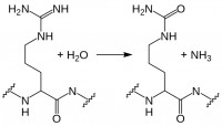
While myelin basic protein, filaggrin, and several histone proteins are naturally citrullinated, other proteins such as fibrin and vimentin can become citrullinated during an inflammatory response.  Citrullination, enzymatic conversion of arginine residues into citrulline, increases protein hydrophobicity, which can change its structure.  In RA, these citrullinated proteins are recognized as “non-self” by immune cells, leading to production of ACPA.  Recent studies suggest these autoantibodies are not merely convenient diagnostic markers resulting from the autoimmune response, but instead may play a role in RA pathogenesis. Current research in this area includes identifying subtypes of RA based on ACPA positivity and specificity, and determining the roles and mechanisms of action for ACPA in RA autoimmunity.

Little is known about the B cells which produce these ACPA.  However, in a recent report in Annals of Rheumatic Disease, Kerkman and her colleagues used B cells isolated from the peripheral blood of ACPA-positive and -negative RA patients, as well as healthy individuals, to examine ACPA production in vitro and identify the ACPA-producing cell populations.

Initially, the authors stimulated peripheral B cells with B cell activating factor (BAFF) and anti-IgM F(ab′)2-fragments to induce ACPA production.  Although total IgG production was equivalent across the cultures, only B cells from ACPA-positive RA patients produced ACPA.  There was good correlation of ACPA titers obtained from in vitro culture with in vivo patient ACPA titers, underscoring the utility of this model system.  Next, the authors examined spontaneous ACPA production in unstimulated peripheral blood mononuclear cells (PBMC) from ACPA-positive RA patients.  In this case, total IgG was up to 100x lower than that seen in their studies with stimulated B cells; however, the amount of ACPA produced was equivalent.

Were ACPA in PBMC cultures generated solely by circulating plasmablasts, or were antigen presenting cells (APC) present in the PBMC population also stimulating production of ACPA by memory or even naïve B cells?  Kerkman et al. used FACS to selectively deplete ACPA-positive RA patient PBMC of plasmablast/plasma cell or naïve/memory populations, as well as to sort the CD19+ B cell subpopulations.  Naïve B cells (CD20+CD27-) did not produce any ACPA, even when stimulated with BAFF and IgM F(ab′)2.  Memory B cells (CD20+CD27+) produced ACPA upon stimulation, indicating CCP-specific memory cells are present in the circulation of ACPA-positive RA patients; however, ACPA production in CD20-depleted PBMC remained essentially unchanged, while unstimulated PBMCs depleted of plasmablasts/plasma cells produced significantly less ACPA.

acpa b cells

This study demonstrates the presence of circulating ACPA-producing plasmablasts/plasma cells in the peripheral blood of patients with ACPA-positive RA.  This is a novel and unexpected finding, since the plasmablast population is typically a transient population within PBMCs following antigen exposure, with antibody production continuing from mature plasma cells in the spleen and lymph nodes.
Circulating ACPA-producing B cells may persist in RA due to plasmablast replication and/or to memory B cell activation in response to persistent systemic citrullinated antigens.  Currently approved RA therapies which target the CD20+ B cell population, such as rituximab, would affect the memory B cell population, but not CD20- plasmablasts.  New therapies targeting circulating plasmablasts/plasma cells in addition to memory B cells could significantly limit ACPA production and subsequent immunological damage in RA, including that due to ACPA-induced TNFα production and complement activation.  Delineating circulating plasmablasts as a major source of ACPA is therefore a step forward in the quest to determine the roles and mechanisms of action for ACPA in RA pathogenesis, and underscores the possibility of developing effective new therapies by targeting specific B cell populations in RA.

Further Reading:

Circulating plasmablasts/plasma cells as a source of anti-citrullinated protein antibodies in patients with rheumatoid arthritis.  Kerkman PF, Rombouts Y, van der Voort EIH, Trouw LA, Huizinga TWJ, Toes REM, Scherer HU.  Ann Rheum Dis 2013 Jul; 72:1259–1263.

The effect of targeted rheumatoid arthritis therapies on anti-citrullinated protein autoantibody levels and B cell responses.  Modi S, Soejima M, Levesque MC.  Clin Exp Immunol 2013 Jul; 173(1):8-17.

B effector cells in rheumatoid arthritis and experimental arthritis.  Finnegan A, Ashaye S, Hamel KM.  Autoimmunity 2012 Aug; 45(5):353-63.

New Research Points the Way Towards Mechanism of Action, Receptor for MS Copolymer Drugs

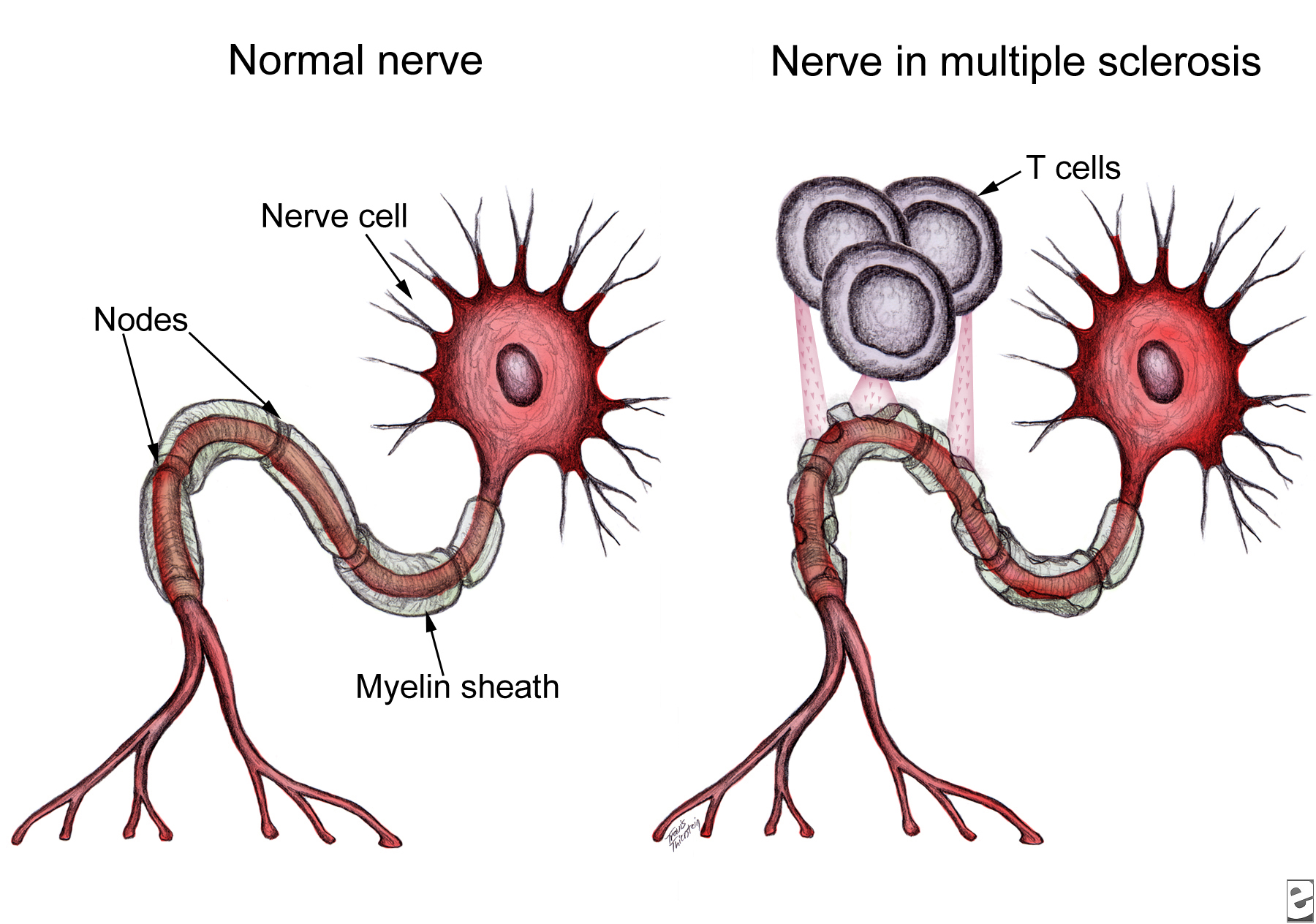
Multiple_sclerosis_T_cellsNeurological damage in multiple sclerosis (MS) is caused by autoreactive immune cells, which attack myelin sheathing on axons of the brain and spinal cord, leading to inflammation and myelin loss.  The MS drug Copaxone is a copolymer of glutamic acid, lysine, alanine, and tyrosine (YEAK) that is thought to impair myelin attack by inhibiting MBP self antigen presentation to autoreactive T cells; a related copolymer in which phenylalanine replaces glutamic acid (YFAK) has been developed based on MBP binding to class II MHC.  However, little is known about these drugs’ molecular targets or mechanisms of actionIn vitro studies suggest IL-10 secretion by B cells or regulatory and Th2-CD4+ T cells is involved.  Recent data demonstrate YEAK and YFAK also have MHC-independent effects on macrophages and dendritic cells, although the receptor which mediates these effects is unknown.  In a recent article in The Journal of Immunology, Koenig and colleagues isolated YEAK- and YFAK-interacting proteins from macrophage lysates and identified structures required for copolymer interaction with cells.

Following incubation with RAW264.7 macrophage lysate, biotinylated copolymers were recovered using avidin-coated beads and the associated cellular proteins were identified using mass spectroscopy.  One high-frequency hit with known surface expression and involvement in immune signaling was gp96.  Cell surface gp96 directly activates innate immune cell cytokine production, acts as a class I MHC antigen chaperone, and has been proposed as a Th2-specific co-stimulatory molecule.  CD91 has been implicated in gp96 stimulation of antigen-presenting cells (APC) and is involved in signaling and endocytosis of several ligands.  App was also identified as a surface protein that interacts with YEAK and YFAK.  A β-amyloid species precursor in Alzheimer’s disease, its function on myeloid cells is not well understood.

Macrophages secrete CCL22, a chemoattractant for regulatory and Th2 T cells, in response to YEAK or YFAK.  Koenig et al. studied this response in wild-type versus gp96-, CD91-, and App-deficient cells and found no impairment in any of the knock-out cell lines, indicating that despite their interactions with YEAK and YFAK, neither gp96, CD91, nor App are involved in cell signaling by these copolymers.

Lysine confers a positive charge on these copolymers, leading Koenig et al. to propose that cellular binding may be mediated by electrostatic interaction rather than conformation.  Indeed, increasing salt concentration reduced protein interactions with biotinylated copolymer.  Importantly, using cell lines lacking specific sulfation enzymes, the authors demonstrated that YEAK and YFAK bind to negatively charged heparan sulfate proteoglycans (HSPG).  This interaction is functional: RAW264.7 cells stimulated with YFAK in the presence of heparin sulfate, a structurally similar competitor of HSPG, did not produce CCL22.

HSPG are glycoproteins which contain one or more covalently attached heparin sulfate (HS) chains.  Membrane HSPG are known to act as co-receptors for many growth factors and could therefore play a role in the cellular effects of YEAK by activating cell signaling through an associated receptor or preventing signaling by that receptor’s natural ligand.  For example, YEAK binding to HS, a co-receptor for gp96 binding to CD91, may alter cellular uptake of gp96-peptide complexes via CD91, affecting self antigen cross-presentation and T cell activation by APC.

Alternatively, HSPG also function as receptors for constitutive as well as ligand-induced endocytosis.  YEAK interaction with HSPG may promote its fluid-phase uptake and delivery to intracellular target(s) in a manner similar to that of cationic cell-penetrating peptides.  In fact, gene ontology term enrichment analysis highlights “RNA binding” as a molecular function of YEAK- and YFAK-interacting proteins identified in this study, supporting a potential cytosolic or nuclear site of action.

Significant work remains to define the receptors and molecular mechanisms of action for these copolymers and aid rational design of future immune-modulating drugs.  The authors’ list of 222 copolymer-interacting proteins and characterization of sulfated glycosaminoglycans as the moieties responsible for functional interaction of these copolymers with innate immune cells serve as a solid foundation for further research in this area.

Further Reading:

Amino acid copolymers that alleviate experimental autoimmune encephalomyelitis in vivo interact with heparan sulfates and glycoprotein 96 in APCs.  Koenig PA, Spooner E, Kawamoto N, Strominger JL, Ploegh HL.  J Immunol.  2013 Jul 1; 191(1):XXX.  Epub ahead of print 2013 June 5.

Heparan sulphate proteoglycans fine-tune mammalian physiology.  Bishop JR, Schuksz M, Esko JD.  Nature. 2007 Apr 26; 446(7139):1030-7.

Interactions between heparan sulfate and proteins – design and functional implications.  Lindahl U, Li JP.  Int Rev Cell Mol Biol. 2009; 276:105-59.

Cell surface heparan sulfate proteoglycans influence MHC class II-restricted antigen presentation.  Léonetti M, Gadzinski A, Moine G.  J Immunol. 2010 Oct 1; 185(7):3847-56.


INTESTINAL BACTERIA LINKED TO LYMPHOMA

The human gut harbors approximately one thousand different bacterial species (intestinal microbiota). Intestinal microbiota number 100 trillion cells; over 90 percent of the cells in the body are bacteria. The composition of each person’s microbiome — the body’s bacterial make-up — is very different, due to the types of bacteria people ingest in their early lives, as well as the effects of diet and lifestyle.

Several studies implicated intestinal bacteria in various cancers. Gram-negative Helicobacter species were found to be associated with liver cancer, colon cancer, and breast cancer. A recent study published in the peer reviewed journal Nature by Yoshimoto et al. (2013) reported that gut bacteria of obese mice unleash high levels of an acid that promotes liver cancer. In rodents, intestinal bacteria influence obesity, intestinal inflammation and certain types of epithelial cancers. However, in human, little is known about the identity of the bacterial species that promote the growth or protect the body from cancer. Therefore, studies are warranted to determine whether differences in peoples’ microbiomes affect their risk for cancer, and whether changing the bacteria can reduce this risk. A clinical trial at the National Cancer Institute (NCI) is currently evaluating the relationship between intestinal bacteria and breast cancer risk (Clinical Trials.gov number: NCT01461070).

intestinal  bacteria

For the first time, a recent study by Yamamoto et al. (2013) demonstrated a relationship between intestinal microbiota and onset of lymphoma (a type of blood cancer of B or T lymphocytes). Yamamoto and colleagues studied mice with ataxia-telangiectasia (A-T), a genetic disease that in humans and mice is associated with a high rate of B-cell lymphoma. These investigators discovered that of mice with A-T, those with certain microbial species lived much longer than those with other bacteria before developing lymphoma, and had less of the gene damage (genotoxicity) that causes lymphoma. A high-throughput sequence analysis of rRNA genes identified the bacteria Lactobacillus johnsonnii in abundance in more cancer-resistant mouse colonies compared to cancer-prone mouse colonies.This study by Yamamoto et al. also created a detailed catalog of bacteria types with promoting or protective effects on genotoxicity (a chemical or other agent that damages cellular DNA, resulting in mutations or cancer) and lymphoma, which could be used in the future to formulate combination therapies that kill the bacteria that promote cancer (such as antibiotics) and increase the presence of the bacteria that protect from cancer (like probiotics).

References:

1.   Ward, J.M., et al., Chronic active hepatitis in mice caused by Helicobacter hepaticus. Am J Pathol, 1994. 145(4): p. 959-68.

2.   Yoshimoto, S., et al., Obesity-induced gut microbial metabolite promotes liver cancer through senescence secretome. Nature, 2013. 499(7456): p. 97-101.

3.   Yamamoto, M.L., et al., Intestinal Bacteria Modify Lymphoma Incidence and Latency by Affecting Systemic Inflammatory State, Oxidative Stress, and Leukocyte Genotoxicity. Cancer Res, 2013. 73(14): p. 4222-4232.