Clinical Significance of CD133 and KLK6 in Malignant Brain Tumors

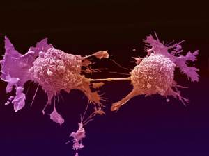
As mentioned in a previous article about Glioblastoma Multiforme (GBM), there are several obstacles that make these lethal brain tumors virtually incurable. Two hallmarks of malignant brain tumors are believed to be the major impediments to effective killing of all tumor cells via the current aggressive course of treatment: glioma cells’ exceptional invasivity and the heterogeneity of GBM, not only among individual patients, but also within a single tumor mass. Thus, these two glioma characteristics have generated great interest in GBM clinical research.

brain tumors,cancer research,glioblastoma,glioma

The malignancy of CNS tumors is categorized by the World Health Organization (WHO) grading system, ranging from grade I- grade IV based on the tumor’s “aggressiveness” (GBM is WHO grade IV astrocytoma).  However, unlike other neoplasms -in which local dissemination is usually limited and metastasis occurs via the vasculature or the lymphatic system– single glioma cells travel for several centimeters through adjacent brain tissue but will almost never establish systemic metastasis. Thus, the WHO tumor grade positively correlates with proliferation rate, rather than tumor invasion. This suggests the involvement of several independent genetic events that eventually lead to glioma progression, which is also consistent with the high levels of GBM heterogeneity. In order to develop a therapeutic agent that targets (finds and destroys) malignant glioma cells, it’s necessary to find a marker that’s expressed exclusively on the surface of these cells. Due to the high heterogeneity of GBM, as well as variations amnog individual patient brain tumors, no such marker has yet been identified.

There are compelling evidences of a subpopulation of malignant cells in GBM, which exhibit stem-cell like characteristics, such as multipotency, the ability to self-renew and invade; these tumor-initiating cells are referred to as cancer stem cells (CSC) and are believed to be responsible for tumor recurrence in GBM patients. Thus, researchers are no longer focusing on a single mutation/marker. Furthermore, it is becoming prominently important to rely on primary GBM patient samples for collecting data, since glioma cell lines do not represent the cellular and molecular components characteristic of the primary GBM.

CD133 (also known as Prominin-1 or AC133) is a penta-spanning membrane protein with two heavily glycosylated extracellular loops, and is recognized as a stem cell marker for certain normal and cancerous tissues. CD133 accumulates near the Golgi and ER and is also expressed on the cell surface. Although many previous studies reported that the presence of CD133+ glioma CSC subpopulation in GBM, drive tumor formation and its rapid proliferation, subsequent conflicting findings showed CD133-negative glioma cells’ ability to self-renew and form tumors in xeno-transplantation assays.

The recent findings by Brescia’s group confirm that cell-surface CD133 expression is a marker for self-renewing and tumor-initiating GBM cells, but a non-essential element for stem cell properties in all GBM cases. They show that even though membrane-bound CD133 were detectable only in a fraction of the patient samples (neurospheres and in freshly dissociated tumors), CD133 mRNA and intracellular CD133 protein were found expressed at high levels in almost all the examined neurosphere samples.

Through clonal analysis, they demonstrated the intrinsic capabilities of sinlge glioma cells and thier progeny: every clone derived from a single CD133-negative cell, contained a mixture of both CD133-negative and CD133-positive cells. Furthermore, they show an interesting interconvertible regulation of cell-surface CD133, through subcellular localization between the cytoplasm and the plasmamembrane. The localization of CD133 is likely determined by the tumor-associated microenvironmental cues.

The findings in this study have significant value in GBM research, taking one step closer to understanding the progression of malignant gliomas. However, the existence of the cytoplasmic CD133 reservoir and the re-cycling of this protein to the plasmamembrane and vise versa, hinders the therapeutic potential of using CD133 as a glioblastoma-targeting marker.

Patient samples,brain tumor,cancer research,GBM

In a separate study published in Neuro-Oncology on the same week, researchers at Mayo Clinic introduced a significant association between malignant gliomas and Kallikrein 6 (KLK6) enzyme. KLK6 is a member of the kallikrein family of secreted serine proteases and has been reported to be elevated within areas of inflammation in CNS, suggesting its regulated expression with T-cell activation. Notably, the serum of Multiple Sclerosis (MS) patients contains elevated KLK6 levels as well. This study shows that as the KLK6 expression levels increase, the post-surgery survival times of GBM patients decrease. They found the highest levels of KLK6 were present in the most severe GBM. These results were obtained by looking at 60 samples of grade IV astrocytomas (thus categorized as GBM), and a less aggressive grade III astrocytomas.


Scarisbrick’s group also showed the possible role of KLK6 in promoting survival of malignant glioma cells, as well as increasing their resistance to apoptosis-inducing agents, such as radiotherapy (RT) and temozolomide (TMZ). The data supporting the pro-survival effect of KLK6 may introduce a new GBM therapeutic strategy, albeit the supporting experiments of these observations were conducted on U251 glioma cell line. If future studies report similar results that confirm the ability of KLK6 enzyme to promote primary patient tumor cells’ survival, then developing a therapeutic agent which targets KLK6’s function is a promising addition to GBM course of treatment ensuing the surgical resection of the tumor, preceding chemo-and radiotherapy.

Further Reading:

CD133 is essential for glioblastoma stem cell maintenance.

CD133 as a marker for regulation and potential for targete therapies in glioblastoma multiforme.
Clinical significance and novel mechanism of action of kallikrein 6 in glioblastoma.
Functional role of kallikrein 6 in regulating immune cell survival.
 

2013 Conferences in Tumor Immunology and Cancer Immunotherapy


Tumor Invasion and Metastasis

January 20 – 23, 2013

Omni San Diego Hotel, San Diego, CA

Online registration is closed, but attendees may register onsite on at the conference registration desk on a first-come, first-served basis beginning from Sunday, January 20 at 4:00 p.m.


Keystone Symposia: Cancer Immunology and Immunotherapy

January 27 – February 1, 2013.

Fairmont Hotel Vancouver, Vancouver, British Columbia, Canada.

Registration is still open until January 27, 2013.


Keystone Symposia: Antibodies as Drugs

January 27 – February 1, 2013.

Fairmont Hotel Vancouver, Vancouver, British Columbia, Canada.

Abstract submission is closed. Registered attendees can bring a poster onsite.

Registration is still open online.


Global Technology Community: The 2nd Novel Immunotherapeutics Summit

January 30 – February 1, 2013.

The Westin Gaslamp Quarter, San Diego, California, USA.

The summit includes a pre-summit workshop and 4 concurrent tracks.

Abstracts can be submitted online, and registration is available for Workshop alone, the Workshop + the Immunotherapetics & Immunomonitoring Track, or the entire Summit.

**Purchase an All Conference Pass for attendance to ALL Global Technology Community Conferences for One Year for $3,995 ($1,990 for Acad/Govt)**

Workshop: Immune Responses in Tumor Microenvironment

January 30, 2013.  This is a half day workshop.

Track: 5th Immunotherapeutics & Immunomonitoring

Track: 11th Cytokines & Inflammation

Track: 2nd Allergy & Respiratory Drug Discovery

Track: Immunogenicity & Immunotoxicity


2013 Tumor Immunology Lab Symposium: Tumor vs Immune System: A Cytokine Battle!

February 6, 2013.

Radboud University Nijmegen Medical Centre, Nijmegen, Netherlands.

Registration is Free, however limited to 150 attendees!


First Symposium of the Cancer Research Center of Lyon

February 13 – 15, 2013.

Convention Center, Lyon, France.

Online registration deadline: February 3, 2013.


Global Technology Community: 2nd Novel Cancer Therapeutics Summit

February 25 – 26, 2013.

Palms Casino Resort / Palms Place, Las Vegas, Nevada, USA.

Abstracts and registration can be submitted online.


IMMUNO 2013: 10th International Conference on New Trends in Immunosuppression and Immunotherapy

March 11 – 12, 2013.

Hotel Fira Palace, Barcelona, Spain.

Registration is still open online.


Arrowhead’s 2nd Annual Cancer Immunotherapy Conference: Stem Cells and Cancer Immunotherapy

April 4-5, 2013

The Washington Post Conference Center, Washington, D.C., USA.

Abstract submissions are due by March 10, 2013 to: poster@arrowheadpublishers.com

Registration and application for Speaking Opportunities can be done online.


Global Technology Community: Cancer Immunotherapy and Immunomonitoring Conference

April 22 – 25, 2013.

Hilton Garden Inn, Krakow, Poland.

Abstracts and registration can be submitted online.


Cancer Immunotherapy Consortium 2013 Scientific Colloquium: Entering the Era of Combination Therapies: Practical Implementation

April 25-27, 201.

Willard Intercontinental, Washington, DC, USA.

Early Registration Deadline: March 15, 2013.


Roche – Nature Medicine Imunology Symposium 2013: Host Immunity to Cancer and Chronic Viral Infections

April 28–30, 2013.

Roche Forum, Buonas, Switzerland.

This is a closed symposium.  Only 50 attendees will be selected to participate in addition to the invited speakers.  Applications for attendance and abstracts can be submitted online.

Application & Abstract Submission Deadline: February 21, 2013.


T cell Function and Modulation Meeting

April 28 – May 1, 2013.

Makena Beach & Golf Resort, Maui (Makena), HI.

Registration can be submitted online and is limited to the first 125 attendees.


CIMT Annual Meeting

May 14 – 16, 2013.

Rheingoldhalle Congress Center, Mainz, Germany

The Association for Cancer Immunotherapy (CIMT) Annual Meeting is the largest meeting in Europe focused on research and development in cancer immunotherapy.

Abstract submission deadline: March 15, 2013.

Early Registration deadline: March 15, 2013.


Cold Spring Harbor Asia Conference: Tumour Immunology and Immunotherapy

October 28 – November 1, 2013.

Suzhou Dushu Lake Conference Center, Suzhou, China.

Abstract submission deadline: August 16, 2013.

Early Registration deadline: August 16, 2013.


Websites that list upcoming Conferences & Events in Immunology, Tumor Immunology, and Cancer Immunotherapy:


The American Association of Immunologists (AAI) Meetings and Events Calendar

Nature Reviews Immunology’s list of conferences

Cancer Immunity Journal’s List of Conferences


New in MS Research: Tip-Dendritic Cells Promote Inflammation

Multiple Sclerosis (MS) is a chronically progressive, neuroinflammatory autoimmune disease of the central nervous system (CNS), believed to be antigen-driven and predominantly T-cell-mediated. MS is characterized by demyelinated areas or patches of sclerosis (plaques) localized within the brain and spinal cord.

The normal physiological state of CNS is considered an anti-inflammatory environment, or “immune privileged”, which is partly due to passive-entry restriction of peripheral immune cells. During MS pathogenesis myelin-specific T-cells overcome these barriers, enter the CNS and recruit inflammatory cells that will eventually target and destroy the myelin protein, which leads to axonal damage.

Cytotoxic T-cells,Multiple Sclerosis,CD4 T-Cells

Although the initial trigger leading to the development of myelin-specific T-cells in MS is still not clear, it has been shown that MS patients’ blood and cerebrospinal fluid (CSF), contain activated myelin‐reactive CD4+ T-cells, whereas only non-activated myelin‐reactive T-cells are present in non-MS samples. 

Both CD4+ and CD8+ T cells have been observed in MS acute and chronic lesions, respectively. Nonetheless, these two T-lymphocytes are activated by different CNS-resident antigen‐presenting cells (APCs) that trigger the recruitment of innate immune cells through presenting myelin antigens to CD4+ and CD8+ T cells, leading to the subsequent “determinant spreading”.

Previous studies have identified cells that present myelin to CD4+ T-cells: once inside the CNS, the CD4 molecule of autoreactive CD4+ T-cells binds to a non-polymorphic site on the major histocompatibility complex (MHC) class II, which is expressed by local myelin-presenting dendritic cells (DCs). In the absence of inflammation in CNS, there is a very low constitutive expression of MHC molecules, which are often present on cells of the lymphoid system.

In contrast CD8 molecule of the CD8+ T-cells binds to the MHC class I molecule, which serves to present specific antigens to the T-lymphocytes’s T-cell receptor (TCR). Thus, CD8+ T-cells (aka. cytotoxic T-cells) are involved in class I‐restricted lysis of antigen‐specific targets. However, until recently the APCs responsible for activating myelin-specific CD8+ T-cells were not known.

In January 2013, Goverman’s group from University of Washington showed that during experimental autoimmune encephalomyelitis (EAE)-an animal model of MS initiated by CD4+ T-cell- Tip-dendritic cells (Tip-DCs) play a major role in activating naive CD8+ T-cells. Based on this study, CD8+ T-cells are presented with MHC class I–restricted myelin basic protein (MBP) and activated by CD11b+ Tip-DCs. In addition, it was reported that under the inflammatory conditions of EAE, oligodendrocytes also presented MBP, to the CD8+ T-cells, which made them recognizable targets of the activated myelin-specific CD8+ T-cells, leading to sustained chronic inflammation (aka. Determinant spreading). This suggests that myelin-specific CD8+ T-cells may be responsible for the ongoing axonal destruction in “slow burning” MS lesions by directly lysing oligodendrocytes.

Tip-Dendritic Cells,CD8+ T-cells,Multiple Sclerosis

Tip-DCs are likely derived from the inflammatory monocytes that have accumulated in the brain and the spinal cord during EAE. Goverman proposed that one possible mechanism of acquiring MBP by Tip-DCs is via phagocytosing and processing myelin debris or dead oligodendrocytes, and then presenting the myelin peptides. Furthermore, they also hypothesized that CD8+ T-cells activated by Tip-DCs may contribute to the immune cascade amplification by secreting additional Interferon-gamma (IFN-γ) within CNS.


Identifying specific DCs involved in antigen-presenting and activation of all T-lymphocytes involved in the neuroinflammatory response is important for the development of potential autoimmune disease therapies that target immunogenic DC functions. Although Goverman’s findings may seem marginal on the surface, it is in fact a big step forward in understanding the etiology of MS; further investigations are required to address the origin of these Tip-DCs and the precise mechanisms through which they become myelin-presenting cells. Also, future studies in human MS are essential to confirm Tip-DCs’ reported functions as well as their interactions with patients’ oligodendrocytes.

Further reading:

MHC class I-restricted myelin epitopes are cross-presented by Tip-DCs that promote determinant spreading to CD8(+) T cells.

Antigen Presentation in the CNS by myeloid dendritic cells drives progression of relapsing experimental autoimmune encephalomyelitis. 

Dendritic cell CNS recruitment correlates with disease severity in EAE via CCL2 chemotaxis at the blood–brain barrier through paracellular transmigration and ERK activation.


High Throughput Systems for Maximizing Human PBMC Assay Potential

Humans are a heterogeneous population and studies comparing populations of humans require a high number of samples for statistical validity.  In addition, human samples such as PBMC are precious in that they represent the immune state of an individual at a point in time.  Thus, when studies are done to analyze a particular state of the immune response in individuals, such as pre- versus post-vaccination, or along the course of a disease state, once used, the samples can never be replaced.  To make the most of human PBMC samples, in particular when patient samples are being used, it is important to not only carefully optimize assays, but additionally be able to maximize the questions that can be addressed with these samples.

Having recently completed a large study involving human patient PBMCs, I encourage the use of high throughput assays systems that allow for a streamlined experimental approach.  All of these assays involve 96-well plate based methods and commercially available kits.


Basic Equipment for 96-well Plate Assays:

Multichannel Pipettes are necessary for quickly performing all 96-well plate assays.  These come in p1000, p200, p20, and p2 volumes.

96 well plate96-well Plates:  Different types of 96-well plates are available for different assay types.  There are various surface coatings including tissue-culture treated polystyrene for cell cultures, uncoated, and others.  Plates can have various plate bottom geometries and optical characteristics.  For instance there are black plates available for light-sensitive assays.  For protocols involving volumes larger then 250ul, there are deep-well plates that carry a 2ml volume per well.

VPscientific multichannelMultichannel Vacuums: Companies such as V&P Scientific offer a multitude of multichannel vacuum manifolds that fit plates of different depths for removing supernatant from wells via vacuum apparatus.  Often these will be the proper length such that they don’t touch the well bottom and work well with removing buffers from centrifuged PBMC cell cultures, such as during washing steps for flow-cytometry.

http://www.vp-scientific.com/wands_&_manifolds.php


PBMC subset Purification:  For magnetic bead based purification of PBMC populations of interest, Stem Cell Technologies offers a 96-well plate EasyPlate™ EasySep™ Magnet that allows separation of up to 1 x 10⁷ cells per well.  Currently only negative or untouched cell isolation methods are supported by this magnetic system due to the larger size of the magnetic beads used in Stem Cell Technologies’ negative isolation kits compared with positive isolation kits.

http://www.stemcell.com/en/Products/All-Products/EasyPlate-EasySep-Magnet.aspx


RNA Isolation:  Qiagen offers two kits for 96-well purification of total RNA from cells.  The RNeasy 96 Kit and RNeasy Plus 96 Kit.  These are 96-well column based platforms which require either a Qiagen vacuum manifold or specialized centrifuge for the protocol.  The RNeasy 96 Kit and RNeasy Plus 96 Kit are similar with the RNeasy Plus 96 Kit utilizing an extra set of steps and columns for elimination of genomic DNA.  The standard RNeasy 96 Kit protocol does however have an optional step for on-column DNAse digestion, however DNAse is not included in the kit.

RNeasy 96 Kit: http://www.qiagen.com/products/rnastabilizationpurification/rneasysystem/rneasy96.aspx

RNeasy Plus 96 Kit:http://www.qiagen.com/products/rneasyplus96kit.aspx


RNA Quantification is much easier if done by 96-well methods than one sample at a time.  Life Technologies’ Quant-iT™ RiboGreen® RNA Assay Kit is extremely sensitive but requires a fluorescence-plate reader.  Thermo Scientific now has a NanoDrop 8000 UV-Vis Spectrophotometer that quantifies nucleic acid concentrations from 96-well plates.

http://products.invitrogen.com/ivgn/product/R11490

http://www.nanodrop.com/productnd8000overview.aspx


In summary, systematic high-throughput protocols can be developed using 96-well systems such as these and many others.  Thus, numerous PBMC samples can be put through multiple experimental procedures in a streamlined manner, maximizing efficiency and minimizing experimental variation.  In this way, multiple questions can easily be simultaneously addressed in precious PBMC samples.

Clinical Trials: What’s New in Oncology?

Novel Immunotherapy Shows Promise for Various Types of Cancers

Since the approval of Provenge and Yervoy immunotherapies, development of anti-cancer immune therapies has gained a lot of momentum. Bristol-Myers Squibb’s and Ono Pharma’s antibodies targeted toward PD-1 and PD-L1 molecules are showing great promise for the treatment of non-small cell lung cancer, melanoma, kidney cancer and ovarian cancer. PD-1 is a molecule found on T-cells, when PD-1’s ligand PD-L1 binds to it T-cells loose activity or die. These targeted antibodies block the binding of ligand to receptor and in doing so maintain anti-tumor T-cell activity.   

Investigational Drug Trastuzamab Emtansine Delays Progression of Advanced HER2-positive Breast Cancer

Trastuzamab emtansine  (T-DM1) is a combination drug containing the trastzamag (Herceptin) antibody attached to chemotherapeutic agent DM1. DM1 is toxic when delivered alone into the bloodstream, combining it with an antibody which has specificity for a given antigen limits its potential wide range toxicity to only cells positive for Her2. Women treated with T-DM1 benefited from a 3 month progression free survival compared to patients treated with lapatinib (Her2/neu and EGFR inhibitor) and chemotherapeutic, capecitabine (DNA synthesis inhibitor) combination treatment.

The FDA is expected to decide on the approval of T-DM1 on Feb. 26, 2013.

Improved Therapy for Rare Form of Brain Cancer
Brain tumor resized 600
Clinical trials conducted on patients with anaplastic oligodendrogliomas, a rare form of brain cancer (anaplastic oligodendrogliomas account for less than 10% of brain cancers) were found to live much longer if treated with a combination of chemotherapy and radiation therapy rather than radiation alone. These findings came after a long term (10 yr) follow up in patients whose tumors had mutations or deletions in both chromosomes 1 and 19 which account for about half of all cases.  Patients who lacked those mutations did not show any benefit to the combination treatment.  This work highlights the importance of genetic screening of a patients cancer and tailoring of cancer treatments.  

Promising New Treatment for Drug-Resistant Leukemia

Chronic Myeloid Leukemia (CML) patients who have failed all therapeutic options now have a new drug option, ponatinib. This drug is efficacious at inhibiting various mutations of the BCR-ABL fusion protein known to cause CML. First and second-generation BCR-ABL inhibitors imatinib, desatinib and nilotinib are effective in the treatment of CML but eventually acquired resistance develops towards these treatments or in some cases there is no response. Resistance to these therapies is largely attributed to mutations on BCR-ABL. Ponatinib is said to overcome these limitations based on its intelligent design that renders it capable of blocking BRC-ABL’s various mutations.

The FDA approved ponatinib on Dec. 14, 2012 for the treatment of CML and Philadelphia chromosome-positive acute lymphoblastic leukemia (ALL). 

Direct conversion or iPSCs: Do all roads lead to Rome?

During development, when cells are programmed to perform specific tasks via terminal differentiation, they don’t undergo fate changes and perform their specific tasks throughout life, a process referred to as phenotypic stability. The idea of reprogramming challenged this concept and introduced the notion that the genomic material from any given cell can be reprogrammed to move back in the developmental cascade and become more unrestricted in terms of what other cell types it could generate in the process. It was initially shown that through Somatic Cell Nuclear Transfer (SCNT), one can reprogram the nucleus of any somatic cell by injecting it into oocytes that were experimentally devoid of their own nucleus. Once the oocyte divides and begins the process of developing, pluripotent cells can be isolated to be used for many purposes (e.g., further differentiation into a specific cell type, such as neurons, expansion, etc.).

stem cells

Over the last decade, induced pluripotent stem cell (iPSC) technology revolutionized the way we now create pluripotent cells from differentiated, somatic cells. By ectopically expressing handful of genes -as low as 2 of them in some cases- differentiated cells can be pushed to express genes that are associated with a more undifferentiated state and eventually transform into “embryonic stem cell like” colonies. Numerous laboratories around the world is utilizing this technology to generate iPSCs for future cell-replacement therapy, studying disease mechanisms, testing drug toxicity and more. However, depending on the cell type that’s being differentiated from the iPSCs for these particular studies, one may not need to generate iPSCs and go straight to the last step of generating a particular cell type (e.g., neurons, beta endocrine cells, etc.) by using direct conversion.

describe the image

In direct conversion (also referred to as transdifferentiation), the ectopic expression of transcription factors converts somatic cells from one lineage to another without reverting the cell all the way back to a fully undifferentiated state. Direct conversion has been around a lot longer than the iPSC technology. For instance, conversion of fibroblasts into muscle cells by overexpression of MyoD [1] or conversion of lymphoid cells into macrophages by expressing Pu-1 [2] were among the first demonstrations that a single transcription factor could directly convert one cell type into another. Note that these initial studies achieved conversion from one cell type to another within the same lineage. More recently, using the iPSC approach, there have been studies that achieved direct conversion from one germ layer to that of another, such as fibroblasts to neurons or liver cells. Furthermore, there have been studies that utilized direct conversion in vivo, such as in vivo conversion of exocrine to endocrine pancreas cells [3], or in vivo conversion of fibroblasts into in functional myocytes in an infarct area [4]. Without a doubt, the success of recent experiments using direct conversion was inspired by Yamanaka’s discovery of iPSC technology, further indicating that the approach has importance beyond just the reprogramming of somatic cells into pluripotent cells.

In direct conversion, one cell type is transformed into another without going through the intermediate pluripotent state (i.e., iPSCs). In specific applications where the pluripotent state is not necessary, this is a welcome change, since the process of creating and maintaining iPSCs are labor intensive and costly. Furthermore, for cell-based replacement therapies, differentiation of cells from iPSCs and transplanting them into recipients carry additional risk factors, such as increased chance of tumor formation, while dealing with pluripotent cells. In the two examples stated above, in which the pancreatic exocrine cells were converted directly into endocrine cells or the resident fibroblasts in the injured heart that were converted into functional cardiomyocytes, there was no need for an intermediate pluripotent state to achieve the goal of transdifferentiation, and the process of conversion took place in vivo. By utilizing the right set of transcription factors, the cells transformed from one lineage into another and achieved the final result of repair and/or restoration of function following disease/injury. Even though, iPSCs would be necessary for many applications, for certain tasks, we can reach the same final aim by using direct conversion without the need for iPSCs. Furthermore, direct lineage conversion could provide important new sources of human cells for creating in vitro disease models and cell-based replacement therapies. While the use of direct conversion gains more popularity, it will be significant to carefully determine the fidelity of reprogramming and to develop methods for robustly and efficiently generating one specific human cell type from another, directly.

References:

[1] Expression of a single transfected cDNA converts fibroblasts to myoblasts. Davis et al., (1987) Cell, 51, 987-1000

[2] Stepwise Reprogramming of B Cells into Macrophages. Xie et al., (2004) Cell, 117, 5, 663-676

[3] In vivo reprogramming of adult pancreatic exocrine cells to beta cells. Zhou et al., (2008) Nature 455, 627-632.

[4] Heart repair by reprogramming non-myocytes with cardiac transcription factors. Song et al., (2012) Nature, 485, 599–604.

Cell-Based Therapies for Malignant Brain Tumors

Malignant gliomas are classified by the World Health Organization (WHO), as grade IV tumors of neuroepithelial tissue and are the most common and deadly intracranial tumors, accounting for more than 70% of all brain tumors. One of the most prominent intrinsic behaviors of Glioblastoma Multiforme (GBM) is its invasiveness within the host’s central nervous system (CNS), in which single glioma cells travel a distance from the tumor mass and invade adjacent brain tissue

Due to the exceptional migratory ability of glioma cells, surgical resection of the tumor is almost always followed by tumor recurrence with foci located as close as 1 centimeter from the resection cavity or as far as the contralateral hemisphere. Despite advancements in surgical techniques and post-operational delivery of chemotherapeutics and radiation, the prognosis for glioma patients remains dismal making these lethal tumors virtually incurable. Although the history of glioma treatment dates back to the 19th century, the median survival of patients remains less than 14 months post-diagnosis. Thus, the only way to cure GBM is by essentially eliminating all glioma cells, including the single cells which have disseminated within the parenchyma, away from the tumor mass.

A promising potential strategy to treat high-grade brain tumors, is through cell-based therapies (CBTs) that incorporate autonomous tracking of tumor cells. Two examples of CBTs, which are currently under investigation in brain tumor clinical trials, include: 1) employing of genetically engineered neural stem cells (NSCs) as target-specific therapeutic-agent delivery vehicles, and 2) the adoptive transfer of tumor-specific, genetically engineered cytotoxic T-lymphocytes (CTLs).


The significance of NSCs and CTLs for treating CNS diseases: 

One anatomical feature unique to the CNS is the presence of the blood brain barrier (BBB), which restricts the access of many compounds including many chemotherapeutic agents into the CNS.  Thus, the BBB prevents effective drug delivery from the circulatory system to the tumor sites within the brain.  Even when the drug is administered intracranially to overcome the limitations presented by the BBB, the densely packed environment of brain’s parenchyma inhibits effective diffusion of the drug throughout the brain and prevents the drug from reaching the tumor cells. These anatomical properties of the CNS are also responsible for inefficient distribution of gene therapy in the brain.

NSCs readily cross the BBB and intrinsically target invasive tumor cells that have migrated away from the tumor mass in vivo. The HB1.F3 NSC line, developed by Dr. Karen Aboody, is one clonally derived human cell line that is particularly well characterized and is used clinically for glioma therapy. Another advantage of this therapeutic model is its efficacy through both intracranial and intravenous administration, without rejection elicited by the host’s immune system against the NSCs; this is due to HB1.F3 NSCs’ low levels of MHC Class I antigen expression and undetectable levels of MHC Class II antigens.

Neural stem cell targeting and killing a brain tumor cell

Previous studies have indicated CTLs’ ability to target and kill GBM, medulloblastomas and therapeutically resistant subpopulations of glioma stem–like cancer-initiating cells (GSC), which express interleukin-13 receptor α2 (IL13Rα2). Although the BBB is not permeable to CTLs, Dr. Christine E. Brown reported a non-toxic strategy of delivering these T-cells to the CNS tumors, by placing a fibrin matrix-embedded with CTLs, in the resection cavity during surgery.

CD4 T-cell

In vivo,secretion of monocyte chemotactic protein-1 (MCP-1) also known as chemokine C-C motif ligand 2 (CCL2), by the cancer cells attract CD4+ and CD8+ T-cells, leading to the subsequent host antitumor immune response. Brown’s group has shown that the same tumor-secreted chemoattractants will recruit genetically engineered CTLs. Since CTLs migrate freely within fibrin matrices, the presence of MCP-1 in the surrounding environment attracts the CTLs to migrate out of the fibrin.  In their in vitro model, the IL13Rα2-specific T-cells successfully migrated out of the fibrin clot and killed the surrounding glioma cells.

Utilization of fibrin matrices allows a safe, non-toxic delivery of CTLs, without causing additional injury (i.e. injury caused by a catheter or needle injection) and inflammation to the brain tissue.

There are other therapeutic approaches for treatment of malignant brain tumors. Following are a list of further readings on the content of this article, as well as other current cancer research studies in regards to GBM:


Auffinger, B. et al. “New Therapeutic Approaches for Malignant Glioma: In Search of the Rosetta Stone.” F1000 Med Rep 4.18 (2012): doi: 10.3410/M4-18.

Brown, C.E., et al. “Stem-like tumor-initiating cells isolated from IL13Rα2 expressing gliomas are targeted and killed by IL13-zetakine-redirected T cells.” Clinical Cancer Research, 18 (8), (2012) pp. 2199-2209

Khosh, N., et al. “Contact and Encirclement of Glioma Cells in vitro is an Intrinsic Behavior of a clonal Human Neural Stem Cell Line.” PLoS ONE7 (12) (2012) . doi:10.1371/ journal.pone. 0051859

Zou, Z., et al. “Cytotoxic T Lymphocyte Trafficking and Survival in an Augmented Fibrin Matrix Carrier.” PLoS ONE, 7(4) (2012). doi:10.1371/journal.pone.0034652

Aboody, K., et al. “Translating stem cell studies to the clinic for CNS repair: current state of the art and the need for a Rosetta stone.” Neuron, 26 May 2011 (Vol. 70, Issue 4, pp. 597-613)

Markers for Identification of Regulatory T cells in Human PBMC

Forkhead box P3 (FoxP3)+ CD4+ T cells, known as regulatory T cells or TREGs, are a class of negative regulatory T cells that function to suppress immune responses, thereby establishing tolerance, preventing autoimmunity, and allowing tumor escapes from immune surveillance.   TREGs are thought to be generated by two major mechanisms.  Natural TREGs are generated through positive selection in the thymus via differential TCR signaling compared with conventional T cells.  Adaptive or converted TREGs are thought be generated in the periphery by conversion of conventional CD4+ T cells via various mechanisms.

TREGs are a heterogeneous population of T cells that function via cell-contact dependent and independent mechanisms to suppress various immune cell types.  Contact-dependent mechanisms of suppression include expression of negative regulatory receptors such as CTLA4, or killing of associated dendritic cells (DCs) through secretion of perforin and granzyme B.  Contact-independant mechanisms of suppression include TREGs secretion of immune suppressive cytokines including IL-10 and TGFb.   High expression of the IL-2 co-receptor CD25 allows TREGs to act as a sink for IL-2 thereby leading to IL-2 deprivation of conventional T cells and inhibition of proliferation.

TREGs are thus an important class of cells and study of these cell populations in human PBMC requires an understanding of the surface and intracellular markers that can be used for flow cytometry analysis and isolation by Fluorescence-activated cell sorting (FACS) or other methods.

Miyara et. al. identified three functionally unique FoxP3+ populations in  freshly isolated CD4+ T cells from human PBMC.  These three populations could be identified by flow cytometry staining of CD45RA, FoxP3, and CD25.  CD25 and FoxP3 expression were highly correlated in the CD4+ population, and I have consistently seen this in my own analyses of unstimulated human PBMC.  The three populations included CD45RA+FoxP3low cells which were CD25++, CD45RAFoxP3high cells which were CD25+++, and CD45RAFoxP3low cells which were CD25 ++.  When these populations were FACS sorted based on CD45RA and CD25 expression, only CD45RA+CD25++ and CD45RACD25+++ cells were functionally suppressive in co-culture experiments with TCR-activated CD25CD45RA+CD4+ responder T cells.  Thus CD45RA+FoxP3lowCD25++ cells and CD45RAFoxP3highCD25+++ cells were denoted as naïve/resting and effector/activated TREGs, respectively.  CD45RAFoxP3low cells in contrast, are likely a heterogeneous mixture of cells and include some cells able to produce IFNg, IL-17, and IL-2 upon PMA+ ionomycin stimulation.  Because dividing effector T cells are able to transiently express FoxP3 at low levels, these cells are likely to be contained in the CD45RAFoxP3low population.  Thus, when using CD25 or FoxP3 to identify TREGs by flow cytometry, CD45RA should be included, and care must be taken with the gating strategies.

Treg Identification FoxP3 CD25 CD45RA resized 600

CD25 in combination with TNFR2 and/or the lack of expression of CD127 have been shown to identify FoxP3+ TREGs that are highly suppressive even in CD25low populations and thus may be excellent markers in particular for FACS sorting of TREGs for functional analyses wherein FoxP3 cannot be utilized as a selection marker.

Several other markers have been used to delineate different populations of TREGs.  The intracellular inhibitory receptor CTLA4, the co-stimulatory receptor ICOS, and the MHC class II cell surface receptor HLA-DR, are co-expressed with FoxP3 in the CD45RAFoxP3high TREG population and may be utilized as specific markers of that population.

Depending on the assay conditions, additional markers may be used to identify TREGs.  LAP, CD121a, and CD121b have been noted as highly specific markers of TREGs but are not expressed in the resting state, becoming transiently induced under assay conditions utilizing TCR stimulation.

This is by no means an exhaustive list of markers that have been used to identify human TREGs in their various functional subsets and states.  The 2011 review in Int Immunopharmacol. by Chen et. al. discusses the usage of these and other markers including CCR6, LAG-3, GARP, CD103, CD39, and CD49d.

In summary, there are multiple combinations of markers that can be used to identify functionally different TREG populations within human PBMC.  The selection of these markers should be considered in the context of the assay type being done and the questions being asked about these heterogeneous populations of cells.

Further Reading:

Regulatory T cells: mechanisms of differentiation and function.  Josefowicz SZ, Lu LF, Rudensky AY.  Annu Rev Immunol. 2012;30:531-64.

Foxp3+ regulatory T cells: differentiation, specification, subphenotypes.  Feuerer M, Hill JA, Mathis D, Benoist C. Nat Immunol. 2009 Jul;10(7):689-95.

Functional delineation and differentiation dynamics of human CD4+ T cells expressing the FoxP3 transcription factor.  Miyara M, Yoshioka Y, Kitoh A, Shima T, Wing K, Niwa A, Parizot C, Taflin C, Heike T, Valeyre D, Mathian A, Nakahata T, Yamaguchi T, Nomura T, Ono M, Amoura Z, Gorochov G, Sakaguchi S.  Immunity. 2009 Jun 19;30(6):899-911.

Resolving the identity myth: key markers of functional CD4+FoxP3+ regulatory T cells.  Chen X, Oppenheim JJ. Int Immunopharmacol. 2011 Oct;11(10):1489-96.

A peripheral circulating compartment of natural naive CD4 Tregs. D. Valmori, A. Merlo, N.E. Souleimanian, C.S. Hesdorffer, M. Ayyoub.  J. Clin. Invest., 115 (2005), pp. 1953–1962.

Activation-induced FOXP3 in human T effector cells does not suppress proliferation or cytokine production.  Allan SE, Crome SQ, Crellin NK, Passerini L, Steiner TS, Bacchetta R, Roncarolo MG, Levings MK. Int Immunol. 2007 Apr;19(4):345-54.

CD127 expression inversely correlates with FoxP3 and suppressive function of human CD4+ T reg cells.   W. Liu, A.L. Putnam, Z. Xu-Yu, G.L. Szot, M.R. Lee, S. Zhu, P.A. Gottlieb, P. Kapranov, T.R. Gingeras, B. Fazekas de St Groth et al.  J. Exp. Med., 203 (2006), pp. 1701–1711

Co-expression of TNFR2 and CD25 identifies more of the functional CD4+FOXP3+ regulatory T cells in human peripheral blood.  Chen X, Subleski JJ, Hamano R, Howard OM, Wiltrout RH, Oppenheim JJ.  Eur J Immunol. 2010 Apr;40(4):1099-106.

Characterization of Myeloid Suppressor Cells in the Tumor Microenvironment

Myeloid suppressor cells (MSCs) such as of macrophages and myeloid derived suppressor cells (MDSCs) are thought to be key players in cancer promotion and resistance to therapy.  MSCs originate from the myeloid lineage in the bone marrow and circulate the bloodstream. They are recruited from the peripheral blood to tissues or tumor sites by cytokines such as colony stimulating factor-1 (CSF-1) where they can differentiate into macrophages or MDSCs. Their normal role is to protect a host from possible autoimmune reactions and to subdue over active immune responses.

Macrophages can exist in various activation states depending on the cues they receive from their environment. Classically activated or M1 macrophages are said to be anti-tumor and pro-inflammatory.  Signaling from cytokines such as GM-CSF, TNF, IFN-γ, or microbial stimuli such as LPS promote an M1 response. M1 macrophages are said to be cytotoxic, and secrete reactive oxygen species (ROS), and thus can damage tissue. Alternatively activated or M2 macrophages are said to be pro-tumorigenic and anti-inflammatory. Cytokines such as IL4, IL-13 and IL-10 are said to promote an M2 activation state, although the designation of IL-10 as an M2 skewing cytokines remains controversial in the field. M2 macrophages downregulate T-cell activity, secrete high levels of growth factors, angiogenenic factors and matrix remodeling enzymes. Thus, macrophages can either promote an anti-tumor inflammatory response or suppress it depending on the cytokines (signals) and they encounter.

The different activation states of macrophages can be characterized by variations in cell surface and intracellular marker expression levels.  Both M1 and M2 macrophages express myeloid markers CD11b and CD33, monocyte marker CD14 as well as macrophage marker, glycoprotein CD68. M1 macrophages express high levels of pro-inflammatory cytokines IL-12, IL-23, and low levels of the anti-inflammatory cytokine IL-10 while the reverse is true for M2 macrophages. The mannose receptor CD206 and scavenger receptor CD163 are expressed at elevated levels in M2 macrophages.  CD68, CD163 and CD206 markers are used largely for immunohistological characterization of macrophages although flow cytometry analysis with these marker is also possible.

Retrospective studies have been carried out on breast, melanoma, pancreatic, and non-small cell lung cancer specimens to name a few, assessing the phenotype of tumor associated macrophages based CD68, CD163, and CD206 expression levels.  In general, a negative or poor prognosis was associated with higher levels of M2 versus M1 macrophages.
It is important to note that variations of the M2 phenotype exist.  Additionally, these phenotypes are quite plastic and it is therefore possible for macrophages to switch between activation states.

MDSCs are a heterogeneous population of immature myeloid cells that share many functions with tumor associated macrophages. Although their most noted function is a strong ability to suppress T-cell proliferation and activity. MDSCs consist of polymorphonuclear (granulocytic) and monocytic cells (PMN and MO-MDSC). PMN-MDSCs do not express HLA-DR while MO-MDSC express low levels of HLA-DR and thus both are poor antigen presenting cells. CD14 and VEGFR1 are markers that can be used to differentiate between PMN and MO-MDSCs populations.  PMN-MDSC phenotype is VEGFR1+CD14 and MO-MDSCs are VEGFR1CD14+.

Based on the pro-tumorigenic properties of MSCs they present novel targets for anti-cancer therapies.  Pre-clinical studies in mouse models suggest blockade of MSC recruitment to tumors in combination with chemotherapies and anti-angiogenic treatments have beneficial effects in delaying the onset cancer cells resistance to therapy.

Further reading:
Transcriptional Profiling of the Human Monocyte-to-Macrophage Differentiation and Polarization: New Molecules and Patterns of Gene Expression

Myeloid Cells in the Tumor Microenvironment: Modulation of Tumor Angiogenesis and Tumor Inflammation

Tumour-associated macrophages are a distinct M2 polarised population promoting tumour progression: Potential targets of anti-cancer therapy

Identification of human stem cell-like memory T cells in PBMC

Memory T cell populations are heterogeneous in phenotype and function and many questions remain as to the mechanisms mediating their long term persistence.  Recent research by several groups have described populations of antigen-experienced T cells within human peripheral blood mononuclear cells (PBMC) that exhibit stem cell-like characteristics: increased self-renewal capacity and the ability to derive the more differentiated central and effector memory and effector populations in vitro and in vivo, and may thus be the cell type mediating memory T cell persistence.

In 2011, Gattinoni et. al. identified a population of stem cell-like memory T cells (TSCM) with surface markers characteristic of naive T cells in human PBMCs.   TSCM cells were CD45RO, CCR7+, CD45RA+, CD62L+, CD27+, CD28+ and IL-7Ra+.  The TSCM population comprised 2-3% of CD8+ and CD4+ T cells in healthy donors.  These TSCM cells could be differentiated from naïve T cells by high expression of CD95 and IL-2Rb, markers which are also expressed by memory T cells.  Furthermore, the TSCM population exhibited a gene expression profile that was intermediate between naïve (TN) and central memory (TCM) cells.

Like memory T cells, these TSCM cells were antigen-experienced and exhibited rapid effector activity upon T cell receptor (TCR) stimulation.  Importantly, they also exhibited the stem-like property of self-renewal in the presence of homeostatic IL-15 signals.  Following TCR stimulation, TSCM cells could differentiate into TCM and effector memory (TEM) T cell subsets, and the authors demonstrated a progressive differentiation pattern of TN à TSCM à TCM à TEM, where no differentiation in the opposite direction was observed following TCR stimulation of sorted TSCM, TCM, and TEM populations.  Human TSCM cells also survived significantly longer and produced more progeny in vivo then either TCM or TEM populations in a NOD.Cg-PrkdcscidIl2rgtm1Wjl/SzJ (NSG) mouse xenograft adoptive transfer model.

One of the most exciting clinical implications of this work however, was the demonstration that TSCM cells exhibited profoundly superior anti-tumor activity than either TCM or TEM  populations in a xenograft mouse tumor model where mesothelin-specific human T cell populations were transferred into NSG mice bearing human mesothelioma M108 tumors.  Thus, TSCM cells may be the most effective cellular subset for use in adoptive T cell therapy in cancer patients.

Subsequent to this finding, a paper was published by Lugli et. al., describing a protocol for identifying and isolating human TSCM cells from PBMCs as well as their in vitro expansion.  The flow cytometry staining panel proposed for TSCM cell identification includes antibodies targeting CD3, CD8, CD4, CD45RO, CCR7, either CD62L, CD27, CD28 or CD45RA, and CD95CD58 and CD122 (IL-2Rb) were also proposed as additional markers for better differentiation of TSCM cells from naïve populations which express these at lower levels.

Identification of human stem cell-like memory T cells in PBMC


Interestingly, the naïve-like TSCM population described by Gattinoni et. al. is not the only memory T cell population demonstrated to have “stem-like” characteristics.

In a quest to understand the mechanism by which patients who have undergone multiple rounds of cytotoxic chemotherapy induced lymphopenia maintain resistance to viral infections, Turtle et. al. described a population of human PBMC CD8+ T cells within the central and effector memory populations that were distinguished by expression of high levels of IL-18Rα and the natural killer (NK) cell receptor CD161.  These cells exhibited a hematopoietic stem cell-like capacity to efflux chemotherapeutic agents mediated by expression of ABCB1, survive chemotherapy, and replenish the virus-specific memory T cell pool in acute myeloid leukemia (AML) patients.

Human memory TH17 cells also have stem cell-like characteristics.  Despite their effector memory-like surface marker phenotype being CD45RO+ CCR7CD62L, compared with TH1 and TH2 subsets, TH17 cells were shown to have increased capacities for proliferation, in vivo persistence, resistance to apoptosis, and higher expression levels of stem-cell associated genes HIF1a, Notch, Bcl2, OCT4, and Nanog.  TH17 cells were able to differentiate into TH1 and TREG subsets.  TH17 cells also express CD161 and thus may overlap with Turtle et. al.’s CD161+ABCB1+ stem-like memory cells.

However, conflicting evidence has been presented as to the identity of CD161+ IL-17 expressing cells and whether or not these cells are in fact Vα7.2+ mucosal associated invariant T cells (MAITs) which are selected by nonpolymorphic MHC class Ib molecules.  MAIT cells however are not known to be virus-specific whereas the CD161+ABCB1+ stem-like memory population identified by Turtle et. al. included influenza and EBV-specific populations.  Thus much remains to be clarified regarding the overlap between CD161+ABCB1+ stem-like memory populations, IL-17 expressing CD4+ (TH17) and CD8+ (TC17) cells which also express CD161, and CD161+ IL-17 expressing MAIT cell populations.

In summary, the ability to identify various stem-like memory CD4 and CD8 human T cell populations in human PBMC using flow cytometry allows for many questions to be addressed about the phenotype, functions, and clinical applications of these cells.

.

Further Reading:

A human memory T cell subset with stem cell-like properties.  Gattinoni L, Lugli E, Ji Y, Pos Z, Paulos CM, Quigley MF, Almeida JR, Gostick E, Yu Z, Carpenito C, Wang E, Douek DC, Price DA, June CH, Marincola FM, Roederer M, Restifo NP. Nat Med. 2011 Sep 18;17(10):1290-7.

Identification, isolation and in vitro expansion of human and nonhuman primate T stem cell memory cells.  Lugli E, Gattinoni L, Roberto A, Mavilio D, Price DA, Restifo NP, Roederer M. Nat Protoc. 2012 Dec 6;8(1):33-42.

A distinct subset of self-renewing human memory CD8+ T cells survives cytotoxic chemotherapy.  Turtle CJ, Swanson HM, Fujii N, Estey EH, Riddell SR. Immunity. 2009 Nov 20;31(5):834-44.

Human TH17 cells are long-lived effector memory cells.  Kryczek I, Zhao E, Liu Y, Wang Y, Vatan L, Szeliga W, Moyer J, Klimczak A, Lange A, Zou W. Sci Transl Med. 2011 Oct 12;3(104):104ra100.

Human MAIT cells are xenobiotic-resistant, tissue-targeted, CD161hi IL-17-secreting T cells.  Dusseaux M, Martin E, Serriari N, Péguillet I, Premel V, Louis D, Milder M, Le Bourhis L, Soudais C, Treiner E, Lantz O. Blood. 2011 Jan 27;117(4):1250-9. doi: 10.1182/blood-2010-08-303339. Epub 2010 Nov 17.

CD161-expressing human T cells.  Fergusson JR, Fleming VM, Klenerman P. Front Immunol. 2011;2:36. doi: 10.3389/fimmu.2011.00036.