Identification of New Potential Drug Targets for Treatment of Lupus

autoantibodies 
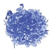
Systemic lupus erythematosus (SLE) is a complex autoimmune disease that afflicts tens of millions of people worldwide.  The most prominent feature is generation of “autoantibodies” to self-proteins and nucleic acids, resulting in immune complex (IC) formation and organ inflammation.  Affected patients may demonstrate rashes, joint pain, anemia, or kidney damage, and untreated complications can often be fatal.  In addition, most SLE patients demonstrate continuously elevated levels of interferon (IFN) α, which is naturally produced by activated plasmacytoid dendritic cells (pDCs) 1.  pDCs are a rare subset of DCs found in the blood and peripheral lymphoid organs that function in host defense by secreting proinflammatory cytokines to initiate the innate immune response.  pDCs are activated following engagement of Toll-like receptors (TLRs), which recognize molecular signatures of bacteria and viruses.  Studies have shown that the frequency of circulating pDCs is significantly reduced in SLE patients, due to increased migration to inflammatory sites in affected organs 2.  Although pDCs have been implicated in contributing to autoimmunity via continuous type I IFN production, their exact role in lupus pathogenesis has not been clearly elucidated.

Recently, in PNAS, Baccala et al. provided direct evidence that in the absence of pDCs, the disease manifestations of Lupus were significantly decreased 3.  Since IRF8 is a hematopoietic cell-specific transcription factor known to be essential for pDC development 4, the authors knocked out IRF8 in NZB mice, a widely used mouse model for SLE.  Appropriately, pDCs were absent in IRF8-deficientNZB mice, and type I IFNs were undetectable even after injection with CpG DNA, a standard method of inducing the interferon pathway.  Interestingly, autoantibody production was almost completely abrogated and kidney disease was drastically improved compared to wild-type NZB mice.  Taken together, their results suggest that without pDCs, SLE disease manifestations are significantly reduced.

Next, the authors sought to examine specifically how pDCs promote systemic autoimmunity.  They used another mouse model with a mutation in Slc15a4, which is characterized by normal development of pDCs but an absence of type I IFN production by pDCs.  It is still unclear how a mutation in Slc15a4 leads to a disruption in proinflammatory cytokine production in pDCs, but since Slc15a4 is a peptide/histidine transporter, others hypothesize that it transports free histidine from the endosome to the cytosol to enable cathepsin-mediated cleavage of endosomal TLRs required for subsequent signaling 5.  Similar to the IRF8-deficient NZB mice, Slc15a4 mice had significantly reduced autoantibodies, decreased kidney disease, and extended survival.  This finding rules out the possibility that pDCs contribute to disease through other functions outside of type I IFN production.

In summary, Baccala et al. provide direct evidence that pDCs contribute to the abnormal manifestations of SLE via hyperproduction of type I IFNs.  Thus, IRF8 and Slc15a4 serve as new potential drug targets for treatment of SLE.  Current therapies involve broad immunosuppressive drugs, which suppress multiple arms of the immune system, increasing a patient’s risk for various infections and cancer.  Specific pharmacologic inhibition of IRF8 or Slc15a4 could prevent Lupus-specific flare-ups, as well as manifestations of other autoimmune diseases.

References

1          Gilliet, M., Cao, W. & Liu, Y. J. Plasmacytoid dendritic cells: sensing nucleic acids in viral infection and autoimmune diseases. Nat Rev Immunol 8, 594-606, doi:10.1038/nri2358 (2008).

2          Ronnblom, L. The type I interferon system in the etiopathogenesis of autoimmune diseases. Ups J Med Sci 116, 227-237, doi:10.3109/03009734.2011.624649 (2011).

3          Baccala, R. et al. Essential requirement for IRF8 and SLC15A4 implicates plasmacytoid dendritic cells in the pathogenesis of lupus. Proc Natl Acad Sci U S A 110, 2940-2945, doi:10.1073/pnas.1222798110 (2013).

4          Tsujimura, H., Tamura, T. & Ozato, K. Cutting edge: IFN consensus sequence binding protein/IFN regulatory factor 8 drives the development of type I IFN-producing plasmacytoid dendritic cells. J Immunol 170, 1131-1135 (2003).

5          Park, B. et al. Proteolytic cleavage in an endolysosomal compartment is required for activation of Toll-like receptor 9. Nat Immunol 9, 1407-1414, doi:10.1038/ni.1669 (2008).

RESISTANCE TO LUNG CANCER THERAPY: A CASE REPORT

A study recently published in The New England Journal of Medicine (Jun 1st, 2013) identified an acquired mutation in the ROS1 kinase domain resulting in resistance to crizotinib in a woman with metastatic lung adenocarcinoma.

Crizotinib is an oral ATP-competitive selective Non Small cell Lung Cancer resized 600inhibitor of the anaplastic lymphoma kinase (ALK) and MET tyrosine kinase that inhibits tyrosine phosphorylation of activated ALK at nanomolar concentrations. In 2011, crizotinib was approved by the U.S. Food and Drug Administration (FDA) for treatment of patients with locally advanced or metastatic non-small-cell lung cancer (NSCLC) that are ALK-positive. Activating mutations or translocations of the ALK gene have been discovered in various types of cancer, including anaplastic large-cell lymphoma, neuroblastoma, inflammatory myofibroblastic tumor, and non–small-cell lung cancer. Because of its role in lung cancer, ALK receptor tyrosine represents a potential therapeutic target.

In addition to ALK mutations or translocations, chromosomal rearrangements in another tyrosine kinase receptor, ROS1, was identified in a molecular subset of NSCLC with distinct clinical characteristics that are similar to those observed in patients with ALK-rearranged NSCLC. Crizotinib was found highly sensitive in lung cancer patients who harbor rearrangements in ALK or ROS1. However, resistance to crizotinib was reported in lung cancer due to secondary mutations in ALK. To overcome this problem a new compound CH5424802 has been identified and is currently in clinical trials (ClinicalTrials.gov number, NCT01588028) for ALK-positive NSCLC.

A 48-year-old woman with metastatic lung cancer and a distant history of light smoking was initially treated with first line of chemotherapy with carboplatin and pemetrexed. Genetic analysis with patient’s cancer cells showed no mutation in oncogenic KRAS or EGFR and no ALK translocations. Additional molecular testing revealed ROS1 rearrangement lead to expression of a fusion protein CD74-ROS1. After three cycles of chemotherapy, marked disease progression was noted and patient’s condition deteriorated. The patient was then enrolled in a clinical trial evaluating the safety and efficacy of crizotinib in cancer patients with ROS1 translocations (ClinicalTrials.gov number, NCT00585195). Computed tomographic scan (CT) obtained two months after treatment noted dramatic response to treatment. However, one month later, while the patient was still taking crizotinib, disease progression was observed and unfortunately the patient expired. Molecular analysis of tumor samples from all sites of disease detected a mutation glycine to arginine Gly2032Arg (G2032R) spanning CD74-ROS1 fusion area that had not been observed in pretreated samples. No other mutation of ROS1 kinase was identified by deep sequencing. Thus this suggested that appearance of G2032R mutation was an early event in crizotinib-resistant tumor cells.

To identify role of G2032R mutation in crizotinib resistance, 293T cells were transfected with either mutated or nonmutated G2032R CD74-ROS1 and subsequently treated with tyrosine kinase inhibitors crizotinib and TAE648. Cells transfected with a mutated form of ROS1 exhibited a half-maximal inhibitory concentration (IC50) value greater than 1000 nM while for nonmutated cells it was approximately 30 nM for crizotinib and 50 nM for TAE648. Crystal structure analysis of ROS1 revealed an arginine at position 2032 resulted in steric interference of crizotinib binding. Collectively, this study reported a mechanism of acquired resistance to crizotinib in a cancer driven by oncogenic ROS1 fusion. Therefore, in the context of these observations, it may be necessary to identify novel compounds that specifically target the G2032R ROS1 mutant to overcome the development of crizotinib resistance in cancers driven by ROS1.


References:

1. Awad MM, Katayama R, McTigue M, et al. Acquired Resistance to Crizotinib from a Mutation in CD74-ROS1. N Engl J Med 2013.

2. Bergethon K, Shaw AT, Ou SH, et al. ROS1 rearrangements define a unique molecular class of lung cancers. J Clin Oncol 2012;30:863-70.

3. Sakamoto H, Tsukaguchi T, Hiroshima S, et al. CH5424802, a selective ALK inhibitor capable of blocking the resistant gatekeeper mutant. Cancer Cell 2011;19:679-90.


describe the image

Arup Chakraborty is postdoctoral research fellow at the National Cancer Institute, Bethesda, MD. He earned a doctoral degree from Texas Tech University, and his primary research interest is in the field of clinical cancer mainly in mechanisms of resistance to molecularly targeted therapies


Computers meet T cells: in silico identification of mutated tumor antigens targeted by T cells

It is well accepted that T cells can recognize and kill tumors that arise in individuals but that tumor cells escape immune surveillance due to the immunosuppressive tumor microenvironment that renders these T cells dysfunctional is less understood.  Only a relatively small number of antigens that T cells recognize for tumor-killing have been identified, and the methods used to identify these antigens are quite cumbersome.  In a recent article in Nature Medicine, Robbins et al. utilize informatics methods to identify mutated tumor antigens in melanoma patients that allowed effective targeting by anti-tumor T cells.

Genome sequencing T cells

In an effort to identify clinically relevant mutated tumor cell epitopes recognized by T cells, Robbins et al. first performed whole-exome sequencing of tumor cells and matched normal cells from melanoma patients who demonstrated tumor regression following adoptive transfer of autologous tumor infiltrating lymphocytes (TILs).  Mutations in tumor cells that resulted in amino acid changes were identified and then screened using an MHC binding algorithm that predicts high affinity binding of peptide sequences to specific HLA alleles.  Candidate peptides of 9-10 amino acids in length were synthesized and pulsed with specific HLA-expressing target cell lines to load the peptides into the MHC complex.  Peptide-pulsed target cells or autologous tumor cell lines were then cultured with autologous TILs from the same donor and IFN-gamma production was assessed as a read out of T cell activation.

Three metastatic melanoma patients were assessed using this methodology.  The first patient was homozygous for HLA-A*0201, and thus mutated melanoma cell line peptides predicted to bind to the HLA-A*0201 allele were identified by the MHC-binding algorithm.  From this donor, 4 out of 55 candidate peptides elicited IFN-gamma responses from autologous T cells cultured with peptide-pulsed target cells.  Two of these mutated peptides were found to correspond to the casein kinase1α1 protein (CSNK1A1), one peptide was mapped to the growth arrest specific 7 gene (GAS7) gene, and the fourth was a fragment of the HAUS augmin-like complex, subunit 3 (HAUS3) protein.  The wild-type versions of each of these peptides bound very poorly (100-10,000 fold less) or not at all to the HLA and were not recognized by T cells.  Two other donors were assessed for predicted binding of mutated peptides to HLA-A*0101 and HLA-A*1101.  Autologous T cell responses were found to be activated in response to mutated peptides from pleckstrin homology domain containing, family M member 2 (PLEKHM2), protein phosphatase 1 regulatory subunit 3B (PPP1R3B), matrilin 2 (MATN2), and cyclin-dependent kinase 12 (CDK12) genes, but not their wild-type counterparts.  Furthermore, tumor lines were validated to express these mutated proteins.

Finally, the authors compared the reactivity of peripheral blood mononuclear cells (PBMCs) drawn before and after adoptive TIL transfer into two of these patients to determine if anti-tumor reactive T cell clones persisted in vivo.  T cells that recognized the same tumor antigens as the TILs were identified post-adoptive transfer at greater levels than prior to adoptive transfer.  Thus, T cells that recognize mutated tumor epitopes may play a clinically relevant role in mediating tumor regression.  Many questions remain, including a direct demonstration that such tumor-reactive TILs are responsible for mediating the observed tumor regression in these patients, and whether further mutation of these residues might facilitate immune escape later it the course of disease. 

Mining exomic sequencing data to identify mutated antigens recognized by adoptively transferred tumor-reactive T cells.  Robbins PF, Lu YC, El-Gamil M, Li YF, Gross C, Gartner J, Lin JC, Teer JK, Cliften P, Tycksen E, Samuels Y, Rosenberg SA. Nat Med. 2013 May 5. doi: 10.1038/nm.3161.

NetMHCpan, a method for quantitative predictions of peptide binding to any HLA-A and -B locus protein of known sequence.  Nielsen M, Lundegaard C, Blicher T, Lamberth K, Harndahl M, Justesen S, Røder G, Peters B, Sette A, Lund O, Buus S. PLoS One. 2007 Aug 29;2(8):e796.

Upcoming Oncology Conferences and Events: Sept-Nov, 2013

This listing includes upcoming Oncology-related conferences from September – November, 2013.


September

Current Trends in Urological Cancer

September 11, 2013

Wolfson Centre, The Medical School, University of Birmingham

Edgbaston, United Kingdom

Advance registration deadline: August 14, 2013

Advances in Ovarian Cancer Research: From Concept to Clinic

September 18-21, 2013

J.W. Marriott Marquis Miami

Miami, FL

Abstract submission deadline: July 8, 2013

Advance registration deadline: August 5, 2013

Cancer Vaccines

September 18-19, 2013

London, United Kingdom

Frontiers in Basic Cancer Research

September 18-22, 2013

Gaylord National Resort and Convention Center

National Harbor, MD

Abstract submission deadline: July 9, 2013

Advance registration deadline: August 6, 2013

Cancer Advance at Harvard Medical School

September 19, 2013

Harvard Medical School

Boston, MA

Clinical Genomics for Cancer Management Conference

September 23-24, 2013

Seaport Hotel

Boston, MA

Abstracts due: August 23, 2013

Advance registration deadline: August 23, 2013

17th ECCO – 38th ESMO – 32nd ESTRO European Cancer Congress

September 27th to October 1st 2013

Amsterdam, Netherlands

Advance registration deadline: Aug 6, 2013

Late Breaking Abstract Submission Deadline: Aug 7, 2013


October

UAE Cancer Congress 2013

October 3-5, 2013

InterContinental Festival City

Dubai, UAE

Abstract Submission Deadline: June 30, 2013

Early Registration Deadline: August 31, 2013

Cancer Epigenomics

October 6-8, 2013

Melia, Sitges, Spain

Abstract submission deadline June 21, 2013

Early Registration Deadline: August 2, 2013

4th International Conference on Stem Cells and Cancer (ICSCC-2013): Proliferation, Differentiation and Apoptosis

October 19-22, 2013

Mumbai, India

Abstract Submission Deadline: June 30, 2013

Early Registration Deadline: June 30, 2013

15th World Conference on Lung Cancer

October 27-30, 2013

Sydney Australia

Abstract Submission Deadline:  June 21, 2013

Early Registration Deadline: August 2, 2013


November

Bioactive Lipids in Cancer, Inflammation and Related Diseases

November 3 – 6, 2013

San Juan, Puerto Rico

Abstract Submission Deadline: August 23, 2013

Early Registration Deadline: August 16, 2013

Pediatric Cancer at the Crossroads: Translating Discovery into Improved Outcomes

November 3-6, 2013

Westin Gaslamp Quarter

San Diego, CA

Abstract submission deadline: August 28, 2013

Advance registration deadline: September 26, 2013

The Translational Impact of Model Organisms in Cancer

November 5-8, 2013

Omni San Diego

San Diego, CA

Abstract submission deadline: August 26, 2013

Early registration deadline: September 23, 2013

Translational Cancer Research for Basic Scientists

November 10-15, 2013

Omni Parker House Hotel

Boston, Massachusetts

Application deadline: May 13, 2013


Websites that list upcoming Conferences & Events in Oncology:

American Association for Cancer Research

Conference Alerts: Academic Conferences Worldwide

Genentech BioOncology

FDA APPROAVES NEW DRUGS FOR MELANOMA

Melanoma is a type of skin cancer that arises from specialized pigmented cells in our body known as melanocytes, which are responsible for the production of melanin (a pigment responsible for skin and hair color). Because most melanoma cells still make melanin, melanoma tumors are usually brown or black. It accounts for 4% of all skin cancers; however, it is responsible for the largest number of skin cancer related deaths in the world. In the U.S, according to the national cancer institute, estimated new cases and deaths from melanoma in 2013 will be 76,690 and 9,480 respectively (for details please refer to my blog titled “targeting B-RAF kinase in melanoma”).

describe the image

BRAF is a serine/threonine protein kinase that activates the mitogen activated protein kinase (MAPK) signaling pathway. Approximately 50% of melanomas harbor activating BRAF mutations among which mutations at codon 600, resulting in substitution of glutamic acid for valine (B-RAFV600E), are the most prevalent. Activated BRAF phosphorylates and activates mitogen-activated protein kinase kinase proteins (MEK1 and MEK2), which then activate downstream MAP kinases. The MAPK pathway is implicated in the regulation of proliferation and survival of tumor cells in many cancers. This suggests that both B-RAF and its downstream MEK kinase could serve as attractive targets in cancer therapeutics. In 2011, the U.S. Food and Drug Administration (FDA) approved vemurafenib for the treatment of V600E B-RAF mutated melanoma patients. As a single agent, vemurafenib resulted in some degree of tumor regression among 90% of melanoma patients early in the course of treatment. Continuing with the effort to target B-RAF and MEK, several studies tested the efficacy of other compounds to inhibit these components of the MAPK pathway. Based on international clincal trials, on May 29th, 2013, the U.S FDA approved two new drugs Tafinlar (dabrafenib) and Mekinist (trametinib) for use in advanced melanomas with B-RAF V600E mutation. Mekinist is also approved for another form of B-RAF mutilated patients, V600K, which accounts for approximately 10% of B-RAF mutated metastatic melanoma. The mutation status of the melanoma patients are detected by an FDA-approved test, such as companion diagnostic assay from bioMerieus S.A., and THxID-B-RAF.

Tafinlar (dabrafenib) is an orally bioavailable B-RAF-inhibitor which selectively binds to and inhibits the activity of mutated B-RAF (V600E). The FDA approval of dabrafenib is based on an open label multicenter phase III study where 250 were randomly assigned to receive either dabrafenib (187 patients) or dacarbazine (63 patients). Dacarbazine is an alkylating agent which is also use to treat malignant melanoma. The study observed a statistically significant increase in progression-free survival (PFS) in patients treated with dabrafenib, compared to dacarbazine. With dabrafenib, the median PFS was 5.1 months and overall response rate was 52%. The most common adverse reactions with dabrafenib were skin-related toxic effects, fever, fatigue, arthralgia, and headache.

Trametinib is an orally bioavailable inhibitor of MEK which specifically binds to and inhibits MEK 1 and 2, resulting in an inhibition of growth factor-mediated cell signaling and cellular proliferation in various cancers. The FDA approval of trametinib is based on the phase 3 open-label trials which randomly assigned 322 patients who had metastatic melanoma with a V600E or V600K BRAF mutation to receive either trametinib or dacarbazine or paclitaxel (a mitotic inhibitor used in cancer chemotherapy). The study observed a statistically significant increase in PFS in trametinib treated patients compared to other treatments. The PFS was 4.8 month for patients treated with trametinib, while with other chemotherapeutic treatments it was 1.5 months. Rash, diarrhea, and peripheral edema were the most common toxic effects noted following trametinib treatment.

GlaxoSmithKline, manufacturer of both new drugs, reported that the products would be available no later than the early part of the third quarter of 2013.

MAPK Pathway Components: Modulators of Ataxin1 Toxicity in SCA1

amyloidWith the increasing prevalence of neurodegenerative disorders in the aging population, it has become more and more important to understand the molecular pathways that regulate and advance these disorders. Due to the high level of complexity of the mammalian brain, it is very difficult to devise improved targeted treatments. The biggest limitation in neurodegenerative disease research being the lack of viable biomarkers for the elder population. Neurodegenerative disorders such as Alzheimer’s, Parkinson’s and polyglutamine diseases, share many pathogenic abnormalities such as the accumulation of misfolded proteins due to mutations rendering them resistant to degradation or over-expression of the wild type form.

In the May 2013 issue of Nature, Dr. Zoghbi and colleagues at the Baylor College of Medicine, devised a strategy to identify therapeutic entry points that influence the levels of disease-driving proteins. They applied their approach to spinocerebellar ataxia type 1 (SCA1), a disease cased by expansion of the polyglutamine tract in ataxin 1 (ATXN1), using modulation of the ATXN1 pathway as a proof-of-principle. This model was chosen for several reasons: (1) neurodegeneration in SCA1 parallels with the levels of the mutant ATXN1 protein; (2) over-expression of wild type ATXN1 results in neurodegeneration; and (3) SCA1’s pathogenic mechanisms are well characterized. In order to identify regulators of ATXN1 levels the authors developed a human medullablastoma-derived cell line containing the transgene glutamine-expanded ATXN1 fused to red fluorescent protein (mRFP-ATXN1(82Q)).  Next, to distinguish modifiers that regulate ATXN1 protein levels from those affecting transgene transcription they included an internal ribosomal entry site followed by yellow fluorescent protein downstream of ATXN1 (mRFP-ATXN1(82Q)-IRES-YFP).  Their screen focused entirely on kinases and kinase like genes based on the fact that ATXN1 phosphorylation is known to be critical for its toxicity and because kinases are pharmacologically targetable. The authors tested 1908 small interfering RNAs (siRNAs) targeting 638 genes and assessing ATXN1 levels as a readout.  Subsequently, 50 siRNAs (corresponding to 45 genes) were selected based on their ability to reduce the ratio of RFP to YFP by 2 standard deviations from the mean.

A parallel genetic screen was performed using the Drosophila SCA1 model that expresses ATXN1(82Q). This model can be identified by an external eye phenotype. Here they screened 704 alleles (337 kinase encoding, including shRNA and loss of function mutations) for those that would modify ATXN1 levels. Based on morphological and histological assessments, they identified 51 alleles (49 genes) that suppressed ATXN1 toxicity in vivo. Additionally, human cell-based screens showed 10 human modifier genes that reduced ATXN1 and it’s associated toxicity, corresponding to 8 Drosophila modifiers.  Network analysis revealed that the MAPK cascade was the most enriched in both Drosophila and human, where 6/10 genes in human belonged to the canonical MAPK pathway (ERK1, ERK2, MED2, MEK3, MEK6, and MSK1).

ATXN1(82Q) is know to impair motor performance, thus, to determine the effects of the MAPK pathway on the central nervous system, a motor performance test was carried out in Drosophila. Decreasing the MEK, ERK1/2, and MSK1 Drosophila homologues by siRNA lead to increased motor performance and lifespan. Decreasing upstream MAPK pathway homologues suppressed ATXN1(82Q) eye defects and improved motor and lifespan phenotypes.  Conversely, constitutively active RAS exacerbated ATXN1 eye degeneration.  In human cells, decreasing HRAS and FNTA lead to decreased ATXN1 protein levels, and decreasing RAS homologues reduced ATXN1 in vivo.

Previous studies by Dr. Zoghbi’s group reported ATXN1 levels were sensitive to S776 phosphorylation.  Hence, they determined that of MAPK kinases implicated here, MSK1 would be able to phosphorylate the consensus sequence associated with S776.  To prove this, they performed an in vitro kinase assay with purified MSK1 and ATXN1 and found robust ATXN1-S776 phosphorylation in both mutant and WT protein forms. Next, cerebellar fractionation assays of WT mice revealed MSK1 was enriched and had increased activity in S776 phosphorylated fractions. Alternatively, immunodepletion of MSK1 from mouse cerebellar extracts lead to decreased S776 phosphorylation.

Next, they sought to determine whether the MAPK pathway could serve as a pharmacological target for SCA1. Human cells expressing ATXN1(82Q) were treated with a PDI84352 (MEK1/2 inhibitor), GW5704 (RAF1 inhibitor), and a Ro31-8220 (MSK1 inhibitor). Pharmacological inhibition of MAPK pathway lead to decreased ATXN1(82Q). Moreover, addition of MAPK inhibitors to cerebellar slices decreased ATXN1 levels.

Lastly, to test the genetic interaction between ATXN1 and MSK1, ATXN1(154Q) knock in mice (Atxn154Q/+) were bred to Msk1+/- Msk2+/- mice. Atxn154Q/+9 week old mice display a motor phenotype that can be quantified using a rotarod test.  Breeding of Atxn154Q/+ Msk1+/- Msk2+/- mice lead to better rotarod performance. Owing to the fact that ATXN1 alterations lead to Purkinje cell degeneration, they next determined whether eliminating one copy of MSK1 could rescue the loss of Purkinje cells in another mouse model of ATXN1(82Q), B05/+.  Indeed, single copy deletion of Msk1 lead to partially suppressed Purkinje loss phenotype and double MSK1 and MSK2 single copy deletion (B05/+Msk1+/- Msk2+/-), lead to decreased levels of ATXN1.

In summary, Dr. Zoghbi’s group have devised a proof-of-principle strategy that opens many new avenues for the identification of modifiers for neurodegenerative diseases. They utilized combined cross-species genetic screens to identify novel modifiers of ATXN1, and validated in human, mouse, and Drosophila models. This study focused on an early event in pathogenesis that could possibly delay disease onset and progression for this class of neurodegenerative disorders. The RAS-MAPK-MSK1 pathway’s role identified here (phosphorylation of S776-ATXN1) provides a novel pharmacological target for SCA1 and more importantly opens new avenues for combination therapies for this disease. Neurodegenerative disease research has primarily focused on developing treatments for advanced symptoms of neurodegeneration. It would be interesting to determine what the therapeutic benefits are of targeting the RAS-MAPK-MSK1 pathway are on a more advanced form of this disease and whether there would be at least partial reversion of motor defects.


References:

Park, J., et al., RAS-MAPK-MSK1 pathway modulates ataxin 1 protein levels and toxicity in SCA1. Nature.

Emamian,E.S.etal. Serine776 of ataxin-1is critical for polyglutamine-induced disease in SCA1 transgenic mice. Neuron 38, 375–387 (2003).

Jorgensen, N. D. et al. Phosphorylation of ATXN1 at Ser776 in the cerebellum. J. Neurochem. 110, 675–686 (2009).

A bifunctional FoxP3+ regulatory T cell subset converts to pro-inflammatory helper T cells

Recently a number of studies have arisen characterizing Tregulatory cellvarious functional subsets of CD4+ FoxP3+ regulatory T cells (TREGS), as well as their plasticity and ability to differentiate into other TH subtypes.  For instance, TREGS that express RORγt were found to be the specific TREG subset that promotes pro-tumor immune functions in colorectal cancer patients.  In a recent article in Immunity, Sharma et al. identify another TREG subset: FoxP3+ TREGS that loose expression of Eos convert to a pro-inflammatory helper subtype that promotes naïve CD8+ T cells differentiation into potent effectors.

Eos is a transcription factor in the Ikaros family, and acts as an obligate co-repressor in complex with FoxP3 to inhibit expression of FoxP3-repressed genes.  In a quest to understand why TREGS in inflammatory environments were observed to become pro-inflammatory without losing FoxP3 expression, Sharma et al. examined the expression of Eos in FoxP3+ TREGS under inflammatory conditions.

Conversion of FoxP3+ TREGS into an inflammatory phenotype was demonstrated by acquired expression of IL-2, IL-17, and CD40L in the draining lymph nodes of a vaccination site compared with FoxP3+ TREGS at distant lymph nodes that did not gain this function.  In these converted inflammatory FoxP3+ TREGS, expression of Eos was rapidly lost.  IL-6 was required for downregulation of Eos, as TREGS in mice lacking IL-6 did not lose Eos expression under the same conditions.  However, IL-6 alone was insufficient for Eos downregulation, which also required interactions with MHC class II on activated dendritic cells.  Loss of Eos expression was furthermore shown to be required for acquisition of the pro-inflammatory phenotype, as TREGS with forced overexpression of Eos did not undergo this conversion.

Interestingly, not all FoxP3+ TREGS were equivalent in their propensity to lose Eos expression and become pro-inflammatory.  Thymic FoxP3+ TREGS were assessed for stability of Eos under treatment with cyclohexamide. CD38+CD69+CD103 TREGS were “Eos-labile” and specifically lost Eos expression within one hour of cyclohexamide treatment, while CD38CD69CD103+ TREGS maintained Eos expression.  Expression of other markers associated with FoxP3+ TREGS including CD25 and CTLA-4 were equivalent between these two phenotypes highlighting the inability of using these TREG markers to discriminate between these populations.  When these FoxP3+ TREGS were sorted into CD38+CD103and CD38CD103+ subsets and transferred into mice, followed by the vaccination schema, only CD38+CD103 TREGS lost Eos expression and gained CD40L and IL-2 expression. The Eos-labile TREGS do however have characteristic suppressive functions when examined in several models including protection from colitis in a Rag-deficient CD45RBHI effector cell-driven autoimmune colitis model and in vitro suppression of T cell proliferation driven by anti-CD3.

Because the Eos-labile subset was observed in the thymus as part of the natural TREG repertoire, the authors examined the signals required for development of this subset.  Again, IL-6 was required as this subset did not arise in IL-6-/- mice.  Epigenetic analysis of DNA methylation patterns comparing these FoxP3+ TREGS subsets revealed distinctive patterns of methylation yet these subsets were still much more closely related to each other as compared with FoxP3 CD4+ T cells.  Future studies will be needed to determine the nature of these epigenetic differences and which signals are controlled by IL-6.

Interestingly, the authors explored the functional contribution of the Eos-labile pro-inflammatory TREGS subset on CD8+ priming in the vaccination model.  Depletion of TREGS resulted in loss of CD8+ T cell proliferation and granzyme B expression as well as loss of CD86 upregulation on DCs, while adding back just the Eos-labile subset or IL-2 plus CD40-agonist antibodies rescued these defects.  The Eos-labile subset did not however, contribute to reactivation of memory CD4+ T cells, and thus these cells appear to play a specific role in the initial priming stages of naïve T cell activation.  Thus, despite having regulatory activity, these cells are critical in priming CD8+ T cell responses by supplying IL-2 and CD40L signals.

However, indoleamine 2,3-dioxygenase (IDO) was able to block Eos downregulation and acquisition of IL-2, IL-17, and CD40L expression.  Importantly, in a murine tumor vaccination model, blocking IDO was important for FoxP3+ inflammatory TREG induction and acquisition of anti-tumor effector CD8+ T cell responses.  The mechanism of IDO inhibition of Eos downregulation was found to be at least in part, dependent on the antagonization of the IL-6-STAT3 pathway by IDO-mediated production of kynurenine-pathway metabolites which activate the aryl hydrocarbon receptor (AhR).  Interestingly, different AhR ligands have been previously shown to differentially regulate induction of TH17 cells vs. TREGS (Quintana et al.), and kyenurine was a TREG inducing AhR ligand (Mezrich et al.).  Additionally, the contrasting effects of IL-6 and IDO will be an important factor in priming immune cell responses.

Overall, this thorough investigation identified the mechanisms that induce and inhibit this newly defined Eos-labile TREG subset that maintains FoxP3 expression and has typical suppressive TREG activity, yet is critically important in priming effector T cell immune responses.  Future studies will be needed to address how these cells balance regulatory and priming activities as well as the relationships between this subset and the many other TREG subsets described.


An inherently bifunctional subset of foxp3(+) T helper cells is controlled by the transcription factor eos.   Sharma MD, Huang L, Choi JH, Lee EJ, Wilson JM, Lemos H, Pan F, Blazar BR, Pardoll DM, Mellor AL, Shi H, Munn DH. Immunity. 2013 May 23;38(5):998-1012. doi: 10.1016/j.immuni.2013.01.013. Epub 2013 May 16.

Eos, goddess of treg cell reprogramming.  Rieder SA, Shevach EM. Immunity. 2013 May 23;38(5):849-50. doi: 10.1016/j.immuni.2013.05.001.

Control of T(reg) and T(H)17 cell differentiation by the aryl hydrocarbon receptor.  Quintana FJ, Basso AS, Iglesias AH, Korn T, Farez MF, Bettelli E, Caccamo M, Oukka M, Weiner HL. Nature. 2008 May 1;453(7191):65-71. doi: 10.1038/nature06880. Epub 2008 Mar 23.

An interaction between kynurenine and the aryl hydrocarbon receptor can generate regulatory T cells.  Mezrich JD, Fechner JH, Zhang X, Johnson BP, Burlingham WJ, Bradfield CA. J Immunol. 2010 Sep 15;185(6):3190-8. doi: 10.4049/jimmunol.0903670. Epub 2010 Aug 18.

Optimizing Assays to Find Rare Antigen-Specific T cells in Cryopreserved PBMCs

Immunomonitoring of T cell based immune responses spans a wide range of therapeutic applications such as infectious and autoimmune diseases and is particularly important for vaccine research. Regardless of the therapeutic application, immunomonitoring can be a daunting task due to the variability of methods and protocols available. There are several commonly used functional assays for the enumeration of antigen specific CD8+ T cells and there is great variability in the protocols that are used for these assays. Thus, making it increasing difficult to thoroughly interpret data obtained from multi-center clinical trials and to compare results between laboratories. In order to address some of the issues associated with immunomonitoring of clinical trials, the Association for Immunotherapy of Cancer (CIMT) formed a CIMT monitoring panel tasked to standardize protocols for assaying T cell antigen immune responses. Thirteen centers from 6 different European countries participated in this study. They were given the same samples and asked to determine the number of antigen specific T cells and assess their antigen specific function using tetramer staining and a functional assay of their choice. Common techniques used for monitoring antigen induced immune responses included ELISPOT assays, HLA-multimer staining and intracellular cytokine staining (ICS).

Pre-tested samples of peripheral blood mononuclear cells (PBMC), synthetic peptides, and PE-conjugated HLA-tetramers were distributed to each center. Using HLA-typed healthy volunteers, PBMCs were isolated by Ficoll density gradient separation. Each sample was tested for T cell reactivity against CMV and influenza. All centers received an HLA-A negative control as well as HLA-A positive samples consisting of a combination of CMV and influenza reactive PBMCs. The study comprised of 2 phases; Phase I consisted of all centers performing the assays with their commonly used protocols, and in Phase II each center received optimized protocols based on the findings from Phase I.

For Phase I’s tetramer-staining assay, the laboratories could choose to stain samples with antibodies (Ab) for CD8+ alone, CD3+CD8+, or CD4+ CD8+ and use their preferred Ab clone, fluorescent dye, and Ab concentration. For the functional assays synthetic peptides were provided and each group could choose either the INF-γ ELISPOT assay, FACS-based intracellular INF-γ staining or both with their antigen concentration of choice ranging from 1-10 g/ml. To reduce variability in FACS analysis, sample plots were provided as well as gate settings and quadrants. Tetramer-staining data reported included; number of viable cells post-thawing, cytometer model, number of lymphocytes and/or CD8+ cells analyzed. Data was presented as percent of tetramer-positive cells among CD8+, CD3+CD8+, or CD4+ lymphocytes depending on what antibody cocktail was chosen. For the functional assays each center reported the type of ELISPOT plates used, reagents and conditions used, and number cells tested.

Tetramer results from the Phase I study showed the number CD8+ cells analyzed significantly affected the sensitivity of tetramer staining. Antigen-specific T cell reactivity when less than 30,000 CD8+ T cells were counted resulted in only 70% responsiveness detected. In contrast, when more than 30,000 CD8+ cells were counted, an 89% response was observed. Although, when antigen-specific T cells were present at high frequencies the number of counted cells did not matter. Interestingly, Ab clone variability, Ab concentration, or cytometer type did not result in any significant differences. Thus, the main factors affecting antigen-specific T cell reactivity by tetramer staining is the number of CD8+ cells used. For Phase II it was then recommended at least 1 x10⁶ PBMCs are used for this assay.

The majority of groups chose the INF-γ ELISPOTas their functional assay. Results showed a large amount of heterogeneity between the centers. Some centers included a resting phase after thawing the cells, of 2-20 hours, resulting in 73% positive reactivity (number of spot forming cells per seeded PBMC). In contrast, not allowing a resting phase resulted in only detecting 30% of the positive cells. Additionally, intra-center replicate reproducibility was significantly affected by the number of replicates used, where duplicates often failed the Student t test and triplicates were sufficient to reach statistical significance. Addition of allogenic-APCs for binging and presentation of the synthetic peptides was found to have a negative effect on detection response (28% of all responses vs. 58%). When looking at the number of cells seeded per well, those with more than 4 x10⁵ PBMC detected 71% positive samples and those with less than 4 x10⁵ only detected 43%. Granted, when antigen specific T cells were available at high frequencies the number of counted cells did not affect the response rates. Consequently, Phase II’s minimum requirements for the INF-γ ELISPOT protocol included: (1) triplicates should be performed for each test antigen (2) avoid using allogenic-APCs (3) include a resting phase (4) use over 4 x 10⁵ PBMCs per well.

Another interesting finding from this study was that lab experience in performing these assays had no effect on the performance of the assays compared to labs that had just adopted the techniques. Further highlighting the importance of developing standardized protocols for immunomonitoring assays. This study did not however, address specific detection limits for the ELISPOT assays, the variability between ELISPOT plate readers, nor serum source effects on background and specificity. In addition, it was not reported whether live/dead cell stains where included in the tetramer assays and how combinations of these may have had an effect on the sensitivity of the assay.

Overall, this study identified several factors that should be generally implemented when performing tetramer staining and INF-γ ELISPOT assays with cryopreserved PBMC samples. Furthermore, these protocol modifications are particularly important when assaying antigen-specific T cell populations present at low frequencies.

Reference:

The CIMT-monitoring panel: a two-step approach to harmonize the enumeration of antigen-specific CD8+ T lymphocytes by structural and functional assays. Britten CM, Gouttefangeas C, Welters MJ, Pawelec G, Koch S, Ottensmeier C, Mander A, Walter S, Paschen A, Müller-Berghaus J, Haas I, Mackensen A, Køllgaard T, thor Straten P, Schmitt M, Giannopoulos K, Maier R, Veelken H, Bertinetti C, Konur A, Huber C, Stevanović S, Wölfel T, van der Burg SH. Cancer Immunol Immunother. 2008 Mar;57(3):289-302. Epub 2007 Aug 25.

A theory of everything: commensal gut bacteria link environmental exposures to sex hormones in modulating autoimmunity

Whether an individual develops autoimmunity depends on how environmental and genetic factors interact to influence immune function. Genome wide association studies have revealed numerous risk alleles associated with diverse autoimmune diseases and small animal models have helped parse the effects of some of these gene variants on specific components of the immune system (see previous post). These approaches, however, largely ignore two of the more perplexing aspects of autoimmunity: (1) the strong female predominance of many autoimmune diseases and (2) the high incidence of autoimmunity in industrialized areas of the world compared to poorer, rural areas. The issue of sexual dimorphism in autoimmunity has led investigators to surmise that sex hormones are potent modulators of immune function. Indeed, altering estrogen, progesterone, or testosterone levels can influence the progression of autoimmune reactions in animals and people, although the specific mechanisms underpinning these phenomena remain controversial1. The increasing incidence of autoimmunity in the industrialized world has given rise to the “hygiene hypothesis”: exposure to specific microbes early in life is necessary for a fully-functioning immune system. These beneficial exposures are lacking in modernized, clean cities which contributes to immunological derangement and self-reactivity. Evidence supporting the hygiene hypothesis has accumulated in recent years to the point that clinical trials are in progress testing whether purposeful infestation with parasitic worms (thought to be the major microbial exposure missing from a modern upbringing) can control inflammatory bowel disease and multiple sclerosis2.

Microbiota

In a recent paper in Science Magazine, Dr. Jayne Danska’s group at the University of Toronto present an intriguing hypothesis that unifies the roles of early-life microbial exposure and sex hormone levels in driving an autoimmune response3. The group, with lead author Dr. Janet Markle, used a well-established mouse model of type-1 diabetes (T1D)—the non-obese diabetic (NOD) mouse—that is genetically predisposed to develop spontaneous, immune-mediated destruction of beta-islets at around 15 weeks of age, causing diabetes. Similar to several human autoimmune diseases, NOD mice have a 2:1 female-to-male sex bias in progressing to diabetes. In agreement with this, Markle et al. found that male mice housed under standard laboratory conditions were protected from developing diabetes compared to female mice. However, if NOD mice were born and raised in germ-free conditions (i.e. their intestines were never colonized by commensal bacteria) males developed the disease at the same rate as females. In addition, the environment in which the mice were raised impacted their levels of sex hormones: male mice raised in germ-free conditions had lower serum testosterone levels than those raised in standard conditions while germ free female mice had higher testosterone levels than those kept in standard cages. These data supported the conclusion that colonization with commensal bacteria was responsible for the protection of male NOD mice against T1D and that bacterial colonization somehow regulated the production and/or use of testosterone.

To get a sense of the extent to which gut microbiota influenced general physiology of adult male and female NOD mice, the authors used mass spectrometry to profile almost 200 unique small-molecule metabolites in serum. They found that male and female NOD mice raised in standard conditions had distinct profiles of serum metabolites. In contrast, there were few detectable differences between the metabolite profile of males and females raised in germ free conditions. This data suggested two hypotheses: (1) male and female mice had different physiologic responses to the same commensal bacteria, or (2) male and female mice had different commensal communities influencing their hormone and metabolite levels. The authors then sequenced bacterial 16S ribosomal RNA from the intestines of NOD mice at various stages of maturation (just after weaning, at puberty, and as adults). While male and female NOD mice had indistinguishable gut microbiota after weaning, sex-based differences in commensal bacteria were apparent at puberty and became even more pronounced in adulthood.

Having established that adult male and female NOD mice have distinct bacterial populations in their intestines, Markle et al. showed that transplanting the “male” gut microbiota into pre-pubescent female NOD mice altered the composition of the recipient’s commensal populations for several weeks and resulted in increased serum testosterone levels. Importantly, transplantation of male commensal bacteria protected the female recipients from T1D. Markers of T1D disease activity, such as inflammation of beta-islets and production of auto-antibodies, were reduced in recipients of male gut bacteria compared to unmanipulated females. This effect was abrogated upon treatment with the anti-androgen Flutamide, indicating that testosterone levels were a critical regulator of autoimmune pathogenesis.

Markle et al. have put forth an interesting model in which sexual maturation results in sex-specific programming of intestinal commensal bacteria. These distinct populations have differential effects on host physiology and hormonal balance which, in turn, modulate immune function. The centrality of gut microbiota to immunity raises the intriguing possibility of treating a dysfunctional immune system by altering the make-up of the intestine’s commensal communities. Such studies are already underway for intestinal disorders using “fecal transplants”, but the concept may extend to more systemic autoimmune disorders4. This approach would benefit from knowing the specific effects of different bacterial species on host physiology in order to identify the critical components of effective therapeutic microbial regimens.  Similarly, it will be interesting to see which of the changes in metabolite and hormone levels that accompany shifts in commensal populations are most impactful on immune function. The metabolite profiling approach shown in this article could be useful for identifying endogenous compounds that serve as immune modulators.

Reference:

1. Sex differences in spontaneous versus induced animal models of autoimmunity. Lee TP, Chiang BL. Autoimmun Rev. 2012 May;11(6-7):A422-9. doi: 10.1016/j.autrev.2011.11.020. Epub 2011 Dec 4.

2. Vaccine against autoimmune disease: can helminths or their products provide a therapy? Zaccone P, Cooke A. Curr Opin Immunol. 2013 Mar 2. pii: S0952-7915(13)00027-7. doi: 10.1016/j.coi.2013.02.006.

3. Sex differences in the gut microbiome drive hormone-dependent regulation of autoimmunity. Markle JG, Frank DN, Mortin-Toth S, Robertson CE, Feazel LM, Rolle-Kampczyk U, von Bergen M, McCoy KD, Macpherson AJ, Danska JS. Science. 2013 Mar 1;339(6123):1084-8. doi: 10.1126/science.1233521. Epub 2013 Jan 17.

4. Stool transplants: ready for prime time? Weissman JS, Coyle W. Curr Gastroenterol Rep. 2012 Aug;14(4):313-6. doi: 10.1007/s11894-012-0263-7.


From bedside to bench: linking autoimmunity-associated gene variants to immune function

Autoimmunity results when the immune system, normally tasked to defend against infections and cancer, attacks the body’s own tissues. There are over 80 clinically-distinct autoimmune diseases that differ in terms of which tissues are targeted and which therapies are most effective. Rheumatoid arthritis (RA) and inflammatory bowel disease (IBD) result in the destruction of joints and the intestinal tract, respectively. These diseases respond well to agents such as adalimumab (Humira) and etanercept (Enbrel) that block the action of TNF-alpha, a cytokine that can promote inflammation. During multiple sclerosis (MS) the immune system attacks the central nervous system resulting in progressive neurologic deficits. Despite being an inflammatory disease, multiple sclerosis is actually worsened through the use of anti-TNF-alpha therapies.

Identifying the fundamental dysfunction at the root of an autoimmune disease would aid in choosing the best of available therapies or devising new ones. Advances in genome sequencing technology have allowed researchers to generate an expanding list of genetic differences present in individuals with various autoimmune diseases compared to healthy people. Although several of these disease-associated gene variants have known roles in the immune system, how they contribute to specific autoimmune processes is largely unknown. There is a need for functional characterization of these gene variants in order to determine how they alter immunity and to stratify them as therapeutic targets.

Dr. David Rawlings’ group at Seattle Children’s Hospital sought to address this challenge in a recent paper published in the May 2013 issue of the Journal of Clinical Investigation. The group, with lead author Dr. Xuezhi Dai, investigated a genetic variant of protein tyrosine phosphatase non-receptor 22 (PTPN22) which had previously been linked to several autoimmune diseases, including type 1 diabetes (T1D), RA, Graves’ Disease, and systemic lupus erythematosus (SLE). PTPN22 encodes an enzyme called LYP, a protein tyrosine phosphatase whose general function is to modulate the intensity of certain signals within cellular signaling networks. The disease-linked variant results in an amino acid switch from arginine to tryptophan at position 620 (LYP-R620W). How LYP or LYP-R620W work to modulate immune activity is incompletely understood.

transgenic mouse

To gain insight into the role of LYP-R620W in autoimmune patients, Dai et al. created a genetically engineered mouse with an analogous arginine to tryptophan switch in the mouse version of LYP (called PEP-R619W). The “knock-in” mice expressing PEP-R619W were viable but had slightly shorter life spans compared to their counterparts with normal PEP. As the engineered mice aged they manifested signs of autoimmunity, such as inflamed lung tissue and blood vessels, as well as signs of chronic kidney damage. In addition, PEP-R619W rendered the mice more susceptible to an experimental form of type 1 diabetes.  These mice also produced numerous auto-antibodies, a hallmark of certain autoimmune diseases.

The PEP-R619W knock-in mouse allowed the authors to look in close detail at the effect of this gene variant on specific immune cell populations. Dai et al. found that the knock-in mice had larger numbers of activated/memory T cells than their normal counterparts, indicating a chronically active immune system. T cells from the knock-in mice were shown to be hyper-responsive to stimulation of their antigen receptors indicating augmentation of the intracellular signals that dictate T cell activation. Similarly, the authors found that knock-in mice had larger numbers of specific B cell populations that occur in active immune states. B cells from the knock-in mice proliferated more than those from normal animals in response to stimulation and were more easily induced to secrete antibody. These findings led the authors to conclude that expression of PEP-R619W results in a lower threshold for activation in both T and B cells which contributed to the autoimmune phenotype. Interestingly, the authors discovered that expression of the disease-linked variant exclusively in B cells was sufficient to generate mice with signs of autoimmunity.

Dai et al. provide a great example of how the tools of bench science can be used to deepen the knowledge gained from analysis of patient specimens. Further determination of PEP/LYP substrate specificity and the dynamics of its phosphatase activity during lymphocyte activation could generate targets for the development of highly selective immune suppressants. In addition, the autoimmune phenotype generated with this knock-in mouse is relatively mild. It would be interesting to see how other disease-linked gene variants would cooperate with PEP-R619W to generate either a more aggressive disease or one that resembles a particular autoimmune syndrome. Finally, the ability of B-cell-specific PEP-R619W expression to stimulate autoimmunity suggests that B cells are a critical component of the autoimmune process in patients with this genetic variant. This model provides the opportunity to compare different therapeutic modalities in the PEP-R619W background (for example, B cell depletion versus anti-TNF agents). Such studies could provide the basis for predicting clinical responses to autoimmune therapies based on genotype.

References:

A disease-associated PTPN22 variant promotes systemic autoimmunity in murine models. Dai X, James RG, Habib T, Singh S, Jackson S, Khim S, Moon RT, Liggitt D, Wolf-Yadlin A, Buckner JH, Rawlings DJ. J Clin Invest. 2013 May 1;123(5):2024-36. doi: 10.1172/JCI66963. Epub 2013 Apr 24.