Highlight: How TNF knocks out Tregs!

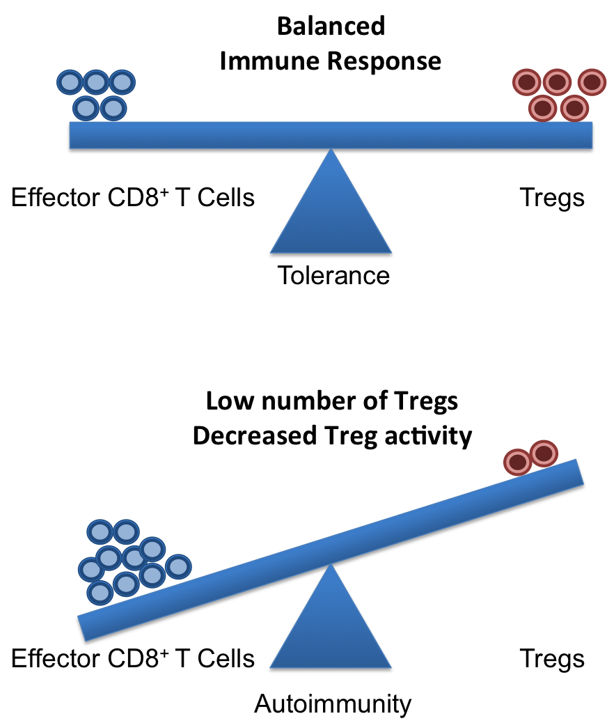
A healthy and functional immune system requires a delicate balance of pro- and contra-inflammatory signals. Whereas, it is important to induce a strong and efficient immune response against pathogens, it is similarly important to dampen these responses after the pathogen is fought off to revert the immune system to a calm steady state. If the balance is disturbed, diseases can on the one hand, become chronic/overwhelming or, on the other hand, inflammatory responses that cannot terminate can result in autoimmune responses.

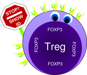
Crucial elements in the regulation of excessive immune responses are regulatory T (Treg) cells. Tregs are known to inhibit the response of other immune cells. Their essential role in limiting overwhelming immune responses is demonstrated by the detrimental consequences of their loss. Mice or humans lacking Tregs develop widespread and lethal autoimmune diseases. Besides several surface markers, Tregs are best characterized by the expression of the transcription factor FoxP3. This factor is essential for Treg function and its artificial expression in other T cells can induce a regulatory potential. Therefore, the expression of FoxP3 is required for a T cell to have regulatory potential (Buckner; Josefowicz et al.). However, it was known for many years that in cases of numerous autoimmune diseases FoxP3+ Tregs could be found in high numbers at the sides of inflammation, but that they did not demonstrate any or not sufficient regulatory activity. This enigmatic observation was so far poorly understood (Buckner; Josefowicz et al.).Treg balance

In the March 2013 issue of Nature Medicine Nie and colleagues shed new light on the underlying mechanism that impairs Treg function at the sites of inflammation. Studying Treg cells from rheumatoid arthritis (RA) patients the authors demonstrated that phosphorylation of FoxP3 of the serine at position 418 (S418) is required for its regulatory action. If FoxP3 lacks this particular phosphorylation the Treg cell is not suppressive! FoxP3 S418 in Tregs is usually phosphorylated and hence Tregs are regulatory by default. However, the authors show that due to the action of the enzyme ‘protein phosphatase 1’ (PP1) FoxP3 can lose its S418 phosphorylation. Intriguingly, the presence of the cytokine TNF lead to an up-regulation of PP1 expression in the Tregs in a dose-dependent manner, and this lead to de-phosphorylation of FoxP3 S418. Treg cells expressing a mutant FoxP3 that replaced the serine at position 418 with an alanine retained their suppressive potential even in the presence of TNF, demonstrating the importance of the phosphorylation of S418. With this finding, the authors were able to link the pro-inflammatory milieu (TNF) to a specific effect inside of the Tregs (de-phosphorylation of S418) that lead to the observed loss of the regulatory function of Treg cells. Importantly, the authors were also able to demonstrate the therapeutic potential of this knowledge. They monitored RA patients that underwent treatment with blocking anti-TNF antibodies (infliximab) and found that Tregs from patient PBMCs restored S418 phosphorylation and regained regulatory potential!

This is the second case for a post-transcriptional regulation of FoxP3 that can influence Treg function. Deacetylation of FoxP3 has been linked to impaired Treg function previously (Tao et al.). Additionally, the work of Nie et al. now adds mechanistic information to previous reports on the negative effect of TNF on Tregs (Valencia et al.; Zanin-Zhorov et al.).

Given the ubiquitous role of TNF during inflammation, it is very likely that the mechanism described by Nie et al. applies to many if not all cases of ongoing inflammation where Treg function is impaired. Furthermore, their data on the effects of anti-TNF antibody treatment in RA suggest a similar therapeutic potential in other autoimmune diseases. Surely, this report will ignite further investigation in this direction and will aid the development of better treatments for patients suffering from autoimmune diseases.

References:

Bromberg, J., 2013. TNF-α trips up Treg cells in rheumatoid arthritis. Nat Med, 19(3), pp.269–270.

Buckner, J.H., 2010. Mechanisms of impaired regulation by CD4(+)CD25(+)FOXP3(+) regulatory T cells in human autoimmune diseases. Nat Rev Immunol, 10(12), pp.849–859.

Josefowicz, S.Z., Lu, L.-F. & Rudensky, A.Y., 2012. Regulatory T cells: mechanisms of differentiation and function. Annual Review of Immunology, 30, pp.531–564.

Nie, H. et al., 2013. Phosphorylation of FOXP3 controls regulatory T cell function and is inhibited by TNF-α in rheumatoid arthritis. Nat Med, 19(3), pp.322–328.

Tao, R. et al., 2007. Deacetylase inhibition promotes the generation and function of regulatory T cells. Nature Medicine, 13(11), pp.1299–1307.

Valencia, X. et al., 2006. TNF downmodulates the function of human CD4+CD25hi T-regulatory cells. Blood, 108(1), pp.253–261.

Zanin-Zhorov, A. et al., 2010. Protein kinase C-theta mediates negative feedback on regulatory T cell function. Science, 328(5976), pp.372–376.


NEW LIPID BIOMARKERS FOR HEPATOCELLULAR CANCER

Hepatocellular carcinoma (HCC) is an aggressive form of primary liver cancer that occurs more frequently in men than women. This malignancy is different from metastatic liver cancer which originates in another organ (such as the breast or colon) and then spreads to the liver. Even though the incidence of this malignancy is exceptionally high in Asia and Africa, the number of new cases in America and Europe is rapidly increasing, making HCC a worldwide health problem. In spite of improvements in treatment, patients with HCC continue to have a poor prognosis, with 5-year survival rates of only 18%. Therefore, in order to formulate sustained therapeutic strategies, detailed understanding of the molecular network of aggressive HCC is required.

describe the image

In addition to significant genomic and proteomic alterations, cancer cells also exhibit highly unique metabolic phenotype which is characterized by increased glucose uptake, enhanced glycolytic activity, decreased mitochondrial activity, low bioenergetic status, and aberrant phospholipid metabolism. This suggests that metabolism may also play a significant role in differentiating normal cells from neoplastic tissues. Several metabolic markers of malignancy are described in particular tumors, such as N-acetyl aspartate and myo-inositol in brain cancers, citrate in prostate cancer, or triglycerides in liposarcomas, based on tissue-specific biochemistry. Cancer metabolite profiling, or cancer metabolomics, is a promising novel approach to help understand the biological events associated with cancer development and progression. A systemic analysis of the pathways in which these genes and biochemical molecules interact may assist in the identification of key biomarkers or drug targets for clinical intervention. Metabolite detection and quantification is usually carried out by nuclear magnetic resonance (NMR) spectroscopy, while mass spectrometry (MS) provides another highly sensitive metabolomics technology.

Using a combination of gene expression and metabolic profile analysis, a recent study by Budhu et al. (2013) reported identification of lipid biomarkers, monounsaturated lipid metabolite (MUPA) and stearoyl-CoA-desaturase (SCD), as key role players in a subset of HCC termed as hepatic stem cell HCC (HpSC-HCC). HpSC-HCC was found to exhibit stem cell–like gene expression traits and associated with poor prognosis as reported by Yamashita and colleagues. By performing metabolomics profiling of tumor and non-tumor tissue samples from 356 patients, Budhu et al. identified 28 metabolites and 169 genes associated with aggressive HCC. Using an integrative data analysis approach to determine gene-metabolite interconnections, this study suggested genes associated with fatty-acid metabolites may play roles in overall survival, stem cell-like HCC and metastasis-related prognosis. Higher expression of one of the genes stearoyl-CoA-desaturase (SCD) was found to be associated with worse survival and disease-free survival. SCD codes for an enzyme responsible for conversion of saturated palmitic acid (SPA) to its monounsaturated form, palmitoleic acid (MUPA). Based on these results, Budhu and colleagues sought to determine the mechanism by which SCD and its related fatty acids, MUPA and SPA, functionally contribute to aggressive HCC and how altering SCD activity may improve this effect. They noted elevated levels of MUPA in aggressive HCCs, and that MUPA enhanced migration and invasion of cultured HCC cells and colony formation by HCC cells, Huh7. Furthermore, HCC cells that had reduced SCD had decreased migration and colony formation in culture and reduced tumorigenicity in mice. Collectively this study suggested that SCD and its related metabolites may be valuable biomarkers and prognostic indicators for molecular re-staging of HCC.


References:

1. Griffin JL, Shockcor JP. Metabolic profiles of cancer cells. Nat Rev Cancer 2004;4:551-61.

2. Griffin JL, Kauppinen RA. A metabolomics perspective of human brain tumours. FEBS J. 2007;274:1132-9.

3. Costello LC, Franklin RB. ‘Why do tumour cells glycolyse?’: from glycolysis through citrate to lipogenesis. Mol Cell Biochem. 2005;280:1-8.

4. Serkova NJ, Glunde K. Metabolomics of cancer. Methods Mol Biol. 2009;520:273-95.

5. Budhu A, Roessler S, Zhao X, et al. Integrated metabolite and gene expression profiles identify lipid biomarkers associated with progression of hepatocellular carcinoma and patient outcomes. Gastroenterology. 2013;144:1066-1075.e1.

6. Yamashita T, Ji J, Budhu A, et al. EpCAM-positive hepatocellular carcinoma cells are tumor-initiating cells with stem/progenitor cell features. Gastroenterology. 2009;136:1012-24.

Considerations for measuring cytokine levels in serum or plasma

Changes in circulating cytokine and chemokine levels have been associated with many human diseases, and thus understanding the relationships between these changes and disease is an important area of medical research.  Circulating levels of these proteins or other chemistries are measured from plasma or serum collected from peripheral blood draws.  It is important to note that the methods of sampling and storage of plasma or serum are critical for accurate measurements.  Here are some important considerations when planning to measure the levels of cytokines and chemokines in serum or plasma.

Blood collection tubes are available in a choice of factors and should blood tubesbe selected based on the analysis being done, as different anti-coagulants support different chemistries.  Plasma is collected from blood drawn into tubes containing anticoagulants, including sodium or lithium heparin which act to inhibit thrombin from blood clotting, or sodium citrate or EDTA which chelate calcium ions to prevent coagulation.  Serum collection tubes contain clot activators, however this method does not allow collection of peripheral blood mononuclear cells (PBMCs) from the same vial, which means that oftentimes, plasma will be the product of choice to maximize the value of blood drawn in a minimal number of tubes from study participants and healthy donors.

luminex service 2Following collection, plasma or serum should be cryopreserved at -80º C.  Cytokines and chemokine levels can be measured by Enzyme-linked immunosorbent assay (ELISA).  However, this method is time consuming and allows measurement of only one factor at a time.  Luminex, a bead-based multiplex assay, can measure up to 100 cytokines, chemokines, or other soluble proteins at a time. Thus, for a given disease cohort, multitudes of measurements can be made from a single small sample of serum or plasma.  Notably, many cytokines and chemokines exist in very low levels in peripheral blood, thus for each cytokine or chemokine to be measured it is important to determine if the detection range of the assay used is sufficient for the known range of circulating levels of that protein.  Also, levels of these proteins may differ depending on whether they were measured in serum or plasma collected in various anticoagulants, so determinations should be done using the most similar methodologies as comparisons.

A methodology paper by de Jager et. al, discusses several important considerations for analyzing cytokine levels from serum or plasma by Luminex.  In this paper, due to unmeasurably low levels of many cytokines, to allow for more dynamic determinations, whole blood was spiked with recombinant cytokines, or treated with LPS for a time period to upregulate expression of cytokines, prior to plasma collection, cryopreservation, and Luminex assays.

One comparison made was the difference in profiles of 15 cytokines in serum, versus plasma from the same donors collected in sodium heparin, EDTA, or citrate.  Overall, cytokine levels were similar with a few exceptions, including IL-6 having the lowest values in serum compared with plasma, while CXCL8 was significantly higher in serum.  The authors concluded that plasma collected in sodium heparin allowed the best measurements overall for the cytokines assessed.  The time it takes to process and store samples after blood collection may also influence cytokine levels and should be done as consistently as possible for the most robust comparisons.

Another hugely important factor is sample storage time.  As with all assays, experimental variation should be minimized, and thus it is common to store plasma or serum samples until the entire cohort has been collected and then analyze all of the samples simultaneously.  This also comes into play when changes in cytokine profiles over time are to be measured from serial samples from an individual. The authors measured cytokine levels from sodium heparin plasma stored at -80º C over time, for up to four years.  Several cytokines including IL-13, IL-15, IL-17 and CXCL8 began to be degraded within one year of storage, while levels of IL-1α, IL-1β, IL-5, IL-6, and IL-10 were degraded by over 50% in 2-3 yearsIL-2, IL-4, IL-12 and IL-18 were much more stable, maintaining their initial levels out to 3 years post initial storage.  Thus, depending on the cytokines being analyzed it is critical to keep these issues in mind.  These are the same issues that are faced with storage of recombinant proteins that are used to generate the ELISA or Luminex standard curves or in other cytokine assays.

Stability of cytokines following several rounds of freeze-thawing were also assessed.  Almost all of the cytokines analyzed with the exception of IL-6 and IL-10 were affected by freeze thawing the samples.  Thus, when storing plasma or serum samples, it is important to freeze the samples in multiple aliquots such that additional assays can be performed while avoiding this issue.

In conclusion, handling and storage of serum and plasma samples as well as the choice of serum versus plasma collected in different anti-coagulants are all important factors to consider when planning for studies that will include measurement of circulating cytokines and chemokines.

Further Reading:

Prerequisites for cytokine measurements in clinical trials with multiplex immunoassays.  de Jager W, Bourcier K, Rijkers GT, Prakken BJ, Seyfert-Margolis V. BMC Immunol. 2009 Sep 28;10:52. doi: 10.1186/1471-2172-10-52.


Deep Brain Stimulation Shows Increased Cerebral Metabolism in Alzheimer’s Patients

Alzheimer’s disease (AD) is the most common form of dementia typically presenting itself after the age of 60, due to protein misfolding in the brain.  According to the National Institute on Aging, currently it is estimated that 5.1 million Americans suffer from AD. Dimentia, is defined as the loss of cognitive function including thinking, remembering, reasoning abilities, and behavioral abilities.  The severity of the disease ranges from pre-dementia (early onset) characterized by small affects on a persons functioning all the way to advanced where a person requires complete dependence on others for daily living.

AD was originally identified by Dr. Alois Alzheimer in 1906 after a brain biopsy of a woman showed abnormal clumps in her brain, now known as Amyloid plaques.  Amyloid plaques originate from amyloid-β (Aβ) deposits which are insoluble fibrous protein aggregates arising from misfolded proteins and polypeptides.

The dominant hypothesis for AD neuropathology is that Aβ plaque formations initiate a cascade that is followed by loss of neurons and synapses in the cerebral cortex and central subcortical regions as well as abnormal levels of neurotransmitters acetylcholine (decreased levels, leading to decreased cognition) and glutamate (increased levels, leading to neuronal over-activation and cell death).  The combined effects being progressive cell death in select regions of the brain. Currently available treatments for AD include drugs that target acetylcholine, glutamate and the amyloid cascade pathway.  These treatments are only efficacious at treating some of the symptoms they don’t stop the underlying decline and death of neurons. Additionally, it is now believed that Aβ plaques are necessary but not sufficient for the cognitive and neurodegenerative effects seen in AD patients. Therefore, the lack of efficacy in currently available treatments and the much needed understanding of the secondary effects due Aβ plaques, shows there is a dire need for safer and more efficacious therapies which can delay and reverse the effects of AD as well as modulate neuronal function in the affected neural circuits.

In a recent TEDx Talk, Dr. Andres M. Lozano from the Toronto Western Research Institute (TWRI) talks about a novel technology used for treating neuronal disorders, deep brain tissue stimulation (DBS). Dr. Lozano’s team have studied the effects of DBS on a variety of neurodegenerative disorders including AD and Parkinson’s disease showing exciting breakthroughs. DBS is surgical procedure where a brain pacemaker is implanted in an affected region of the brain.  The pacemaker can then be controlled externally and directed to send electrical impulses to a specific location.

The human brain consumes about 25% of the total body glucose levels.  In AD patients, it is well known that glucose uptake/metabolism is significantly impaired. Using positron emission tomography (PET) scans with the radiotracer [18F]-2-deoxy-2-fluoro-D-glucose (measures regional cerebral glucose metabolism) Dr. Lozano’s team found that DBS resulted in increased glucose metabolism in AD patients after 1 yr of treatment (http://clinicaltrials.gov/show/NCT00658125). More importantly, the increased metabolism correlated with better outcomes in global cognition, memory, and quality of life.  Based on preclinical studies, it is believed that DBS functions by inducing the generation of new neurons through electrical impulses.  Although, the effects of DBS on Aβ breakdown remain unclear and are under investigation in preclinical models.

It is important to note that not all patients displayed the same level of benefit from DBS.  In addition, all patients in this study had relatively mild AD and higher basal levels of cognitive function and glucose compared to more advanced patients. Therefore, it is possible that the effects of BDS in advanced AD patients will not be as beneficial, although this remains to be determined. It will be interesting to see what the long term effects of DBS will be and whether BDS will be sufficient to keep AD progression at bay.

Further Reading:

Increased Cerebral Metabolism After 1 Year of Deep Brain Stimulation in Alzheimer Disease. Gwenn S. Smith, Adrian W. Laxton, David F. Tang-Wai, Mary Pat McAndrews, Andreea Oliviana Diaconescu, Clifford I. Workman, Andres M. Lozano. Arch Neurol. 2012;69(9):1141-1148. doi:10.1001/archneurol.2012.590.

Memory rescue and enhanced neurogenesis following electrical stimulation of the anterior thalamus in rats treated with corticosterone. Clement Hamani, Scellig S. Stone, Ariel Garten, Andres M. Lozano, Gordon Winocur. Experimental Neurology.Nov;232(1):100-104. doi: 10.1016/j.expneurol.2011.08.023.

Identification of a new HSC viral transduction enhancer, Vectofusin-1

HSC gene therapy is an emerging therapeutic option for several disorders of the blood and immune system.  Ex vivo cell therapies are based on the ability to isolate CD34+ cells from a patient or a normal donor, expansion ex vivo with genetic modification, and systemic administration into the patient following myeloablative treatment.  An efficient method for gene transfer into HSCs is required for successful gene therapy.  Lentiviral vectors (LVs) have emerged as a robust and versatile tool for ex vivo and in vivo gene delivery into multiple cell types, including HSCs.  LVs can either be pseudotyped with viral envelope glycoproteins that confer a broad tropism, such as the vesicular stomatitis virus G (VSV-G) protein, or those that confer a specific HSC tropism, including gibbon ape leukemia virus (GALVTR), feline endogenous retrovirus RD114 (RD114TR), or amphotropic murine leukemia virus (MLV-A) proteins.  However, viral envelopes vary in transduction efficiency.  Thus, transduction protocols often involve the addition of factors to enhance viral entry, including cationic polymers (polybrene) 1 or fibronectin fragments (Retronectin) 2.

gene therapy

Recently, in Molecular Therapy-Nucleic Acids, Fenard et al identified another viral entry enhancer, Vectofusin-1 3.  Vectofusin-1 is a synthetic, histidine-rich cationic amphipathic peptide derived from the LAH4 peptide family.  LAH4 peptides and their derivatives are known to be efficient DNA transfection agents 4.  In this study, the authors examined whether Vectofusin-1 would also enhance gene transfer of LVs into CD34+ cells derived from human umbilical cord blood.  Indeed, Vectofusin-1 significantly increased the transduction efficiency of LVs pseudotyped with various envelopes (VSV-G, GALVTR, RD114TR, MLV), with transduction levels ranging from 50-80% compared to undetectable transduction levels in its absence.  In addition, the increased transduction efficiency was not cytotoxic.  Addition of Vectofusin-1 during transduction of CD34+ cells did not negatively affect subsequent myeloerythroid differentiation in colony-forming cell (CFC) assays in vitro, or hematopoietic reconstitution in immunodeficient BALB-Rag/γC mice in vivo.  The mechanism for the increased transduction efficiency was attributed to insertion of the peptide in the viral and cellular membranes, resulting in an enhancement in both adhesion and fusion of the viral particles with the cell’s plasma membrane.

In short, the authors demonstrated that Vectofusin-1 is a promising LV entry enhancer that can be potentially used in ex vivo transduction of HSCs for subsequent use in clinical applications.  Addition of Vectofusin-1 to the transduction medium had similar effects as the commonly used Retronectin, although the latter is used to coat plates, suggesting a different mechanism of action.  Future experiments will determine whether Vectofusin-1 and Retronectin can be used together to synergistically enhance HSC transduction.


References:

1          Davis, H. E., Morgan, J. R. & Yarmush, M. L. Polybrene increases retrovirus gene transfer efficiency by enhancing receptor-independent virus adsorption on target cell membranes. Biophys Chem 97, 159-172 (2002).

2          Pollok, K. E. & Williams, D. A. Facilitation of retrovirus-mediated gene transfer into hematopoietic stem and progenitor cells and peripheral blood T-lymphocytes utilizing recombinant fibronectin fragments. Curr Opin Mol Ther 1, 595-604 (1999).

3          Fenard, D. et al. Vectofusin-1, a new viral entry enhancer, strongly promotes lentiviral transduction of human hematopoietic stem cells. Mol Ther Nucleic Acids 2, e90, doi:10.1038/mtna.2013.17 (2013).

4          Kichler, A., Leborgne, C., Marz, J., Danos, O. & Bechinger, B. Histidine-rich amphipathic peptide antibiotics promote efficient delivery of DNA into mammalian cells. Proc Natl Acad Sci U S A 100, 1564-1568, doi:10.1073/pnas.0337677100 (2003).

RORγt+ TREGS: A unique subset of TREGS that specifically promote Colorectal Cancer

tregThe role of CD4+ FoxP3+ regulatory T cells (TREGS) in colorectal cancer (CRC) has continued to be unclear.  TREGS act to suppress inflammatory mechanisms that are associated with tumor progression and can thus act to suppress the development of cancer.  However, TREGS also function to inhibit anti-tumor T cell responses, thereby promoting cancer escape from immune surveillance.  Many studies have been published on the frequencies of TREGS in the peripheral blood and tumors of CRC patients, but there is yet to be a consensus regarding the relationship between TREGS and disease outcome.  In a report by Blatner et. al, the expression of RORγt in a subset of CD4+ FoxP3+ T cells was found to specifically mediate pathogenic pro-tumor activity compared with RORγtCD4+FoxP3+ TREGS in CRC patients.

CD4+ FoxP3+ cells have been classified into three functional populations based on the expression of CD45RA and FoxP3: CD45RA+FoxP3int, CD45RAFoxP3int, and CD45RAFoxP3high.  The CD45RAFoxP3high population exhibits the most suppressive activity of these subsets.  In the study by Blatner et. al, the CD45RAFoxP3high population was found to be specifically expanded in peripheral blood mononuclear cells (PBMCs) and within the tumor of CRC patients and increased with cancer stage.  Because IL-17 expressing CD4+ FoxP3+ cells have been described in the gut and enhanced in patients with CRC and Crohn’s disease, the authors examined CD4+ FoxP3+ populations for expression of the TH17 transcription factor, RORγt. 

In CRC patients, a large fraction of all three subsets of TREGS in PBMCs and in the tumor were found to express RORγt.  Interestingly, when TREG populations were sorted from healthy donors versus CRC patients, CRC patient TREGS  retained suppressive activity over T cell proliferation but had lost their ability to suppress mast cell degranulation.  Expression of IL-17 was also found in a large percentage of CRC TREGS, in a fashion mutually exclusive from IL-10 expression.

To further explore the role of RORγt in CRC, APC∆468 polyposis-prone mice were crossed with mice deficient in RORγt.  RORγt-/-APC∆468 mice were highly resistant to polyp development, had reduced expansion of splenic proinflammatory macrophages, myeloid-derived suppressor cells (MDSCs) and polyp-associated mast cells, compared with RORγt+APC∆468 mice.  Interestingly, the effect of RORγt deficiency in APC∆468 mice was not phenocopied by the loss of IL-17.  Instead, although IL-17 deficiency reduced the frequency of polyps, mast cell recruitment to polyps was enhanced, and ultimately IL-17-/-APC∆468 mice developed invasive lesions.

Overall, this study revealed several fascinating points: CD4+FoxP3+RORγt+ cells appear to be a pathogenic TREG subset that have lost their anti-inflammatory properties and are specifically expanded in CRC patients where they assist in disease progression.  The function of RORγt was not synonymous with IL-17 in TREGS, indicating that other effects of RORγt contribute to the role of these cells in tumor pathogenesis.  Thus, the roles and relationships between FoxP3, RORγt, and IL-17 in TREGS deserve further attention in CRC pathogenesis.  Hopefully, a clearer understanding of this newly identified subset of RORγt+  TREGS and their role in CRC progression will enable much improved methodology for targeting specific TREGS populations in CRC and other disease settings.

Further Reading:

Expression of RORγt marks a pathogenic regulatory T cell subset in human colon cancer.  Blatner NR, Mulcahy MF, Dennis KL, Scholtens D, Bentrem DJ, Phillips JD, Ham S, Sandall BP, Khan MW, Mahvi DM, Halverson AL, Stryker SJ, Boller AM, Singal A, Sneed RK, Sarraj B, Ansari MJ, Oft M, Iwakura Y, Zhou L, Bonertz A, Beckhove P, Gounari F, Khazaie K. Sci Transl Med. 2012 Dec 12;4(164):164ra159. doi: 10.1126/scitranslmed.3004566.

Translational mini-review series on Th17 cells: induction of interleukin-17 production by regulatory T cells.  Afzali B, Mitchell P, Lechler RI, John S, Lombardi G. Clin Exp Immunol. 2010 Feb;159(2):120-30. doi: 10.1111/j.1365-2249.2009.04038.x. Epub 2009 Nov 11.

Inflammation-driven reprogramming of CD4+ Foxp3+ regulatory T cells into pathogenic Th1/Th17 T effectors is abrogated by mTOR inhibition in vivo.  Yurchenko E, Shio MT, Huang TC, Da Silva Martins M, Szyf M, Levings MK, Olivier M, Piccirillo CA. PLoS One. 2012;7(4):e35572. doi: 10.1371/journal.pone.0035572. Epub 2012 Apr 24.

In colorectal cancer mast cells contribute to systemic regulatory T-cell dysfunction.  Blatner NR, Bonertz A, Beckhove P, Cheon EC, Krantz SB, Strouch M, Weitz J, Koch M, Halverson AL, Bentrem DJ, Khazaie K. Proc Natl Acad Sci U S A. 2010 Apr 6;107(14):6430-5. doi: 10.1073/pnas.0913683107. Epub 2010 Mar 22.


A NEW GENOME-DRIVEN CLASSIFICATION OF ENDOMETRIAL CANCER

Endometrial cancer (EC) is the seventh most commonly diagnosed cancer among women, with 189,000 new cases and 45,000 deaths occurring worldwide each year. In the United States, it is the fourth most commonly diagnosed cancer among women. According to the national Cancer Institute (NCI, USA), in 2013 approximately 50,000 women will be diagnosed with endometrial cancer, with more than an estimated 8,000 deaths from the disease.describe the image

Endometrial cancers are classified into two types: endometrioid (type I) and serous (type II). Type I EC is a less severe form. Risk factors include obesity, anovulation, nulliparity, and exogenous estrogen exposure. This type of EC commonly express both estrogen and progesterone receptors. Clinically, type I EC is more often a low-grade tumor with a favorable prognosis.

On the contrary, type II EC is a more life-threatening form and not associated with estrogen exposure. Clinically, this type of EC is marked by an aggressive clinical course, and has a tendency for early spread and poor prognosis. Endometroid (type I) tumors are treated with adjuvant radiotherapy, whereas serous (type II) tumors are treated with chemotherapy. Even though EC is one of the most common pelvic gynecologic malignancies in the world, to date no targeted therapies are available to treat patients.

Therefore, in order to formulate an efficient treatment plan, detailed genomic characterization of primary and metastatic endometrial cancers are required. Several studies have reported numerous genetic changes associated with endometrial cancer. Type I endometrial carcinomas involve mutations in PTEN, KRAS, FGFR2, PIK3CA and β-catenin, as well as defects in DNA mismatch repair. Type II endometrial carcinomas frequently show aneuploidy and TP53, PIK3CA, and PPP2R1A gene mutations. Using whole exome DNA sequencing on 13 primary serous EC patients, a study by Bell and colleagues (2012) identified high frequency somatic mutations in CHD4, FBXW7, and SPOP genes (associated with chromatin-remodeling and ubiquitin ligase complex). These mutations may play a significant role as driver mutations (gene mutations implicated in cancer initiation and progression) in serous EC.

To better understand the molecular alterations associated with endometrial cancer, a recent study was performed by The Cancer Genome Atlas Research Network (TCGA) using integrated genomic and proteomic analysis appearing in a recent issue of Nature journal (May 2nd, 2013).

Using a multiplatform analysis approach on 373 endometrial carcinomas including low-grade endometroid, high-grade endometroid, and serous carcinomas this study provided key molecular insight into the classification of endometrial cancer. This new study classified endometrial cancer into four new categories:

* The POLE group contained ultrahigh mutation rates in the POLE gene (involved in cellular metabolism) and frequent activation of the WNT/CTNNB1 signaling pathway

* The hypermutated microsatellite instability group showed a high mutation rate, as well as few copy number alterations, and reduced expression of DNA mismatch repair gene MLH1

* The copy-number low group showed increased expression of progesterone receptor and DNA repair protein RAD50

* The copy-number high group composed of mostly serous tumors and serous-like endometroid tumors and exhibited increased transcriptional activity of cell cycle related genes (MYC, CCNE1, PIK3CA, CDKN2A etc.) and a mutation in tumor suppressor gene TP53.

In addition, this study also observed compelling similarities in the molecular phenotype between 25% of high-grade endometroid tumors and uterine serous carcinoma, suggesting that this genome-based molecular characterization may benefit these patients. Overall, this new molecular characterization might facilitate the discovery of effective, targeted treatments as well as may affect post-surgical adjuvant treatment for women with endometrial cancer.

References:

Bansal N, Yendluri V, Wenham RM (2009) The molecular biology of endometrial cancers and the implications for pathogenesis, classification, and targeted therapies. Cancer Control 16: 8-13.

Hecht JL, Mutter GL (2006) Molecular and pathologic aspects of endometrial carcinogenesis. J Clin Oncol 24: 4783-4791.

Kandoth C, Schultz N, Cherniack AD, Akbani R, Liu Y, Shen H, Robertson AG, Pashtan I, Shen R, Benz CC, Yau C, Laird PW, Ding L, Zhang W, Mills GB, Kucherlapati R, Mardis ER, Levine DA, Network CGAR (2013) Integrated genomic characterization of endometrial carcinoma. Nature 497: 67-73.

Kuhn E, Wu RC, Guan B, Wu G, Zhang J, Wang Y, Song L, Yuan X, Wei L, Roden RB, Kuo KT, Nakayama K, Clarke B, Shaw P, Olvera N, Kurman RJ, Levine DA, Wang TL, Shih IM (2012) Identification of molecular pathway aberrations in uterine serous carcinoma by genome-wide analyses. J Natl Cancer Inst 104: 1503-1513.

Le Gallo M, O’Hara AJ, Rudd ML, Urick ME, Hansen NF, O’Neil NJ, Price JC, Zhang S, England BM, Godwin AK, Sgroi DC, Hieter P, Mullikin JC, Merino MJ, Bell DW, Program NISCNCS (2012) Exome sequencing of serous endometrial tumors identifies recurrent somatic mutations in chromatin-remodeling and ubiquitin ligase complex genes. Nat Genet 44: 1310-1315.

Potential of BDNF in Treating Neurodegenera­tive Disorders

Neurodegenerative diseases are characterized by usually fatal and progressive nervous system dysfunction caused by the death of neurons in the brain and spinal cord. In terms of human suffering and economic cost, neurodegenerative disorders carry an immense disease burden. However, despite extensive clinical research, especially in developing disease-modifying therapeutics, there is no effective medicine that halts or even slows any neurodegenerative disease. Currently in the United States, over 5 million Americans suffer from Alzheimer’s disease (AD), 1 million from Parkinson’s (PD), 400,000 from multiple sclerosis (MS), 30,000 from amyotrophic lateral sclerosis (ALS), and 30,000 from Huntington’s disease (HD). Thus, modification of current therapeutic research strategies and a more aggressive approach is a goal of increasing urgency.

clinical trials,alzheimer's,parkinson's,huntington's,clinical research,phase III

Thus far, the majority of clinical research for treatment of neurodegenera­tive diseases has utilized disease-modifying therapeutics, which either prevent or target elimination of the pathogenetic causes or neurotoxins resulting from the disease. The basis for this approach revolves around several characteristics implicated in neurodegenerative diseases, such as accumulation of neurotoxic substances, autophagy and inflamma­tion, as well as aggregation of misfolded proteins in neurodegenerative disorders, such as amyloid-β (Aβ) aggregates in AD or the mutant Huntington protein in HD, which take place prior to neuronal death. However, data obtained from several Phase III clinical trials indicate low efficacy of these treatments, specially in advanced stages of most neurodegenera­tive disorders; this is mainly due to the poor understanding of the underlying mechanisms of these disorders, hence the lack of knowledge of whether the targeted disease-characteristics are the cause or a symptom of the disease. Furthermore, the inability in early and accurate diagnosis of most neurodegenerative disorders impedes the early evaluation of therapeutic efficacy of new therapeutics.

Although the underlying cellular processes contributing to HD, PD and AD differ, one common denominator in all these neurodegenerative diseases is the presence of inadequate neuronal communication, induced by the loss of synapses. Neuronal communication is carried out via synaptic transmission at neuronal synapses. A change in the properties of synaptic transmission due to brain’s ability to dynamically reorganize itself by forming new neuronal synapses is referred to as synaptic plasticity; compensation for injury as well as adjustment of neural activity in response to new stimuli or changes in their environment are among the most critical known functions of synaptic plasticity. Thus, degeneration of synapses leads to the loss of synaptic plasticity, preventing neuronal stimulation and eventual cell death.

Alzheimer's Disease,Parkinson's,Huntington's,BDNF

According to a recent Review article published by scientists from GlaxoSmithKline in the advanced online edition of the Nature Reviews Neuroscience, Lu and colleagues present a compelling notion in treatment of neurodegenerative disorders; they propose a synaptic repair strategy targeting pathophysiology, which directly underlies the clinical syndromes. Unlike neuronal loss, synapse loss is reversible and synaptic dysfunction has the ability to be repaired, which allows the potential of neuronal repair prior to neuronal death. Furthermore, synaptic repair approach can be utilized for any neurological disease, regardless of the type or origin of the toxic byproduct. Lu’s group has proposed utilization of the synaptogenic molecule brain-derived neurotrophic factor (BDNF) as a potential synaptic repair therapeutic agent.

BDNF, an abundant neurotrophin expressed throughout the central nervous system, binds to NTRK2/TRKB and has been identified as one of the key neural signals regulating neuronal survival, neurogenesis and the only neurotrophin factor associated with synaptic plasticity in humans. Furthermore, in addition to its neuroprotective attribute, previous studies have demonstrated the key role of BDNF in cognitive functions, and synaptic deficit repair despite the presence of accumulated toxic proteins.

In this review, Lu’s team suggest potential routes of activating the BDNF pathway, as well as the importance of developing of a more reliable technique for quantification of synaptic changes in clinical settings, as essential tools in building effective disease-modifying therapies for neurodegenerative disorders. Although the notion of synaptic repair is an attractive one, the utilization of this strategy is still not feasible for the late stages of the disease, in which the irreversible neuronal death has already occurred. Furthermore, since most neurodegenerative diseases are diagnosed after the onset of neuronal death, emphasis on late stage treatment must remain a priority in neurodegenerative clinical research.


Further Reading:

BDNF-based synaptic repair as a disease-modifying strategy for neurodegenerative diseases

Using Application Settings to Standardize Flow Cytometry Results Across Experiments and Instruments

While many fluorescence-based flow flow cytometrycytometry assays can be run without concern for hitting the exact same fluorescence intensity target values across different experiments, there are assays that require standardization such that assays run on different days, or even different flow cytometers will render results that can be directly compared.  BD Biosciences has developed a protocol for enabling “Application Settings” on machines running the FACSDiva V6 software, to create settings that allow for standardized consistent fluorescence intensity target values to be obtained across experiments run on different days or instruments.

The ability to collect replicable fluorescence intensity values across assays is invaluable when assays require directly comparable results.  This can easily be envisioned for use in the clinical setting where specifically defined expression levels of a given marker may be used for prognostic or diagnostic indications or therapeutic responses.  Other examples include assays where the number of samples is too large for an experiment to be logically performed on a single day, or time series experiments where samples will be assayed for the same parameters and assessed for their changes over time.

Creating an optimized Application Settings for a given antibody staining panel requires several steps and the understanding of several principles of flow cytometry, and a thorough reading of the protocol referenced at the end of the article is recommended.  Here I will discuss the basics for generating Application Settings and using them on the same cytometer.  For a more detailed explanation of the procedure and how to apply this to additional instruments which have the exact same laser and detector configurations, please refer to BD’s protocol.

In the first step of this protocol, the user runs the standard Cytometer Setup and Tracking (CS&T) software using the CS&T beads.  This will determine the optimized photomultiplier (PMT) detector voltages for the CS&T beads to minimize electronic noise while maintaining the brightest fluorescence staining in the linear range of the detector.  However, these voltages are optimized for these beads and not for cells.  Thus, once the CS&T report is generated, the next step is to optimize the settings for the cells and stains of interest.

In the next step of this protocol, the objective is to adjust the voltage settings to optimize the balance between the electronic noise and the linear range for each detector using the same cells and fluorescent antibody stains for which the application settings are being created.  The negatively stained cell populations are desired to be located above the noise on the low end, and the positively stained cell populations need to be within the linear range of the measurement.

The electronic noise robust standard deviation (rSDEN) is determined by the CS&T software and indicated in the CS&T report for each detector.  To determine the optimal location between the noise and background for the negatively stained cell populations, a calculation of 2.5 x rSDEN is made for each detector.  Then the voltages are adjusted while running the negatively stained cells to place them at this median fluorescence intensity (MFI) value for every detector being used.

After adjusting for all of the negative populations, the positively stained cells must be assessed to ensure that the cells are still within the detector’s linear range.  The CS&T report also generates a linearity max channel value for each detector.  Thus, the MFI of the positively stained population should be below this value, and allow for any anticipated increases in expression to remain below this value.  Remaining within the linear range is more important than having the negative populations at the 2.5 x rSDEN levels.  Thus, when there is very bright staining, this is important to keep in mind and the voltage should be lowered accordingly.

Finally, after the user has adjusted the voltages to the optimal settings for each detector, the application settings can be saved using the FACSDiva V6 software under: Cytometer Settings -> Application Settings -> Save.

Once the application settings have been created, they can be applied in any future experiments.  To use them, following daily CS&T cytometer setup and performance checks, open a new experiment.  Apply the Use CS&T Settings selection.  Then open the application settings under: Cytometer Settings -> Application Settings -> Apply.  Now consistent MFIs should be obtained for cells run on different experimental days.  One final note is that additional considerations need to be taken if the baseline target values change over time, or a new lot of CS&T beads is used for the daily cytometer setup.  The BD protocol discusses what to do in these instances.

Protocol:

Standardizing Application Setup Across Multiple Flow Cytometers Using BD FACSDiva™ Version 6 Software.  Ellen Meinelt, Mervi Reunanen, Mark Edinger, Maria Jaimes, Alan Stall, Dennis Sasaki, Joe Trotter.

Upcoming Immunology Conferences: July – September, 2013

I previously posted on  Immunology Conferences: March- June, 2013 and 2013 Conferences in Tumor Immunology and Cancer Immunotherapy

This listing will include upcoming Immunology-related conferences from July – September, 2013.


July:

Frontiers in Immunology Researchdescribe the image

July 1 – 4, 2013.

Monte Carlo, Monaco


14th International TNF Conference

July 7 – 10, 2013.

Loews Le Concorde, Quebec, Canada.

Travel grant application deadline: April 5, 2013.

Early Registration deadline: April 5, 2013.


British Society of Allergy & Clinical Immunology Annual Meeting: Allergy Across the Ages

July 8 – 10, 2013.

The International Centre, Telford, UK.

Abstract submission deadline: April 8, 2013.

BSACI membership deadline: June 10, 2013.

Travel fellowship deadline: May 20, 2013.

Early Registration deadline: May 26, 2013.


FASEB Conference: Autoimmunity

July 7 – 12, 2013

Saxtons River, Vermont, USA.

Early Registration deadline: June 3, 2013.


AAI Introductory Course in Immunology

July 13 – 18, 2013.

University of Pennsylvania, Philadelphia, PA, USA.

An intensive introductory immunology course.

Registration deadline: June 28, 2013.


FASEB Conference: Molecular Mechanisms of Lymphocyte Development and Function

July 14 – 19, 2013.

Steamboat Springs, Colorado, USA.

Early Registration deadline: June 3, 2013.


16th International Congress of Mucosal Immunology (ICMI 2013)

July 17 – 20, 2013.

Westin Bayshore Vancouver, Vancouver, Canada.

Late Breaking Abstract Submission Deadline: April 15, 2013.


The American Society for Virology 32nd Annual Scientific Meeting

July 20 – 24, 2013.

Pennsylvania State University, State College, PA, USA.

Early Registration Deadline: May 31, 2013.

T Follicular Helper Cells: Basic Discoveries and Clinical Applications

July 21 – 26, 2013.

The Chinese University of Hong Kong, Hong Kong, China.

Short-talk abstracts deadline: May 15, 2013.

Application deadline: June 23, 2013.

AAI Advanced Course in Immunology
July 28 – August 2, 2013.

Seaport World Trade Center, Boston, MA, USA.

Registration deadline: July 12, 2013.


August:

FASEB Conference: Gastrointestinal Tract XV: Epithelia, Microbes, Inflammation and Cancer

August 11–15, 2013.

Steamboat Springs, Colorado, USA

Registration deadline: July 3, 2013.


14th European Meeting on Complement in Human Disease (EMCHD)

August 17-21, 2013.

Jena, Germany.

Early registration deadline: June 21, 2013.


15th International Congress of Immunology

August 22-27, 2013.

MiCo – Milano Congressi, Milan, Italy.

Late Abstract Deadline: June 30, 2013.

Early Registration Deadline: April 15, 2013.


Immune-related Pathologies: Understanding Leukocyte Signalling and Emerging therapies (IMPULSE 2013)

August 31– September 3, 2013.

Mátraháza, Hungary

Abstract Submission Deadline: June 15, 2013.

Early Registration Deadline: June 15, 2013.


September:

ESF-EMBO Symposium: B Cells From Bedside To Bench And Back Again

September 2–7, 2013.

Pultusk, Poland.

Application Deadline: June 3, 2013.


7th Leukocyte signal Transduction Conference

September 8 – 13, 2013.

Grecotel Kos Imperial Hotel, Kos, Greece.

Early Registration Deadline: June 15, 2013.

Abstract Submission Deadline: June 15, 2013.

Travel Award Application Deadline: June 30, 2013.

2nd International Conference on ImmunoMetabolism: Molecular and Cellular Immunology of Metabolism

September 15–20, 2013.

Sheraton Conference Center, Rhodes, Greece.

Abstract Submission Deadline: June 15, 2013.

Early Registration Deadline: June 15, 2013.

Travel Award Application Deadline: June 30, 2013.


Cytokines 2013: From Molecular Mechanisms to Human Disease

September 29 – October 3, 2013

Hyatt Regency San Francisco, San Francisco, CA, USA.

Early Registration Deadline: May 7, 2013.

Abstract Submission Deadline: May 7, 2013.

A free iPhone/iPad App for the meeting is now available!

The Android version will be out very soon.


3rd International Lymphoid Tissue Meeting

September 15–17, 2013.

Rotterdam, The Netherlands.

Abstract Submission Deadline: July 1, 2013.

Early Registration Deadline: July 15, 2013.


Websites that list upcoming Conferences & Events in Immunology, Tumor Immunology, and Cancer Immunotherapy:

The American Association of Immunologists (AAI) Meetings and Events Calendar

Nature Reviews Immunology’s list of conferences

Cancer Immunity Journal’s List of Conferences

FASEB Scientific Research Conferences Calendar