NEW IMAGING TECHNIQUE DISTIGUISHES TUMORS FROM NORMAL TISSUE

Even though the main objective of tumor surgery is to remove tumors as much as possible without disturbing the adjacent normal tissues, the task is very challenging in the operating room as neoplastic tissue is hard to distinguish from the adjacent healthy tissue. Thus, the portion of tumor still remained in the body after surgery causes recurrence, treatment failure, and poor outcome.

Surgery is an important treatment modality for brain tumors. Therefore, distinguishing normal tissue from tumor is extremely important for brain tumor surgery owing to the risk of damaging functional brain structures. Removal of healthy tissue can cause neurologic problems, but leaving tumor tissue behind can allow the cancer to grow and spread again. This is a major problem with glioblastoma multiforme (GBM), the most common form of malignant brain tumor in adults. Glioblastoma tumors grow quickly and are difficult to treat. The tumors infiltrate normal brain tissue and can’t be easily singled out. Therefore, tools designed to safely maximize the removal of tumor tissue are warranted. So far, experimental attempts to tell the difference between tumors and normal tissue during surgery have had limited success until recently, a new study by Ji et al. (2013) reported successful separation of  tumor-infiltrated brain tissue from surrounding healthy tissue in mice using stimulated Raman scattering (SRS) microscopy. Ji and colleagues provided evidence that SRS microscopy can be used to delineate tumor tissue in a human GBM xenograft mouse model, both ex vivo and in vivo, and in human brain tumor surgical specimens.

brain.scan_

Raman spectroscopy is a technique to study the interactions (vibrational, rotational, and other low-frequency modes) between matter and radiation in a system. It is named after the Indian noble laureate Dr. C.V. Raman (1930) who described the effect of light impinges upon a molecule and its interactions with the electron cloud and the bonds of that molecule. Chemical bonds in molecules have their own sets of vibration frequencies, and produce unique patterns of scattered light called Raman spectra. These spectra can be used as fingerprints to identify and differentiate different molecules in a complex environment. Developed on the concept of Raman spectroscopy, SRS is now emerging as an imaging technique to image biological tissues based on the intrinsic vibrational spectroscopy of their molecular components such as lipids, proteins, and DNA. Being free from the drawbacks of the dye-based methods, this label-free imaging technique exhibits high chemical selectivity enabling its use in complex biological applications including brain imaging.

To this end, Ji and colleagues used SRS microscopy to the problem of distinguishing protein-rich glioblastomas from more lipid-rich surrounding tissue and showed that it can be used to detect glioma ex vivo in human GBM xenograft mice, with results that correlated with the interpretation of hematoxylin and eosin (H&E)–stained slides by a surgical pathologist. Most importantly, this study demonstrated that SRS microscopy can detect extensive tumor infiltration in regions that appear grossly normal under standard bright-field conditions. This study suggests that SRS holds promise for improving the accuracy and effectiveness of cancer surgery. However, several challenges remain to be overcome including making a handheld surgical device with motion correction to acquire images from within a surgical cavity.

Reference:

Ji M, Orringer DA, Freudiger CW, Ramkissoon S, Liu X, Lau D, et al. Rapid, label-free detection of brain tumors with stimulated Raman scattering microscopy. Sci Transl Med. 2013;5:201ra119.


BLOCKADE OF CTLA-4 AND PD-1 ENHANCED TUMOR REGRESSION IN MELANOMA

One of the primary roles of the immune system is the specific identification and elimination of tumor cells on the basis of their expression of tumor-specific antigens or molecules induced by cellular stress. This process is referred to as tumor immune surveillance. In this process the immune system recognizes malignant and/or pre-malignant cells and removes them. However, tumor cells do escape from tumor immune surveillance, and therefore, therapies targeted to enhance antitumor immunity is currently in development.

Blockade of immune checkpoints  is the most promising approach to activate therapeutic antitumour immunity. Immune checkpoints refer to a group of inhibitory pathways connected into the immune system that are important for maintaining self-tolerance. In peripheral tissues immune surveillance also modulates the duration and amplitude of physiological immune responses in order to minimize collateral tissue damage. Studies have suggested that tumor cells adopt many immune-checkpoint pathways as a major mechanism of immune resistance. Immune checkpoint receptors cytotoxic T-lymphocyte-associated antigen 4 (CTLA-4, also known as CD152) and programmed death 1 (PD-1) receptor appear to play important roles in antitumor immunity and have been most actively studied in the context of clinical cancer immunotherapy.

monoclonal3CTLA-4 is expressed on T cells and down modulates the amplitude of T cell activation. Several preclinical studies demonstrated significant antitumor responses following blockade of CTL4-A with limited immune toxicities. This led to the development of two fully humanized  CTLA-4 antibodies ipilimumab and tremelimumab. In clinical trials, ipilimumab demonstrated survival benefits for patients with metastatic melanoma, and was approved by the US Food and Drug Administration (FDA) for the treatment of advanced melanoma in 2010.

On the other hand, PD-1limits T cell effector functions within tissues. Tumor  cells block antitumor immune responses in the tumor microenvironment by upregulating ligands (PDL1 and PDL2) for PD1. Several studies detected increased PD1 expression by tumor infiltrating lymphocytes and the increased expression of PD1 ligands in melanoma, ovarian, lung, renal-cell cancers and in lymphomas. This provided an important rationale to target PD1 in order to enhance antitumor immunity. The fully human antibody nivolumab was found to produce durable objective responses in patients with melanoma, renal-cell cancer, and non-small-cell lung cancer.

Even though individual blocking of CTLA-4 and PD-1 have shown substantial clinical antitumor activity, studies suggest that blocking a single inhibitory receptor only leads to up-regulation of the unblocked pathway. Therefore, in order  to enhance antitumor immunity within the tumor microenvironment it appears to require simultaneous blockade of multiple negative co-stimulatory receptors. In preclinical studies, concurrent inhibition of CTLA-4 and PD-1 resulted in more pronounced antitumor activity than blockade of either pathway alone. On the basis of these observations, a phase I study was conducted to investigate the safety and efficacy of combined inhibition of CTLA-4 and PD-1in advanced melanoma patients and published recently in The New England Journal of Medicine (July 11, 2013). In their study, Wolchok and collagues (2013) treated 53 patients concurrently, and 33 patients sequentially with nivolumab and ipilimumab. Rapid responses were observed in concurrent-regimen cohorts as compared with sequential-regimen cohorts. The objective response rate in the concurrent-regimen cohorts was 40% along with 53% patients exhibited tumor regression of 80% or more. The objective response rate in the sequenced-regimen cohorts was 20% and 13% patients had tumor regression of 80% or more. In both groups, treatment related adverse events were managed with the use of immunosuppressants.

Collectively this study suggested that combined blockade of CTLA-4 and PD-1 would be more effective to enhance antitumor immunity compared to single inhibition of either CTLA-4 or PD-1.

References:

1.  Swann, J.B. and M.J. Smyth, Immune surveillance of tumors. J Clin Invest, 2007. 117(5): p. 1137-46.

2.   Pardoll, D.M., The blockade of immune checkpoints in cancer immunotherapy. Nat Rev Cancer, 2012. 12(4): p. 252-64.

3.   Topalian, S.L., et al., Safety, activity, and immune correlates of anti-PD-1 antibody in cancer. N Engl J Med, 2012. 366(26): p. 2443-54.

4.   Wolchok, J.D., et al., Nivolumab plus ipilimumab in advanced melanoma. N Engl J Med, 2013. 369(2): p. 122-33.

INTESTINAL BACTERIA LINKED TO LYMPHOMA

The human gut harbors approximately one thousand different bacterial species (intestinal microbiota). Intestinal microbiota number 100 trillion cells; over 90 percent of the cells in the body are bacteria. The composition of each person’s microbiome — the body’s bacterial make-up — is very different, due to the types of bacteria people ingest in their early lives, as well as the effects of diet and lifestyle.

Several studies implicated intestinal bacteria in various cancers. Gram-negative Helicobacter species were found to be associated with liver cancer, colon cancer, and breast cancer. A recent study published in the peer reviewed journal Nature by Yoshimoto et al. (2013) reported that gut bacteria of obese mice unleash high levels of an acid that promotes liver cancer. In rodents, intestinal bacteria influence obesity, intestinal inflammation and certain types of epithelial cancers. However, in human, little is known about the identity of the bacterial species that promote the growth or protect the body from cancer. Therefore, studies are warranted to determine whether differences in peoples’ microbiomes affect their risk for cancer, and whether changing the bacteria can reduce this risk. A clinical trial at the National Cancer Institute (NCI) is currently evaluating the relationship between intestinal bacteria and breast cancer risk (Clinical Trials.gov number: NCT01461070).

intestinal  bacteria

For the first time, a recent study by Yamamoto et al. (2013) demonstrated a relationship between intestinal microbiota and onset of lymphoma (a type of blood cancer of B or T lymphocytes). Yamamoto and colleagues studied mice with ataxia-telangiectasia (A-T), a genetic disease that in humans and mice is associated with a high rate of B-cell lymphoma. These investigators discovered that of mice with A-T, those with certain microbial species lived much longer than those with other bacteria before developing lymphoma, and had less of the gene damage (genotoxicity) that causes lymphoma. A high-throughput sequence analysis of rRNA genes identified the bacteria Lactobacillus johnsonnii in abundance in more cancer-resistant mouse colonies compared to cancer-prone mouse colonies.This study by Yamamoto et al. also created a detailed catalog of bacteria types with promoting or protective effects on genotoxicity (a chemical or other agent that damages cellular DNA, resulting in mutations or cancer) and lymphoma, which could be used in the future to formulate combination therapies that kill the bacteria that promote cancer (such as antibiotics) and increase the presence of the bacteria that protect from cancer (like probiotics).

References:

1.   Ward, J.M., et al., Chronic active hepatitis in mice caused by Helicobacter hepaticus. Am J Pathol, 1994. 145(4): p. 959-68.

2.   Yoshimoto, S., et al., Obesity-induced gut microbial metabolite promotes liver cancer through senescence secretome. Nature, 2013. 499(7456): p. 97-101.

3.   Yamamoto, M.L., et al., Intestinal Bacteria Modify Lymphoma Incidence and Latency by Affecting Systemic Inflammatory State, Oxidative Stress, and Leukocyte Genotoxicity. Cancer Res, 2013. 73(14): p. 4222-4232.


A NEW MOLECULAR TARGET FOR BREAST CANCER THERAPY

Over expression of estrogen receptor (ER) has been implicated in over 70% of breast cancers. Thus therapy targeting ER directly or indirectly is the most important modality in the two-thirds of patients with an ER-positive early breast cancer. The mainstay of endocrine therapy targeting ER in postmenopausal women that are currently available includes selective ER modulators such as tamoxifen and raloxifene, and the ‘third-generation’ aromatase inhibitors (AIs), anastrozole, exemestane and letrozole (click here for more information: http://www.cancer.gov/cancertopics/understandingcancer/targetedtherapies/breastcancer_htmlcourse/page2).

Even though endocrine therapy is the most effective treatment for ER-positive metastatic breast cancer, its effectiveness is limited by high rates of innate (intrinsic) and acquired resistance during treatment. Only about 30% of patients with metastatic disease have objective regression of tumor with initial endocrine treatment, while another 20% have prolonged stable disease.Estrogen_Receptor_Positive_Breast_Cancer-3

Even though mutations of ER are rarely reported, other mechanisms such as ER-phosphorylation has been implicated in resistance to tamoxifen.  In addition, several clinical studies suggested potential mechanisms of resistance to endocrine therapy. Some of the mechanisms implicated include loss of ER, loss of progesterone receptor (PR), upregulation of HER-2, and response to sequential endocrine therapy.

Using a high throughput screening, a recent study by Stebbing et al.  identified a regulator of ER-α, Lemur tyrosine kinase 3 (LMTK3), and noted that LMTK3 gene amplification in both circulating free DNA and primary tumors are predictive of resistance to tamoxifen. Using an orthotopic breast cancer model with tamoxifen-resistant breast cancer cells BT474 that overexpress LMTK3, Stebbing and his colleagues noted that tamoxifen treatment along with LMTK3 knock-down resulted in significant inhibition of tumor growth compared to untreated control mice. To evaluate the clinical relevance of this observation, levels of LMTK3 were determined by immunohistochemistry in tumor samples from ER-positive breast cancer patients treated with endocrine therapy. High levels of LMTK3 were observed in non-responders compared to responders suggesting the association of LMTK3 in limiting efficacy of endocrine therapy. To identify genes and signaling pathways affected by LMTK3, a genome-wide gene expression analysis was performed using BT747 cells. One gene whose expression was found to be significantly regulated by LMTK3 was HSPB8 (heat shock 22kD protein 8). Both overexpression of HSPB8 in breast cancer and potential involvement in tamoxifen resistance have been reported by other studies. Taken together, these results suggests that LMTK3 can contribute to tamoxifen resistance.

ER targeted therapy has improved the quality of life and survival of millions of women around the world, however, resistance to therapy continues to be a major problem. Identification of the role LMTK3 in resistance would facilitate to formulate strategies to overcome this problem.

Further Reading:

http://www.cancer.gov/cancertopics/understandingcancer/targetedtherapies/breastcancer_htmlcourse/page2.

Ali S, Coombes RC. Endocrine-responsive breast cancer and strategies for combating resistance. Nat Rev Cancer. 2002;2(2):101-112.

Osborne CK, Schiff R. Mechanisms of endocrine resistance in breast cancer. Annu Rev Med. 2011;62:233-247.

Stebbing J, Filipovic A, Lit LC, et al. LMTK3 is implicated in endocrine resistance via multiple signaling pathways. Oncogene. 2013;32(28):3371-3380.


FIRST TARGETED THERAPEUTIC APPROACH FOR CHRONIC LYMPHOCYTIC LEUKEMIA

Chronic lymphocytic leukemia (CLL) is a slow-growing cancer in which a large number of immature lymphocytes (white blood cells) are found mostly in the blood and bone marrow. It is the most common leukemia in the Western world with incidence rates as high as ~4 per 100,000 individuals in the USA. According to the National Cancer Institute (NCI) it is estimated that in 2013, approximately 15,680 people (9,720 men and 5,960 women) will be diagnosed with CLL and 4,580 men and women will die of CLL. Even though few durable remissions were noted following treatment with chemotherapeutic agents such as chlorambucil, cyclophosphamide, and fludarabine, in the majority of cases these agents are effective for palliation but do not improve survival. An alternative treatment option using chemoimmunotherapy (combination of a chemotherapeutic agent with an anti-CD20 antibody rituximab) was found to have limited efficacy and increased toxicity. In addition, treatment options for CLL are further limited by lack of common genetic target. Nonetheless, many studies reported the association of B-cell receptor (BCR) signaling in the survival of CLL tumor-cells. A downstream component of BCR signaling, a receptor tyrosine kinase, Burton’s tyrosine kinase (BTK) was noted for activation of the Akt, ERK, NF-κB pathways associated with CLL-cell survival.

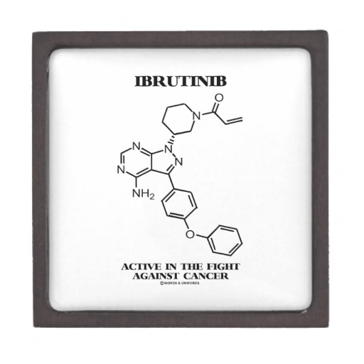
Bruton’s tyrosine kinase is essential for B-cell development and function. BTK deficiency in man or mice results in the B-cell specific immunodeficiencies X-linked agammaglobulinemia (XLA) or x-linked immune deficiency (xid), respectively. It is also implicated in the pathogenesis of B-cell cancers. Studies suggest that the levels of BTK represent a rate-limiting step in BCR signaling and thereby B-cell activation and survival. Therefore, inhibition of BTK in CLL could serve as an effective treatment strategy. In vitro studies reported that following inhibition of BTK with selective inhibitor CLL cells lose their resistance to apoptosis. Preclinical studies also demonstrated that that BTK-deficiency completely abrogated CLL development in mice.IBRUTINIBWith the accumulating evidence of the role of BCR pathway involving BTK in CLL, first targeted therapeutic approach for CLL was tested clinically with BTK inhibitors. A study published recently in The New England Journal of Medicine (July 4, 2013) by Byrd et al. reported a high frequency of durable remissions in patients with relapsed or refractory CLL with a BTK inhibitor, ibrutinib. A phase I study of ibrutinib (previously known as PCI-32765) showed mild-to-moderate toxicity and clinical antitumor activity in patients with relapsed or refractory B-cell cancers; 11 of the 16 patients in the study had CLL or small lymphocytic lymphoma. These preliminary results prompted the initiation of a phase Ib–II study of ibrutinib in CLL; this study involved two different therapeutic doses in patients with relapsed or refractory disease.

In the phase Ib-II multicenter study of ibrutinib, Byrd et al. (2013) assessed the safety, efficacy, and pharmacokinetics of this inhibitor in patients with CLL or small lymphocytic lymphoma (ClinicalTrials.gov number NCT01105247). Among 85 patients enrolled in this study, 51 received 420 mg and 34 received 840 mg ibrutinib orally once daily. In both cohorts the overall response rate was 71%. This treatment resulted in durable response were the 26-month estimated progression-free survival was 75% and the rate of overall survival was 83%. The pharmacodynamic study showed that ibrutinib was able to successfully inhibit BTK. However, disease progression was noted in 13% patients during follow-up. The most common toxicities observed during ibrutinib treatment were diarrhea, fatigue, and upper respiratory tract infection.

As compared to other single agent therapies for relapsed CLL this targeted therapy of BTK inhibition exhibited more durable responses. The durable remissions observed in this study suggest that many patients may be treated successfully with ibrutinib.

References:

1. Gribben, J.G. and S. O’Brien, Update on therapy of chronic lymphocytic leukemia. J Clin Oncol, 2011. 29(5): p. 544-50.

2.  Advani, R.H., et al., Bruton tyrosine kinase inhibitor ibrutinib (PCI-32765) has significant activity in patients with relapsed/refractory B-cell malignancies. J Clin Oncol, 2013. 31(1): p. 88-94.

3. Byrd, J.C., et al., Targeting BTK with ibrutinib in relapsed chronic lymphocytic leukemia. N Engl J Med, 2013. 369(1): p. 32-42.


FDA APPROVED NEW DRUG TO TREAT ADVANCED PROSTATE CANCER

Prostate cancer (PCa) is the second most common cause of male cancer-related death.  According to the National Cancer Institute, in the United States in 2013 the estimated new cases of prostate cancer would be 238,590 and deaths would be 29,720.

Bone metastases are a serious problem in men with advanced PCa. Bone metastases increase the risk of skeletal-related events (SREs) which include pathological fractures, spinal cord compression, bone pain. Both bone metastases and SREs are associated with an unfavorable prognosis and greatly affect quality of life.

Numerous studies have shown the importance of androgens (steroid hormones) in the development of PCa (although the exact role of androgen in PCa development is yet to be determined). Therefore, continuous androgen deprivation has been the standard therapy for metastatic hormone-sensitive disease. Despite a high response rate, resistance to androgen-deprivation therapy occurs in most patients, resulting in a median survival of 2.5 to 3 years. Thus, resulting in the development of castration resistant (CRPC) or hormone-refractory (HRPC) stage. Standard chemotherapy has not proven to be very effective in cases of metastatic CRPC, with a 10–20% response rate and approximately one-year median survival. In the United States docetaxel and cabazitaxel are the only Food and Drug Administration (FDA)-approved chemotherapies for the treatment of metastatic CRPC. Even though these drugs palliate symptoms, the overall survival benefit is moderate. In addition, a cellular immunotherapeutic agent sipuleucel-T (Provenge; Dendreon Corp) has been shown to increase overall survival period by 4.1 months on average but not progression-free survival time for patients with metastatic CRPC.

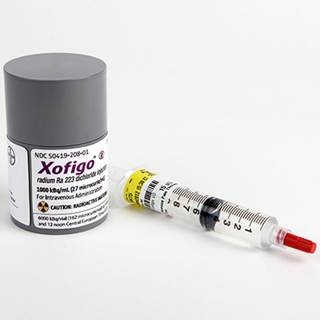
On May 15th, 2013, the U.S. FDA approved Xofigoradium Ra 223 dichloride (Xofigo®; Bayer HealthCare Pharmaceuticals) to treat men with metastatic castration-resistant prostate cancer with bone metastases after receiving medical or surgical treatment. The efficacy of Xofigo® was evaluated in a single clinical trial (Phase 3 ALSYMPCA trial) of 809 men with metastatic castration-resistant prostate cancer. Compared to the controls (patients received placebo plus standard care) with median survival of 11.2 months, patients who received Xofigo® lived a median of 14 months. The side effects noted during the clinical trials among patients treated with Xofigo® were nausea, diarrhea, vomiting, and swelling of the leg, foot, or ankle.

The alpha particle-emitting pharmaceutical Xofigo® is a radio-therapeutic drug. It mimics calcium and forms complexes with the bone mineral hydroxyapatite at areas of increased bone turnover, such as bone metastases. This drug is administered as an intravenous injection.

Overall, Xofigo® was found to extend the survival of men with metastatic prostate cancer and expected to be available in the clinic within a few weeks.


References:

1.         Yagoda A, Petrylak D: Cytotoxic chemotherapy for advanced hormone-resistant prostate cancer. Cancer 1993, 71(3 Suppl):1098-1109.

2.         Harrison MR, Wong TZ, Armstrong AJ, George DJ: Radium-223 chloride: a potential new treatment for castration-resistant prostate cancer patients with metastatic bone disease. Cancer Manag Res 2013, 5:1-14.

3.         Karantanos T, Corn PG, Thompson TC: Prostate cancer progression after androgen deprivation therapy: mechanisms of castrate resistance and novel therapeutic approaches. Oncogene 2013.

RESISTANCE TO ANTI-EGFR THERAPIES IN COLORECTAL CANCER

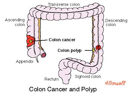
Colorectal cancer (CRC) originates in the tissues of the colon (the longest part of the large intestine), rectum and appendix, and is also known as colon cancer. Most CRCs are adenocarcinomas (cancers that begin in cells that make and release mucus and other fluids). According to the National Cancer Institute (NCI) the estimated new cases of colorectal cancer in the United States in 2013 will be 102,480.

Based on the genetics and etiology of the disease, CRC is usually classified into three specific types: sporadic, inherited, or familial.

Sporadic colorectal carcinomas: describe the imageAccount for approximately 70% of CRC. Sporadic carcinomas are devoid of any familial or inherited predisposition and are common in persons over 50 years of age.

Inherited colorectal carcinomas: This group of CRC includes those in which colonic polyps (an extra piece of tissue that grow in the colon) are a major manifestation of disease and those in which they are not. The nonpolyposis predominant syndromes include hereditary nonpolyposis CRC (HNPCC) (Lynch syndrome I) and the cancer family syndrome (Lynch syndrome II).

Familial colorectal carcinomas: This is the least understood pattern of CRC. In affected families, CRC develops too frequently to be considered sporadic but not in a pattern consistent with an inherited syndrome. Up to 25% of all cases of CRC may fall into this category.

Several studies suggested that like many other types of cancers accumulation of genetic changes were also associated with the development of CRC. Each of these event confers selective growth advantage, ultimately results in uninhibited cell growth, proliferation, and clonal tumor development. Two major mechanisms of genomic alterations that have been implicated in CRC development and progression are chromosomal instability and microsatellite instability. In addition, genes which have been implicated in the tumorigenesis of CRC include p53, p16, p14, APC, β-catenin, E-cadherin, Transforming Growth Factor (TGF)β, SMADs, MLH1, MSH2, MSH6, PMS2, AXIN, STK11, PTEN, DCC, and KRAS. Among these, oncogenic mutation of KRAS is considered a standard molecular biomarker that predicts the clinical benefit for targeted inhibition with epidermal growth factor receptor (EGFR) inhibitors. The EGFR-targeted monoclonal antibodies cetuximab and panitumumab are effective only in a subset of metastatic CRC, and 50% patients who initially respond to cetuximab or panitumumab develop resistance through KRAS mutations. Emergence of secondary resistance to anti-EGFR antibodies has also been implicated through expression of EGFR ligands, HER2 amplification, and deregulation of EGFR recycling process. Altogether, these account for 70-80% of the cases of resistance to anti-EGFR antibodies. This suggests that there might be additional mechanisms of resistance to these agents in CRC.

A recent study by Bardelli et al. (Cancer Discovery, June 6, 2013), the authors addressed the molecular basis of resistance to anti-EGFR therapy in CRC patients who did not develop KRAS mutations. In their study, Bardelli and colleagues identified amplification of the MET proto-oncogene responsible for acquired resistance. Presence of the MET amplicon was detected 3 months after therapy initiation in circulating cell-free DNA of CRC patients. The role of MET amplification in limiting the efficacy of anti-EGFR antibodies was further verified in preclinical CRC models and in patient-derived colorectal cancer xenografts. Marked tumor regression was observed in these models when tumors were treated with a MET inhibitor JNJ-38877605 combined with cetuximab. Therefore, collectively this study suggests that a CRC patient population developing resistance through MET amplification could benefit from combined treatment of MET inhibitor with anti-EGFR monoclonal antibody.

REFERENCES:

Bardelli A, Corso S, Bertotti A, Hobor S, Valtorta E, Siravegna G, Sartore-Bianchi A, Scala E, Cassingena A, Zecchin D, Apicella M, Migliardi G, Galimi F, Lauricella C, Zanon C, Perera T, Veronese S, Corti G, Amatu A, Gambacorta M, Diaz LA, Jr., Sausen M, Velculescu VE, Comoglio P, Trusolino L, Di Nicolantonio F, Giordano S, Siena S (2013) Amplification of the MET Receptor Drives Resistance to Anti-EGFR Therapies in Colorectal Cancer. Cancer Discov 3: 658-673.

Center MM, Jemal A, Smith RA, Ward E (2009) Worldwide variations in colorectal cancer. CA Cancer J Clin 59: 366-378.

Misale S, Yaeger R, Hobor S, Scala E, Janakiraman M, Liska D, Valtorta E, Schiavo R, Buscarino M, Siravegna G, Bencardino K, Cercek A, Chen CT, Veronese S, Zanon C, Sartore-Bianchi A, Gambacorta M, Gallicchio M, Vakiani E, Boscaro V, Medico E, Weiser M, Siena S, Di Nicolantonio F, Solit D, Bardelli A (2012) Emergence of KRAS mutations and acquired resistance to anti-EGFR therapy in colorectal cancer. Nature 486: 532-536.

Sameer AS (2013) Colorectal cancer: molecular mutations and polymorphisms. Front Oncol 3: 114.

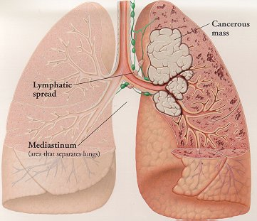
RESISTANCE TO LUNG CANCER THERAPY: A CASE REPORT

A study recently published in The New England Journal of Medicine (Jun 1st, 2013) identified an acquired mutation in the ROS1 kinase domain resulting in resistance to crizotinib in a woman with metastatic lung adenocarcinoma.

Crizotinib is an oral ATP-competitive selective Non Small cell Lung Cancer resized 600inhibitor of the anaplastic lymphoma kinase (ALK) and MET tyrosine kinase that inhibits tyrosine phosphorylation of activated ALK at nanomolar concentrations. In 2011, crizotinib was approved by the U.S. Food and Drug Administration (FDA) for treatment of patients with locally advanced or metastatic non-small-cell lung cancer (NSCLC) that are ALK-positive. Activating mutations or translocations of the ALK gene have been discovered in various types of cancer, including anaplastic large-cell lymphoma, neuroblastoma, inflammatory myofibroblastic tumor, and non–small-cell lung cancer. Because of its role in lung cancer, ALK receptor tyrosine represents a potential therapeutic target.

In addition to ALK mutations or translocations, chromosomal rearrangements in another tyrosine kinase receptor, ROS1, was identified in a molecular subset of NSCLC with distinct clinical characteristics that are similar to those observed in patients with ALK-rearranged NSCLC. Crizotinib was found highly sensitive in lung cancer patients who harbor rearrangements in ALK or ROS1. However, resistance to crizotinib was reported in lung cancer due to secondary mutations in ALK. To overcome this problem a new compound CH5424802 has been identified and is currently in clinical trials (ClinicalTrials.gov number, NCT01588028) for ALK-positive NSCLC.

A 48-year-old woman with metastatic lung cancer and a distant history of light smoking was initially treated with first line of chemotherapy with carboplatin and pemetrexed. Genetic analysis with patient’s cancer cells showed no mutation in oncogenic KRAS or EGFR and no ALK translocations. Additional molecular testing revealed ROS1 rearrangement lead to expression of a fusion protein CD74-ROS1. After three cycles of chemotherapy, marked disease progression was noted and patient’s condition deteriorated. The patient was then enrolled in a clinical trial evaluating the safety and efficacy of crizotinib in cancer patients with ROS1 translocations (ClinicalTrials.gov number, NCT00585195). Computed tomographic scan (CT) obtained two months after treatment noted dramatic response to treatment. However, one month later, while the patient was still taking crizotinib, disease progression was observed and unfortunately the patient expired. Molecular analysis of tumor samples from all sites of disease detected a mutation glycine to arginine Gly2032Arg (G2032R) spanning CD74-ROS1 fusion area that had not been observed in pretreated samples. No other mutation of ROS1 kinase was identified by deep sequencing. Thus this suggested that appearance of G2032R mutation was an early event in crizotinib-resistant tumor cells.

To identify role of G2032R mutation in crizotinib resistance, 293T cells were transfected with either mutated or nonmutated G2032R CD74-ROS1 and subsequently treated with tyrosine kinase inhibitors crizotinib and TAE648. Cells transfected with a mutated form of ROS1 exhibited a half-maximal inhibitory concentration (IC50) value greater than 1000 nM while for nonmutated cells it was approximately 30 nM for crizotinib and 50 nM for TAE648. Crystal structure analysis of ROS1 revealed an arginine at position 2032 resulted in steric interference of crizotinib binding. Collectively, this study reported a mechanism of acquired resistance to crizotinib in a cancer driven by oncogenic ROS1 fusion. Therefore, in the context of these observations, it may be necessary to identify novel compounds that specifically target the G2032R ROS1 mutant to overcome the development of crizotinib resistance in cancers driven by ROS1.


References:

1. Awad MM, Katayama R, McTigue M, et al. Acquired Resistance to Crizotinib from a Mutation in CD74-ROS1. N Engl J Med 2013.

2. Bergethon K, Shaw AT, Ou SH, et al. ROS1 rearrangements define a unique molecular class of lung cancers. J Clin Oncol 2012;30:863-70.

3. Sakamoto H, Tsukaguchi T, Hiroshima S, et al. CH5424802, a selective ALK inhibitor capable of blocking the resistant gatekeeper mutant. Cancer Cell 2011;19:679-90.


describe the image

Arup Chakraborty is postdoctoral research fellow at the National Cancer Institute, Bethesda, MD. He earned a doctoral degree from Texas Tech University, and his primary research interest is in the field of clinical cancer mainly in mechanisms of resistance to molecularly targeted therapies


FDA APPROAVES NEW DRUGS FOR MELANOMA

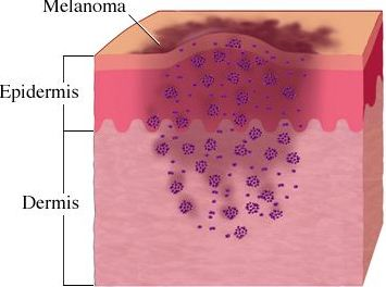
Melanoma is a type of skin cancer that arises from specialized pigmented cells in our body known as melanocytes, which are responsible for the production of melanin (a pigment responsible for skin and hair color). Because most melanoma cells still make melanin, melanoma tumors are usually brown or black. It accounts for 4% of all skin cancers; however, it is responsible for the largest number of skin cancer related deaths in the world. In the U.S, according to the national cancer institute, estimated new cases and deaths from melanoma in 2013 will be 76,690 and 9,480 respectively (for details please refer to my blog titled “targeting B-RAF kinase in melanoma”).

describe the image

BRAF is a serine/threonine protein kinase that activates the mitogen activated protein kinase (MAPK) signaling pathway. Approximately 50% of melanomas harbor activating BRAF mutations among which mutations at codon 600, resulting in substitution of glutamic acid for valine (B-RAFV600E), are the most prevalent. Activated BRAF phosphorylates and activates mitogen-activated protein kinase kinase proteins (MEK1 and MEK2), which then activate downstream MAP kinases. The MAPK pathway is implicated in the regulation of proliferation and survival of tumor cells in many cancers. This suggests that both B-RAF and its downstream MEK kinase could serve as attractive targets in cancer therapeutics. In 2011, the U.S. Food and Drug Administration (FDA) approved vemurafenib for the treatment of V600E B-RAF mutated melanoma patients. As a single agent, vemurafenib resulted in some degree of tumor regression among 90% of melanoma patients early in the course of treatment. Continuing with the effort to target B-RAF and MEK, several studies tested the efficacy of other compounds to inhibit these components of the MAPK pathway. Based on international clincal trials, on May 29th, 2013, the U.S FDA approved two new drugs Tafinlar (dabrafenib) and Mekinist (trametinib) for use in advanced melanomas with B-RAF V600E mutation. Mekinist is also approved for another form of B-RAF mutilated patients, V600K, which accounts for approximately 10% of B-RAF mutated metastatic melanoma. The mutation status of the melanoma patients are detected by an FDA-approved test, such as companion diagnostic assay from bioMerieus S.A., and THxID-B-RAF.

Tafinlar (dabrafenib) is an orally bioavailable B-RAF-inhibitor which selectively binds to and inhibits the activity of mutated B-RAF (V600E). The FDA approval of dabrafenib is based on an open label multicenter phase III study where 250 were randomly assigned to receive either dabrafenib (187 patients) or dacarbazine (63 patients). Dacarbazine is an alkylating agent which is also use to treat malignant melanoma. The study observed a statistically significant increase in progression-free survival (PFS) in patients treated with dabrafenib, compared to dacarbazine. With dabrafenib, the median PFS was 5.1 months and overall response rate was 52%. The most common adverse reactions with dabrafenib were skin-related toxic effects, fever, fatigue, arthralgia, and headache.

Trametinib is an orally bioavailable inhibitor of MEK which specifically binds to and inhibits MEK 1 and 2, resulting in an inhibition of growth factor-mediated cell signaling and cellular proliferation in various cancers. The FDA approval of trametinib is based on the phase 3 open-label trials which randomly assigned 322 patients who had metastatic melanoma with a V600E or V600K BRAF mutation to receive either trametinib or dacarbazine or paclitaxel (a mitotic inhibitor used in cancer chemotherapy). The study observed a statistically significant increase in PFS in trametinib treated patients compared to other treatments. The PFS was 4.8 month for patients treated with trametinib, while with other chemotherapeutic treatments it was 1.5 months. Rash, diarrhea, and peripheral edema were the most common toxic effects noted following trametinib treatment.

GlaxoSmithKline, manufacturer of both new drugs, reported that the products would be available no later than the early part of the third quarter of 2013.

NEW LIPID BIOMARKERS FOR HEPATOCELLULAR CANCER

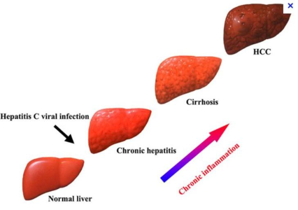
Hepatocellular carcinoma (HCC) is an aggressive form of primary liver cancer that occurs more frequently in men than women. This malignancy is different from metastatic liver cancer which originates in another organ (such as the breast or colon) and then spreads to the liver. Even though the incidence of this malignancy is exceptionally high in Asia and Africa, the number of new cases in America and Europe is rapidly increasing, making HCC a worldwide health problem. In spite of improvements in treatment, patients with HCC continue to have a poor prognosis, with 5-year survival rates of only 18%. Therefore, in order to formulate sustained therapeutic strategies, detailed understanding of the molecular network of aggressive HCC is required.

describe the image

In addition to significant genomic and proteomic alterations, cancer cells also exhibit highly unique metabolic phenotype which is characterized by increased glucose uptake, enhanced glycolytic activity, decreased mitochondrial activity, low bioenergetic status, and aberrant phospholipid metabolism. This suggests that metabolism may also play a significant role in differentiating normal cells from neoplastic tissues. Several metabolic markers of malignancy are described in particular tumors, such as N-acetyl aspartate and myo-inositol in brain cancers, citrate in prostate cancer, or triglycerides in liposarcomas, based on tissue-specific biochemistry. Cancer metabolite profiling, or cancer metabolomics, is a promising novel approach to help understand the biological events associated with cancer development and progression. A systemic analysis of the pathways in which these genes and biochemical molecules interact may assist in the identification of key biomarkers or drug targets for clinical intervention. Metabolite detection and quantification is usually carried out by nuclear magnetic resonance (NMR) spectroscopy, while mass spectrometry (MS) provides another highly sensitive metabolomics technology.

Using a combination of gene expression and metabolic profile analysis, a recent study by Budhu et al. (2013) reported identification of lipid biomarkers, monounsaturated lipid metabolite (MUPA) and stearoyl-CoA-desaturase (SCD), as key role players in a subset of HCC termed as hepatic stem cell HCC (HpSC-HCC). HpSC-HCC was found to exhibit stem cell–like gene expression traits and associated with poor prognosis as reported by Yamashita and colleagues. By performing metabolomics profiling of tumor and non-tumor tissue samples from 356 patients, Budhu et al. identified 28 metabolites and 169 genes associated with aggressive HCC. Using an integrative data analysis approach to determine gene-metabolite interconnections, this study suggested genes associated with fatty-acid metabolites may play roles in overall survival, stem cell-like HCC and metastasis-related prognosis. Higher expression of one of the genes stearoyl-CoA-desaturase (SCD) was found to be associated with worse survival and disease-free survival. SCD codes for an enzyme responsible for conversion of saturated palmitic acid (SPA) to its monounsaturated form, palmitoleic acid (MUPA). Based on these results, Budhu and colleagues sought to determine the mechanism by which SCD and its related fatty acids, MUPA and SPA, functionally contribute to aggressive HCC and how altering SCD activity may improve this effect. They noted elevated levels of MUPA in aggressive HCCs, and that MUPA enhanced migration and invasion of cultured HCC cells and colony formation by HCC cells, Huh7. Furthermore, HCC cells that had reduced SCD had decreased migration and colony formation in culture and reduced tumorigenicity in mice. Collectively this study suggested that SCD and its related metabolites may be valuable biomarkers and prognostic indicators for molecular re-staging of HCC.


References:

1. Griffin JL, Shockcor JP. Metabolic profiles of cancer cells. Nat Rev Cancer 2004;4:551-61.

2. Griffin JL, Kauppinen RA. A metabolomics perspective of human brain tumours. FEBS J. 2007;274:1132-9.

3. Costello LC, Franklin RB. ‘Why do tumour cells glycolyse?’: from glycolysis through citrate to lipogenesis. Mol Cell Biochem. 2005;280:1-8.

4. Serkova NJ, Glunde K. Metabolomics of cancer. Methods Mol Biol. 2009;520:273-95.

5. Budhu A, Roessler S, Zhao X, et al. Integrated metabolite and gene expression profiles identify lipid biomarkers associated with progression of hepatocellular carcinoma and patient outcomes. Gastroenterology. 2013;144:1066-1075.e1.

6. Yamashita T, Ji J, Budhu A, et al. EpCAM-positive hepatocellular carcinoma cells are tumor-initiating cells with stem/progenitor cell features. Gastroenterology. 2009;136:1012-24.