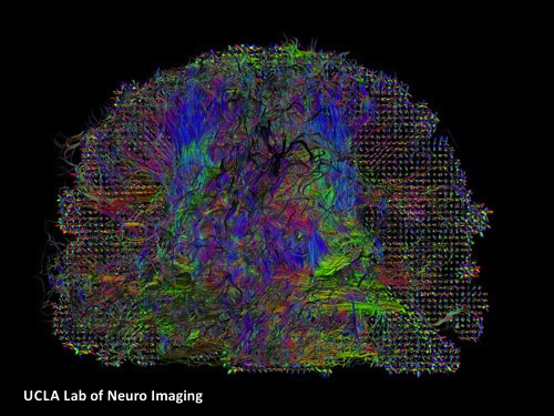
Klotho’s Potential to Reverse MS Demyelination

Klotho is a type I transmembrane protein, expressed in the brain, kidneys and reproductive organs; it is named after “Clotho”, a goddess from Greek mythology who “spins the thread of life” (the length of the threat is determinant of how long a certain individual will live). This denomination is due to the direct positive correlation between Klotho’s expression levels and life span/anti-aging phenotypes.

Klotho protein exists in two forms, membrane Klotho and secreted Klotho, and each form is associated with distinct functions. Some of Klotho’s age-suppressing functions include regulation of fibroblast growth factor-23 (FGF23) signaling, inhibition of insulin/insulin-like growth factor (IGF-1) and Wnt signaling pathways, and regulation of multiple cell-surface ion channels and growth factor receptors.

In contrast to other organ systems, the downstream effects of Klotho have not been as extensively studied within the central nervous system (CNS). This is surprising, considering that not only Klotho is present in serum and cerebrospinal fluid (CSF), but also is highly expressed in the hippocampus, choroid plexus and neurons. Disruption of the myelin sheath, either by activated pro-inflammatory cells or by protein defection within the oligodendrocytes has been previously described in aging brain, but the underlying factor stimulating the disruption was not clear. However, In 2008, Abraham’s group reported the significance of Klotho in age-related cognitive decline (ARCD), showing reduced expression of Klotho in regions of brain white matter as a function of age.

Multiple Sclerosis,White matter,Brain,Alzheimer's,cognition


In January 2013, Abraham’s group reported their new findings regarding the effects of Klotho in oligodendrocyte maturation and CNS myelination, and its relation to ARCD. They showed Klotho‘s role in inducing oligodendrocytic progenitor cells (OPCs) maturation, by enhancing the expression of myelin proteins, such as myelin-associated glycoprotein (MAG), myelin basic protein (MBP), oligodendrocyte-specific protein (OSP/Claudin11), and proteolipid protein (PLP). Based on their in vivo studies, the loss of Klotho expression is correlated to defects in myelin that result in similar progressive axonal degeneration observed in hypomyelinating and demyelinating diseases, such as multiple sclerosis (MS).

Previous studies have shown Klotho’s role in facilitating removal of reactive oxygen species (ROS) and increasing resistance to oxidative stress. Furthermore, Nagai’s team observed impaired cognitive function in Klotho-deficient mice, as well as improved cognition upon treatment with the α-tocopherol antioxidant. Thus, Abraham’s group concluded that Klotho protein may function as a neuroprotective factor in the CNS through its antioxidative stress effect.

Together, these results provide strong evidence for Klotho protein as a key player, not only in myelin maintenance, but also in supporting oligodendrocyte and OPC function in the CNS; this makes Klotho an important member of the family of proteins responsible for neuron-oligodendrocyte communication. Abraham’s group hypothesized that downregulation of Klotho may be accountable for the observed white matter myelin degeneration-mediated ARCD, hence increasing Klotho protein expression can potentially prevent damage/protect myelin in the aging brain.

Brain,memory loss,Alzheimer's,MS

Abraham’s new findings are an exciting and important initial step towards development of new neuroprotective therapeutic strategies, such as induction of endogenous remyelination, for treatment of CNS diseases characterized by oligodendrocyte cell loss, such as MS and Schizophrenia. Additionally, early defects of insulin/IGF-1 receptor signaling in Alzheimer’s disease (AD), including the deficit of glucose metabolism that anticipates cognitive decline, may be partially due to deficiencies in Klotho levels. Further investigation on the precise mechanisms involved in Klotho’s regulation within the CNS seems promising for the future of neurodegenerative disease therapy.


Further Readings:

The Antiaging Protein Klotho Enhances Oligodendrocyte Maturation and Myelination of the CNS

Gene Profile Analysis Implicates Klotho As An Important Contributor To Aging Changes In Brain White Matter Of The Rhesus Monkey

The Putative Role of The Antiageing Protein Klotho in Cardiovascular and Renal Disease

Mesenchymal Stem Cells in the Brain

Mesenchymal stem cells (MSCs) are multipotent cells present in, and can be isolated from a variety of adult tissues, such as bone marrow, umbilical cord blood or adipose tissue. MSCs have a number of advantageous characteristics that have made them a promising candidate for use in the new generation of cell-replacement therapy (CRT), even for central nervous system (CNS) disorders. Major obstacles to CRT in CNS disorders include successful delivery of therapeutic/stem cells to the damaged area/lesions within the CNS, host’s immune response against the allogenic cells and the possibility of ectopic tissue formation. MSCs exhibit unique characteristics which can overcome these obstacles, such as MSCs’ capacity to differentiate into multiple tissue-specific lineage cells, their role as progenitor-cell bioreactors of soluble factors that promote tissue regeneration from the damaged tissue and their immunomodulation capacity. Moreover, MSCs are considered immunoprivileged, meaning they have low expression of class II Major Histocompatibilty Complex (MHC-II) and other immune-stimulatory molecules on their cell surface. The focus of this discussion is on the utilization of MSCs in CNS-disease therapy, in particular multiple sclerosis (MS) and ischemic stroke (IS) .

Bone marrow derived (BM-MSCs) and adipose derived mesenchymal stem cells (AD-MSCs) have shown promising efficacy in an experimental autoimmune encephalomyelitis (EAE) preclinical model of MS, as well as the permanent middle cerebral artery occlusion (pMCAO) model of IS, respectively.

IS occurs as a result of an obstruction within a blood vessel supplying blood to a particular region of the brain, causing neuronal and astroglial damage within the immediate region. Replacement of these cells along with repairing the damaged tissue has been of great interest, turning IS clinical research towards stem cell therapy.

Stroke,stem cell therapy,regenerative therapy,cell replacement,CNS

According to the recent study conducted by Gutierrez-Fernandez’s group, AD-MSCs are as restorative as BM-MSC in promoting recovery, repair and brain protection in IS rat models. They showed significant functional recovery, decreased apoptosis and increased expression of neurogenesis, synaptogenesis, angiogenesis and oligodendrogenesis markers, following the intravenous administration of allogenic AD-MSC and BM-MSC. Although these results suggest a less invasive route for administration of therapeutic cells, further studies addressing the fate of the administered cells are required, prior to clinical consideration of this method.

MS is a chronic, immune-mediated demyelinating disease of the CNS, characterized by demyelinated plaques within the brain and spinal cord. MS plaque formation consists of immune-cell infiltration, damage to oligodendrocytes and their subsequent failure to remyelinate, degeneration of axons, and ultimately astrocytosis. Up to date, there is no cure for MS; the current disease modifying therapies utilized to reduce the frequency/severity of relapses are limited to immunomodulatoion and are only partially effective. Thus, MS researchers have turned their attention to discovering potential therapeutics that will not only stop the autoimmune attacks, but also replace the destroyed CNS cells with properly functioning ones, through CRT.

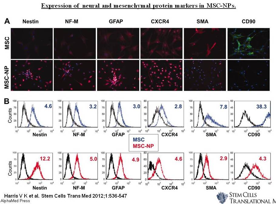
Nonetheless, several recent studies have reported promising results regarding autologous, culture-expanded MSC transplantation in MS models. Based on Harris’s publication in Stem Cells Translational Medicine, intrathecal delivery of Bone marrow mesenchymal stem cell-derived neural progenitors (MSC-NPs) is a promising strategy for cell-based therapy in MS. They show that MSC-NPs derived from both, MS patients and healthy controls, uniformly displayed properties that support MSCs’ therapeutic potential in the CNS, regardless of the donor disease status. Like MSCs, MSC-NPs secrete immunomodulatory factors (such as cytokines and growth factors, including TGF-β, IL-6, IL-10, HGF, heme oxygenase-1, and nitric oxide) which inhibited T-cell proliferation and promoted naïve CD4+ T-cell polarization into FoxP3+ T cells. MSC-NPs also exhibit trophic effects similar to MSCs, by secreting trophic factors that promote oligodendroglial differentiation of neural stem cells (HGF, IGF-1, SDF1α, and VEGF). Furthermore, it was reported that MSC-NPs are neuroectodermally committed and have reduced capacity for mesodermal differentiation (reduced risk of abnormal tissue formation), which makes them a more suitable candidate for cell-based therapy in CNS injury.

Stem Cell Therapy,Multiple sclerosis,MS,Cell replacement therapy

One of the significant aspects of this study is the possibility of utilizing MS-patient’s own cells as the therapeutic cell source; not only because of the reduced immune response, but also due to the evidence of genetic stability of the adult stem cells present in these patients. This suggests several promising genetic and cellular therapeutic strategies to be investigated in the near future.

Further readings:

Characterization of autologous mesenchymal stem cell-derived neural progenitors as a feasible source of stemcells for central nervous system applications in multiple sclerosis.

Effects of intravenous administration of allogenic bone marrow- and adipose

tissue-derived mesenchymal stem cells on functional recovery and brain repair
markers in experimental ischemic stroke

Mesenchymal stem cell transplantation in multiple sclerosis.

Immunosuppressive properties of mesenchymal stem cells: advances and applications.

Clinical Significance of CD133 and KLK6 in Malignant Brain Tumors

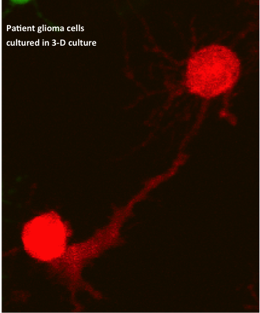
As mentioned in a previous article about Glioblastoma Multiforme (GBM), there are several obstacles that make these lethal brain tumors virtually incurable. Two hallmarks of malignant brain tumors are believed to be the major impediments to effective killing of all tumor cells via the current aggressive course of treatment: glioma cells’ exceptional invasivity and the heterogeneity of GBM, not only among individual patients, but also within a single tumor mass. Thus, these two glioma characteristics have generated great interest in GBM clinical research.

brain tumors,cancer research,glioblastoma,glioma

The malignancy of CNS tumors is categorized by the World Health Organization (WHO) grading system, ranging from grade I- grade IV based on the tumor’s “aggressiveness” (GBM is WHO grade IV astrocytoma).  However, unlike other neoplasms -in which local dissemination is usually limited and metastasis occurs via the vasculature or the lymphatic system– single glioma cells travel for several centimeters through adjacent brain tissue but will almost never establish systemic metastasis. Thus, the WHO tumor grade positively correlates with proliferation rate, rather than tumor invasion. This suggests the involvement of several independent genetic events that eventually lead to glioma progression, which is also consistent with the high levels of GBM heterogeneity. In order to develop a therapeutic agent that targets (finds and destroys) malignant glioma cells, it’s necessary to find a marker that’s expressed exclusively on the surface of these cells. Due to the high heterogeneity of GBM, as well as variations amnog individual patient brain tumors, no such marker has yet been identified.

There are compelling evidences of a subpopulation of malignant cells in GBM, which exhibit stem-cell like characteristics, such as multipotency, the ability to self-renew and invade; these tumor-initiating cells are referred to as cancer stem cells (CSC) and are believed to be responsible for tumor recurrence in GBM patients. Thus, researchers are no longer focusing on a single mutation/marker. Furthermore, it is becoming prominently important to rely on primary GBM patient samples for collecting data, since glioma cell lines do not represent the cellular and molecular components characteristic of the primary GBM.

CD133 (also known as Prominin-1 or AC133) is a penta-spanning membrane protein with two heavily glycosylated extracellular loops, and is recognized as a stem cell marker for certain normal and cancerous tissues. CD133 accumulates near the Golgi and ER and is also expressed on the cell surface. Although many previous studies reported that the presence of CD133+ glioma CSC subpopulation in GBM, drive tumor formation and its rapid proliferation, subsequent conflicting findings showed CD133-negative glioma cells’ ability to self-renew and form tumors in xeno-transplantation assays.

The recent findings by Brescia’s group confirm that cell-surface CD133 expression is a marker for self-renewing and tumor-initiating GBM cells, but a non-essential element for stem cell properties in all GBM cases. They show that even though membrane-bound CD133 were detectable only in a fraction of the patient samples (neurospheres and in freshly dissociated tumors), CD133 mRNA and intracellular CD133 protein were found expressed at high levels in almost all the examined neurosphere samples.

Through clonal analysis, they demonstrated the intrinsic capabilities of sinlge glioma cells and thier progeny: every clone derived from a single CD133-negative cell, contained a mixture of both CD133-negative and CD133-positive cells. Furthermore, they show an interesting interconvertible regulation of cell-surface CD133, through subcellular localization between the cytoplasm and the plasmamembrane. The localization of CD133 is likely determined by the tumor-associated microenvironmental cues.

The findings in this study have significant value in GBM research, taking one step closer to understanding the progression of malignant gliomas. However, the existence of the cytoplasmic CD133 reservoir and the re-cycling of this protein to the plasmamembrane and vise versa, hinders the therapeutic potential of using CD133 as a glioblastoma-targeting marker.

Patient samples,brain tumor,cancer research,GBM

In a separate study published in Neuro-Oncology on the same week, researchers at Mayo Clinic introduced a significant association between malignant gliomas and Kallikrein 6 (KLK6) enzyme. KLK6 is a member of the kallikrein family of secreted serine proteases and has been reported to be elevated within areas of inflammation in CNS, suggesting its regulated expression with T-cell activation. Notably, the serum of Multiple Sclerosis (MS) patients contains elevated KLK6 levels as well. This study shows that as the KLK6 expression levels increase, the post-surgery survival times of GBM patients decrease. They found the highest levels of KLK6 were present in the most severe GBM. These results were obtained by looking at 60 samples of grade IV astrocytomas (thus categorized as GBM), and a less aggressive grade III astrocytomas.


Scarisbrick’s group also showed the possible role of KLK6 in promoting survival of malignant glioma cells, as well as increasing their resistance to apoptosis-inducing agents, such as radiotherapy (RT) and temozolomide (TMZ). The data supporting the pro-survival effect of KLK6 may introduce a new GBM therapeutic strategy, albeit the supporting experiments of these observations were conducted on U251 glioma cell line. If future studies report similar results that confirm the ability of KLK6 enzyme to promote primary patient tumor cells’ survival, then developing a therapeutic agent which targets KLK6’s function is a promising addition to GBM course of treatment ensuing the surgical resection of the tumor, preceding chemo-and radiotherapy.

Further Reading:

CD133 is essential for glioblastoma stem cell maintenance.

CD133 as a marker for regulation and potential for targete therapies in glioblastoma multiforme.
Clinical significance and novel mechanism of action of kallikrein 6 in glioblastoma.
Functional role of kallikrein 6 in regulating immune cell survival.
 

New in MS Research: Tip-Dendritic Cells Promote Inflammation

Multiple Sclerosis (MS) is a chronically progressive, neuroinflammatory autoimmune disease of the central nervous system (CNS), believed to be antigen-driven and predominantly T-cell-mediated. MS is characterized by demyelinated areas or patches of sclerosis (plaques) localized within the brain and spinal cord.

The normal physiological state of CNS is considered an anti-inflammatory environment, or “immune privileged”, which is partly due to passive-entry restriction of peripheral immune cells. During MS pathogenesis myelin-specific T-cells overcome these barriers, enter the CNS and recruit inflammatory cells that will eventually target and destroy the myelin protein, which leads to axonal damage.

Cytotoxic T-cells,Multiple Sclerosis,CD4 T-Cells

Although the initial trigger leading to the development of myelin-specific T-cells in MS is still not clear, it has been shown that MS patients’ blood and cerebrospinal fluid (CSF), contain activated myelin‐reactive CD4+ T-cells, whereas only non-activated myelin‐reactive T-cells are present in non-MS samples. 

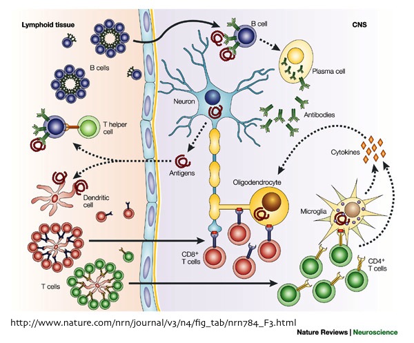
Both CD4+ and CD8+ T cells have been observed in MS acute and chronic lesions, respectively. Nonetheless, these two T-lymphocytes are activated by different CNS-resident antigen‐presenting cells (APCs) that trigger the recruitment of innate immune cells through presenting myelin antigens to CD4+ and CD8+ T cells, leading to the subsequent “determinant spreading”.

Previous studies have identified cells that present myelin to CD4+ T-cells: once inside the CNS, the CD4 molecule of autoreactive CD4+ T-cells binds to a non-polymorphic site on the major histocompatibility complex (MHC) class II, which is expressed by local myelin-presenting dendritic cells (DCs). In the absence of inflammation in CNS, there is a very low constitutive expression of MHC molecules, which are often present on cells of the lymphoid system.

In contrast CD8 molecule of the CD8+ T-cells binds to the MHC class I molecule, which serves to present specific antigens to the T-lymphocytes’s T-cell receptor (TCR). Thus, CD8+ T-cells (aka. cytotoxic T-cells) are involved in class I‐restricted lysis of antigen‐specific targets. However, until recently the APCs responsible for activating myelin-specific CD8+ T-cells were not known.

In January 2013, Goverman’s group from University of Washington showed that during experimental autoimmune encephalomyelitis (EAE)-an animal model of MS initiated by CD4+ T-cell- Tip-dendritic cells (Tip-DCs) play a major role in activating naive CD8+ T-cells. Based on this study, CD8+ T-cells are presented with MHC class I–restricted myelin basic protein (MBP) and activated by CD11b+ Tip-DCs. In addition, it was reported that under the inflammatory conditions of EAE, oligodendrocytes also presented MBP, to the CD8+ T-cells, which made them recognizable targets of the activated myelin-specific CD8+ T-cells, leading to sustained chronic inflammation (aka. Determinant spreading). This suggests that myelin-specific CD8+ T-cells may be responsible for the ongoing axonal destruction in “slow burning” MS lesions by directly lysing oligodendrocytes.

Tip-Dendritic Cells,CD8+ T-cells,Multiple Sclerosis

Tip-DCs are likely derived from the inflammatory monocytes that have accumulated in the brain and the spinal cord during EAE. Goverman proposed that one possible mechanism of acquiring MBP by Tip-DCs is via phagocytosing and processing myelin debris or dead oligodendrocytes, and then presenting the myelin peptides. Furthermore, they also hypothesized that CD8+ T-cells activated by Tip-DCs may contribute to the immune cascade amplification by secreting additional Interferon-gamma (IFN-γ) within CNS.


Identifying specific DCs involved in antigen-presenting and activation of all T-lymphocytes involved in the neuroinflammatory response is important for the development of potential autoimmune disease therapies that target immunogenic DC functions. Although Goverman’s findings may seem marginal on the surface, it is in fact a big step forward in understanding the etiology of MS; further investigations are required to address the origin of these Tip-DCs and the precise mechanisms through which they become myelin-presenting cells. Also, future studies in human MS are essential to confirm Tip-DCs’ reported functions as well as their interactions with patients’ oligodendrocytes.

Further reading:

MHC class I-restricted myelin epitopes are cross-presented by Tip-DCs that promote determinant spreading to CD8(+) T cells.

Antigen Presentation in the CNS by myeloid dendritic cells drives progression of relapsing experimental autoimmune encephalomyelitis. 

Dendritic cell CNS recruitment correlates with disease severity in EAE via CCL2 chemotaxis at the blood–brain barrier through paracellular transmigration and ERK activation.


Cell-Based Therapies for Malignant Brain Tumors

Malignant gliomas are classified by the World Health Organization (WHO), as grade IV tumors of neuroepithelial tissue and are the most common and deadly intracranial tumors, accounting for more than 70% of all brain tumors. One of the most prominent intrinsic behaviors of Glioblastoma Multiforme (GBM) is its invasiveness within the host’s central nervous system (CNS), in which single glioma cells travel a distance from the tumor mass and invade adjacent brain tissue

Due to the exceptional migratory ability of glioma cells, surgical resection of the tumor is almost always followed by tumor recurrence with foci located as close as 1 centimeter from the resection cavity or as far as the contralateral hemisphere. Despite advancements in surgical techniques and post-operational delivery of chemotherapeutics and radiation, the prognosis for glioma patients remains dismal making these lethal tumors virtually incurable. Although the history of glioma treatment dates back to the 19th century, the median survival of patients remains less than 14 months post-diagnosis. Thus, the only way to cure GBM is by essentially eliminating all glioma cells, including the single cells which have disseminated within the parenchyma, away from the tumor mass.

A promising potential strategy to treat high-grade brain tumors, is through cell-based therapies (CBTs) that incorporate autonomous tracking of tumor cells. Two examples of CBTs, which are currently under investigation in brain tumor clinical trials, include: 1) employing of genetically engineered neural stem cells (NSCs) as target-specific therapeutic-agent delivery vehicles, and 2) the adoptive transfer of tumor-specific, genetically engineered cytotoxic T-lymphocytes (CTLs).


The significance of NSCs and CTLs for treating CNS diseases: 

One anatomical feature unique to the CNS is the presence of the blood brain barrier (BBB), which restricts the access of many compounds including many chemotherapeutic agents into the CNS.  Thus, the BBB prevents effective drug delivery from the circulatory system to the tumor sites within the brain.  Even when the drug is administered intracranially to overcome the limitations presented by the BBB, the densely packed environment of brain’s parenchyma inhibits effective diffusion of the drug throughout the brain and prevents the drug from reaching the tumor cells. These anatomical properties of the CNS are also responsible for inefficient distribution of gene therapy in the brain.

NSCs readily cross the BBB and intrinsically target invasive tumor cells that have migrated away from the tumor mass in vivo. The HB1.F3 NSC line, developed by Dr. Karen Aboody, is one clonally derived human cell line that is particularly well characterized and is used clinically for glioma therapy. Another advantage of this therapeutic model is its efficacy through both intracranial and intravenous administration, without rejection elicited by the host’s immune system against the NSCs; this is due to HB1.F3 NSCs’ low levels of MHC Class I antigen expression and undetectable levels of MHC Class II antigens.

Neural stem cell targeting and killing a brain tumor cell

Previous studies have indicated CTLs’ ability to target and kill GBM, medulloblastomas and therapeutically resistant subpopulations of glioma stem–like cancer-initiating cells (GSC), which express interleukin-13 receptor α2 (IL13Rα2). Although the BBB is not permeable to CTLs, Dr. Christine E. Brown reported a non-toxic strategy of delivering these T-cells to the CNS tumors, by placing a fibrin matrix-embedded with CTLs, in the resection cavity during surgery.

CD4 T-cell

In vivo,secretion of monocyte chemotactic protein-1 (MCP-1) also known as chemokine C-C motif ligand 2 (CCL2), by the cancer cells attract CD4+ and CD8+ T-cells, leading to the subsequent host antitumor immune response. Brown’s group has shown that the same tumor-secreted chemoattractants will recruit genetically engineered CTLs. Since CTLs migrate freely within fibrin matrices, the presence of MCP-1 in the surrounding environment attracts the CTLs to migrate out of the fibrin.  In their in vitro model, the IL13Rα2-specific T-cells successfully migrated out of the fibrin clot and killed the surrounding glioma cells.

Utilization of fibrin matrices allows a safe, non-toxic delivery of CTLs, without causing additional injury (i.e. injury caused by a catheter or needle injection) and inflammation to the brain tissue.

There are other therapeutic approaches for treatment of malignant brain tumors. Following are a list of further readings on the content of this article, as well as other current cancer research studies in regards to GBM:


Auffinger, B. et al. “New Therapeutic Approaches for Malignant Glioma: In Search of the Rosetta Stone.” F1000 Med Rep 4.18 (2012): doi: 10.3410/M4-18.

Brown, C.E., et al. “Stem-like tumor-initiating cells isolated from IL13Rα2 expressing gliomas are targeted and killed by IL13-zetakine-redirected T cells.” Clinical Cancer Research, 18 (8), (2012) pp. 2199-2209

Khosh, N., et al. “Contact and Encirclement of Glioma Cells in vitro is an Intrinsic Behavior of a clonal Human Neural Stem Cell Line.” PLoS ONE7 (12) (2012) . doi:10.1371/ journal.pone. 0051859

Zou, Z., et al. “Cytotoxic T Lymphocyte Trafficking and Survival in an Augmented Fibrin Matrix Carrier.” PLoS ONE, 7(4) (2012). doi:10.1371/journal.pone.0034652

Aboody, K., et al. “Translating stem cell studies to the clinic for CNS repair: current state of the art and the need for a Rosetta stone.” Neuron, 26 May 2011 (Vol. 70, Issue 4, pp. 597-613)

The BBB’s Role in Multiple Sclerosis & Alzheimer’s Disease

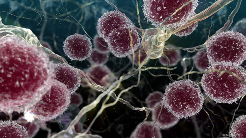
The blood-brain barrier (BBB) is a highly complex structure of the cerebral vasculature; it functions as a selective filter for certain molecules entering and exiting the brain. Through regulating the exchange of very specific molecules and cells between the central nervous system’s Cerebral Spinal Fluid (CSF) and the peripheral blood of the circulatory system, the BBB protects the brain and maintains the scrupulous environment required for neurons and other glial cells to function properly. The significant consequence of BBB disruption is the increased permeability, leading to extravasation of circulating peripheral blood mononuclear cells (PBMC), along with other unspecific molecules. Thus, BBB dysfunction plays a major role in a wide range of neurologic conditions, including Multiple Sclerosis (MS) and Alzheimer’s disease (AD).

MS is associated with the infiltration of CD4+ and CD8+ T-cells and B-cells within the acute inflammatory lesions or the areas of demyelination. The presence of these immune cells at these locations, indicate alterations in BBB structure, which allowed their crossing into the central nervous system (CNS).

During MS, perivascular lesions are formed by T-cells entering the CNS and releasing cytokines that cause inflammation of the endothelial lining (cells forming the blood vessels). This inflammatory response stimulates vasculature in MS lesions to express several cell adhesion molecules (CAMs). The interaction between these CAMs and their leukocytic integrins, permit immune cells to adhere to the inflamed cerebral vasculature, cross the BBB, and proceed to infiltrate the parenchyma. This inflammatory reaction is a vicious cycle; as more immune cells are activated within the CNS, the inflammation amplifies, leading to further BBB damage and recruitment of additional lymphocytes, such as B-cells and cytotoxic T-cells, responsible for the eventual activation of associated macrophages to destroy Myelin. In addition, it has been reported that the firm attachment of monocytes to endothelial cells, stimulates monocytes to secrete reactive oxygen species (ROS) that may further increase BBB permeability to T-cells  and macrophages.

The initial stimulant of MS is not yet clear. Nonetheless, many recent studies suggest that BBB disruption is an early event in MS lesion formation, followed by the massive infiltration of immune cells, which proceed to destroy the myelin and damage the oligodendrocytes.  

T-cells,Multiple Sclerosis,MS,Immune response

Several cerebrovascular abnormalities, such as damaged endothelial and pericyte, microvascular degeneration, reduced glucose transport across the BBB, and abnormal expression of inflammatory markers in the cerebral vasculature have been described in Alzheimer’s disease patients. These observations are associated to the deposition of the β-amyloid peptide (Aβ) in the walls of the BBB vasculature which lead to the loss of certain tight junction proteins that result in disruption of the BBB and eventual cerebral neuroinflammation through activating microglia

The two pathological hallmarks of AD are: 1) the increased production and accumulation of amyloid-β peptides (Aβ)- derived from amyloid precursor protein (APP)- forming neuritic/senile plaques in the brain tissue, 2) the intraneuronal neurofibrillary tangles (NFTs) composed of hyperphosphorylated tau protein, which lead to the loss of synapses and neurons in affected regions. Consequently, these factors in addition to a reduction of Aβ clearance from the brain, which leads to its extracellular accumulation, and its direct toxic effects, induce the subsequent activation of microglial cellsthis inflammatory response results in the release of numerous inflammatory neurotoxic cytokines that will disrupt the neurons’ cytoskeleton, leading to their dysfunction and eventual cellular death.

Alzheimer's disease associated senile plaques

Recent studies suggest that transforming growth factor-β1 (TGF-β1) may play a role in BBB dysfunction in AD pathogenesis. TGF-β1 has a constitutive role in the suppression of inflammation and control microglial activation in the CNS. These observations indicate a significant impairment of TGF-β1 signaling in AD brain. Furthermore, astrocytes may be reactivated by TGF-β1, which would result in further production of Aβ and undergo the aggravating astrogliosis. 

Although it is not yet clear whether these vascular changes are an initial cause for development of AD or if they take place during the later stages of the disease, most Alzheimer’s  patients exhibit vascular pathology and develop  intracerebral hemorrhage and cerebral infracts 


In conclusion, there is strong evidence pointing to the involvement of the BBB dysfunction in many neurologic disorders including MS and AD. Moreover, one common denominator of the BBB malfunction in these diseases is the close association of the astrocytes activated by the interactions between the immune cells and the cerebral vasculature, leading to further increased permeability of BBB. However, further investigations must focus on identifying the mechanisms responsible for the initial cause of the the inflammatory cascade, and the destructive interactions between reactivated astorcytes and endothelial cells lining the BBB. 


Further Reading:

Current and future treatments for Alzheimer’s disease. Yiannopoulou KG, PapageorgiouSG. (2013) Ther Adv Neurol Disord, 6(1) 19–33.

Inflammatory events at blood–brain barrier in neuroinflammatory and neurodegenerative disorders: Implications for clinical disease. de Vries HE, Kooij G, Frenkel D, Georgopoulos S, Monsonego A, Janigro D. (2012) Epilepsia, 53(Suppl. 6):45–52.

New concepts in the immunopathogenesis of multiple sclerosis. Hemmer B, Archelos JJ, Hartung HP(April 2002) Nature Reviews Neuroscience, 3291-301.  

Neuroinflammation and blood–brain barrier changes in capillary amyloid angiopathy.Carrano A, Hoozemans JJ, van der Vies SM, van Horssen J, de Vries HE, Rozemuller AJ. (2012) Neurodegener Dis, 10:329– 331.