Techniques in Cytology – Cytospin: Distinguishing Benign Cells from Malignant Cells

Cytology is a key component in diagnosis and screening of diseases such as cancer. It assesses single cells and clusters of cells from sources such as malignant effusions and peripheral blood. Effusions are fluids that leak from blood and lymph vessels and aggregate in tissues and cavities within the body. This is a common problem in cancer patients and can be a reservoir of malignant cells. However, the total number of cells in effusions is small in comparison to the volumes of fluids that are produced. Therefore, in order to collect these cells for evaluation, they must be concentrated.

Cytospin is a cytology method that is specifically designed to concentrate cells such as these that are found in small numbers. After the cytospin is completed, other cytology methods such as immunocytochemistry can be preformed to evaluate the cells. An example is a recent study from Ikeda et al., in which immunocytochemical staining of cytospins from malignant effusions suggested that EMA, IMP3, and GLUT-1 might be helpful in distinguishing malignant cells from benign cells (1). The cytospin process is a simple procedure. Cells are washed in a serum and/or albumin based PBS or culture media solution. The cells are resuspended in up to 500 µl of this solution. A cytofunnel is attached to a glass slide and slide carrier. The entire apparatus is inserted into a cytocentrifuge and the cell suspension is added.

The cells can be spun at various speeds and times depending on the cell type. Eight hundred rpm for up to 5 minutes is a good place to start. The volume to cell ratio must be dilute enough to ensure formation of a monolayer of cells for the best assessment of the cells.

In the Ikeda et al. study, cytology was used to differentiate benign mesothelial cells from malignant mesothelial cells (1). What are mesothelial cells and why is it important to distinguish between benign and malignant forms? These are cells that make up the epithelial lining of the mesothelium which covers the surface of the peritoneal, pericardial, pleural cavities as well as the organs within the cavities (2). The mesothelium has many functions. It is a protective barrier against physical damage and invading organisms as well as being a frictionless interface for free movement of organs and tissue (2). Other functions include roles in fibrinolysis and coagulation, initiation and resolution of inflammation and tissue repair, antigen presentation, transport of fluid and cells, and tumor cell adhesion and growth (2). Reactive mesothelial cells (benign cells) appear in instances when there is a pathogenic infection or physical trauma. Malignant mesothelioma is a rare cancer of mesothelial cells. In effusions, it is sometimes difficult to distinguish benign cells from malignant cells especially when there are few cell numbers. However, immunocytochemistry performed on cytospin cells helps to differentiate these cells. Biomarkers such as epithelial membrane antigen (EMA), glucose transporter-1 (GLUT-1), and insulin-like growth factor-II mRNA-binding protein 3 (IMP3) are useful in separating benign cells from malignant cells. EMA is a large cell surface glycoprotein, also known as MUC1, expressed by glandular and ductal epithelial cells as well as some hematopoietic cells. It has a protective and regulatory role by acting as a barrier to the apical surface of epithelial cells. GLUT-1 is a protein that assists in the transport of glucose across the plasma membrane of cells. It is decreased when glucose levels are high and increased when levels are low. IMP3 binds to sequences in the 3’-UTR of CD44 mRNA. CD44 has a role in cell migration, cell adhesion, and cell-cell interactions. In the Ikeda study, EMA staining intensity on cytospins of malignant mesothelioma cells was strong but staining in reactive cells was weak. EMA stained both the cytoplasm and the membrane of the cells. GLUT-1 stained the membrane of the cytospin cells and expression was highly sensitive in malignant cells. IMP3 stained the cytoplasm of the cytospin cells and expression was lowest in malignant cells. Although these markers help to differentiate malignant from reactive cells, they should not be used as standalone markers. This is because there is no individual biomarker that is exclusively sensitive and specific enough to discriminate between these cells.

Overall, the cytospin technique is a quick method to collect and concentrate fluids that contain a low number of cells. When performing immunocytochemistry after cytospins one can use the same staining protocol used to stain samples from tissue blocks. Cytospins can also be used to take a closer look at cellular staining from cells that have been processed for flow cytometry. This is an excellent method to provide a first look at the immunopathology of a cell. For subsequent postings, the cytospin process will be dissected by discussing key steps and methods of troubleshooting.

Further Reading:

1. Ikeda, K., Tate, G., Suzuki, T., Kitamura, T. & Mitsuya, T. Diagnostic usefulness of EMA, IMP3, and GLUT-1 for the immunocytochemical distinction of malignant cells from reactive mesothelial cells in effusion cytology using cytospin preparations. Diagn Cytopathol 39, 395-401, doi:10.1002/dc.21398 (2011).

2. Mutsaers, S. E. The mesothelial cell. Int J Biochem Cell Biol 36, 9-16 (2004).

Progranulin Antibodies a Common Link in Vasculitis, Lupus, and RA

Patients with autoimmune rheumatic diseases (ARD) such as rheumatoid arthritis (RA) and systemic lupus erythematosus (SLE) have a significantly increased risk of developing cardiovascular disease (CVD) and often develop CVD earlier than those without underlying autoimmunity, although it is not clear whether CVD is a general consequence of RA and SLE or only affects a subgroup of patients.  Control of autoimmune inflammation by disease-modifying anti-rheumatic drugs (DMARD), especially those that target immune factors also involved in vasculitis (e.g., T and B cells), is believed to have a protective effect.  One area of current research is focused on identifying commonalities across multiple ARD that suggest specific mechanisms of ARD-related CVD in order to develop diagnostics, preventatives, and treatments for those at greatest risk.

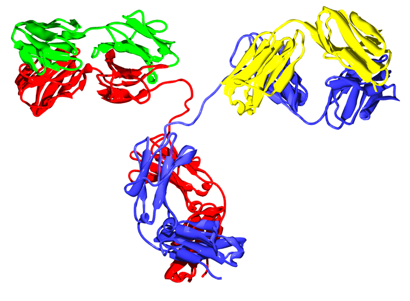
IgG2 antibody
IgG2 antibody

A recent article in the Journal of Autoimmunity suggests anti-progranulin antibodies as one potential mechanism.  Thurner and colleagues used a protein macro-array to screen serum from patients with anti-neutrophil cytoplasmic antibody (ANCA)-associated systemic vasculitides for novel autoantibodies specific to these diseases.  Of the six candidate autoantigens reactive with pooled vasculitis patient serum, progranulin was the only autoantigen appearing in every one of the vasculitides studied.  However, extended screenings showed that a positive progranulin antibody titer was not specific for vasculitides; although the prevalence was low in healthy controls (1/97 or 1%) and patients with melanoma (0/98) or sepsis (0/22), progranulin antibodies were also detectedin serum from patients with RA (16/44 or 36%) and SLE (39/91 or 43%).

Progranulin, also called proepithelin, granulin-epithelin precursor, or acrogranin, is a glycoprotein secreted by epithelial cells, neurons, and certain leukocytes.  In addition to growth factor-like activity, progranulin has immunomodulatory effects in vitro and in vivo.  Full-length progranulin decreases oxidant production by activated neutrophils, blocks TNFα-induced immune responses via binding to TNFR-1 and -2, and promotes up-regulation of IL-4, IL-5, and IL-10.  Progranulin deficiency in mice results in greater inflammation in collagen-induced arthritis (CIA) and collagen antibody-induced arthritis models of human RA; treatment of either progranulin-deficient or wild-type mice with recombinant human progranulin ameliorates CIA inflammation.

Progranulin is cleaved by several proteases into mature granulins.  Neither recombinant nor proteolytically released granulins antagonize TNFα.  Rather, granulins increase expression of pro-inflammatory cytokines IL-1β, IL-8, and TNFα.  SLPI and apolipoprotein A-I binding to progranulin protects it from cleavage by matrix metalloproteinases and other proteases.  However, during inflammation, neutrophils and macrophages release serine proteases that increase progranulin digestion.  In the context of ongoing inflammation in ARD, this may result in increased cleavage of anti-inflammatory progranulin to pro-inflammatory granulin.

Thurner et al. are the first to report the presence of neutralizing anti-progranulin antibodies in RA, SLE, and small- and medium-vessel vasculitides, which may represent a pro-inflammatory mechanism common to several autoimmune diseases.  Their findings provide additional support for exploration of the progranulin/granulin pathway as a therapeutic target and suggest the potential use of anti-progranulin antibodies as a diagnostic and/or prognostic tool in ARD.  Further studies using sera of patients with known autoimmune disease states are needed to confirm these findings and address the additional questions raised, such as –  What causes the failure of self-tolerance to progranulin and the generation of anti-progranulin antibodies, as seen in ~20-40% of the patients in this study?  Are these anti-progranulin antibodies common to all autoimmune diseases?  Could the development of progranulin-neutralizing antibodies even become a biomarker in ARD, for example as a predictor of responsiveness to DMARD therapy, or an indicator of future progression to ARD-related CVD?  We await the results of these and other studies in this area with great interest.

Further Reading:

Progranulin antibodies in autoimmune diseases.  Thurner L, Preuss KD, Fadle N, Regitz E, Klemm P, Zaks M, Kemele M, Hasenfus A, Csernok E, Gross WL, Pasquali JL, Martin T, Bohle RM, Pfreundschuh M.  J Autoimmun. 2013 May; 42:29-38.

Insights into the role of progranulin in immunity, infection, and inflammation.  Jian J, Konopka J, Liu C.  J Leukoc Biol. 2013 Feb; 93(2):199-208.

Cardiovascular disease in autoimmune rheumatic diseases.  Hollan I, Meroni PL, Ahearn JM, Cohen Tervaert JW, Curran S, Goodyear CS, Hestad KA, Kahaleh B, Riggio M, Shields K, Wasko MC.  Autoimmun Rev. 2013 Aug; 12(10):1004–1015.

PD-1 re-expression is differentially regulated by IL-12 vs IFNα in CD8 T cells

TUMOR_immunotherapyDownregulation of immune functions following responses to pathogen infections is critical for limiting damage to the host by the immune system.  T cell activity is known to be downregulated by a variety of negative regulatory mechanisms including negative checkpoint regulatory proteins, a family of CD28-related molecules.  PD-1 is one such molecule that is transiently expressed on activated T cells.   The ligands for PD-1 are PD-L1 and PD-L2, members of the B7 family of molecules which are upregulated on antigen presenting cells and tumor cells.  Interaction of PD-1 with its ligand leads to inhibition of TCR-mediated signaling via recruitment of SHP1 and SHP2 phosphatases to the TCR synapse.  In the August 2013 edition of The Journal of Immunology, Gerner et al., demonstrate that CD8 T cells initially activated in the presence of IL-12 and IFNα differentially re-express PD-1 upon antigen restimulation.

Cytokines play roles in regulation of nearly every aspect of immune responses.  The cytokine milieu present during T cell activation directs differentiation into the different functional classes of CD4 T helper or CD8 T cells.  This study sought to determine the differences in anti-tumor CD8 T cell effector functions mediated when T cells are activated in the presence of various cytokines.  IL-12 and IFNα activate both overlapping and distinct gene programs and promote cytotoxic CD8 T cell responses.  Thus, these cytokines were chosen for comparison in this study.

In this system, CD8+ OT-1 cells were activated ex-vivo in the presence of either IL-12 or IFNα, and transferred into B16-OVA tumor-bearing mice.  T cells activated in the presence of IL-12 were found to mediate tumor-growth inhibition significantly better than if they had been activated in the presence of IFNα.  Over time in tumor-bearing mice, transferred IFNα-matured OT-1 cells were observed to decline in number and lost the ability to produce IFNγ ex vivo upon restimulation, indicating these cells may be exhausted.

Because PD-1 is known to be a marker and mediator of T cell exhaustion, PD-1 expression was examined.  Initial induction levels of PD-1 were comparable on OT-1 cells following ex vivo activation with IFNα or IL-12.  Following transfer into tumor-bearing mice, PD-1 levels declined over time on both types of cells isolated from the spleen and on IL-12 matured cells isolated from the tumor.  However, PD-1 expression was high on transferred IFNα-matured cells when isolated from the tumor.  Similar results were seen when cells were transferred into mice that subsequently received an injection of the OVA peptide.  Thus, CD8+ T cells matured in the presence of IFNα appear to re-express significantly higher levels of PD-1 upon antigen restimulation than IL-12 matured T cells.

PD-1 and PD-L1 targeting with inhibitory antibodies have emerged as promising avenues in tumor immunotherapy.  In this study, anti-PD-1 antibody administration had no additional anti-tumor effect in mice that received IL-12-matured T cells, while in mice that received IFNα-matured T cells, anti-PD-1 antibodies led to inhibition of tumor-growth to a level similar to that in mice that had received IL-12-matured T cells.  Thus, the relatively poor ability of IFNα-matured T cells to efficiently inhibit tumor growth appears to be largely due to PD-1 upregulation.  Finally, when T cells were matured with both IL-12 and IFNα, the effect of IL-12 was dominant.

Many questions remain regarding the mechanisms mediating PD-1 re-expression in IFNα vs. IL-12 matured T cells.  However, since IL-12 activity was dominant over IFNα on regulating PD-1 expression, IL-12 administration during immunotherapy regimens may enhance anti-tumor T cell responses by blocking the mechanisms by which IFNα enhances PD-1 re-expression.

Further Reading:

Cutting Edge: IL-12 and Type I IFN Differentially Program CD8 T Cells for Programmed Death 1 Re-expression Levels and Tumor Control.  Gerner MY, Heltemes-Harris LM, Fife BT, Mescher MF. J Immunol. 2013 Aug 1;191(3):1011-5. doi: 10.4049/jimmunol.1300652. Epub 2013 Jun 26.

CD20-Negative Circulating Plasmablasts Are Target for New B Cell Therapies in Anti-CCP Positive RA

Diagnosis of rheumatoid arthritis (RA) is based on meeting several of the criteria established by the American College of Rheumatology and the European League Against Rheumatism.  One of these criteria is the presence of anti-citrullinated protein antibodies (ACPA).  ACPA seropositivity is currently tested using a filaggrin-derived peptide (anti-cyclic citrullinated peptide [anti-CCP]) ELISA, although other ELISAs in development such as mutated citrullinated vimentin (MCV) show promising results.

citrullination
Citrullination

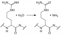
While myelin basic protein, filaggrin, and several histone proteins are naturally citrullinated, other proteins such as fibrin and vimentin can become citrullinated during an inflammatory response.  Citrullination, enzymatic conversion of arginine residues into citrulline, increases protein hydrophobicity, which can change its structure.  In RA, these citrullinated proteins are recognized as “non-self” by immune cells, leading to production of ACPA.  Recent studies suggest these autoantibodies are not merely convenient diagnostic markers resulting from the autoimmune response, but instead may play a role in RA pathogenesis. Current research in this area includes identifying subtypes of RA based on ACPA positivity and specificity, and determining the roles and mechanisms of action for ACPA in RA autoimmunity.

Little is known about the B cells which produce these ACPA.  However, in a recent report in Annals of Rheumatic Disease, Kerkman and her colleagues used B cells isolated from the peripheral blood of ACPA-positive and -negative RA patients, as well as healthy individuals, to examine ACPA production in vitro and identify the ACPA-producing cell populations.

Initially, the authors stimulated peripheral B cells with B cell activating factor (BAFF) and anti-IgM F(ab′)2-fragments to induce ACPA production.  Although total IgG production was equivalent across the cultures, only B cells from ACPA-positive RA patients produced ACPA.  There was good correlation of ACPA titers obtained from in vitro culture with in vivo patient ACPA titers, underscoring the utility of this model system.  Next, the authors examined spontaneous ACPA production in unstimulated peripheral blood mononuclear cells (PBMC) from ACPA-positive RA patients.  In this case, total IgG was up to 100x lower than that seen in their studies with stimulated B cells; however, the amount of ACPA produced was equivalent.

Were ACPA in PBMC cultures generated solely by circulating plasmablasts, or were antigen presenting cells (APC) present in the PBMC population also stimulating production of ACPA by memory or even naïve B cells?  Kerkman et al. used FACS to selectively deplete ACPA-positive RA patient PBMC of plasmablast/plasma cell or naïve/memory populations, as well as to sort the CD19+ B cell subpopulations.  Naïve B cells (CD20+CD27-) did not produce any ACPA, even when stimulated with BAFF and IgM F(ab′)2.  Memory B cells (CD20+CD27+) produced ACPA upon stimulation, indicating CCP-specific memory cells are present in the circulation of ACPA-positive RA patients; however, ACPA production in CD20-depleted PBMC remained essentially unchanged, while unstimulated PBMCs depleted of plasmablasts/plasma cells produced significantly less ACPA.

acpa b cells

This study demonstrates the presence of circulating ACPA-producing plasmablasts/plasma cells in the peripheral blood of patients with ACPA-positive RA.  This is a novel and unexpected finding, since the plasmablast population is typically a transient population within PBMCs following antigen exposure, with antibody production continuing from mature plasma cells in the spleen and lymph nodes.
Circulating ACPA-producing B cells may persist in RA due to plasmablast replication and/or to memory B cell activation in response to persistent systemic citrullinated antigens.  Currently approved RA therapies which target the CD20+ B cell population, such as rituximab, would affect the memory B cell population, but not CD20- plasmablasts.  New therapies targeting circulating plasmablasts/plasma cells in addition to memory B cells could significantly limit ACPA production and subsequent immunological damage in RA, including that due to ACPA-induced TNFα production and complement activation.  Delineating circulating plasmablasts as a major source of ACPA is therefore a step forward in the quest to determine the roles and mechanisms of action for ACPA in RA pathogenesis, and underscores the possibility of developing effective new therapies by targeting specific B cell populations in RA.

Further Reading:

Circulating plasmablasts/plasma cells as a source of anti-citrullinated protein antibodies in patients with rheumatoid arthritis.  Kerkman PF, Rombouts Y, van der Voort EIH, Trouw LA, Huizinga TWJ, Toes REM, Scherer HU.  Ann Rheum Dis 2013 Jul; 72:1259–1263.

The effect of targeted rheumatoid arthritis therapies on anti-citrullinated protein autoantibody levels and B cell responses.  Modi S, Soejima M, Levesque MC.  Clin Exp Immunol 2013 Jul; 173(1):8-17.

B effector cells in rheumatoid arthritis and experimental arthritis.  Finnegan A, Ashaye S, Hamel KM.  Autoimmunity 2012 Aug; 45(5):353-63.

New Research Points the Way Towards Mechanism of Action, Receptor for MS Copolymer Drugs

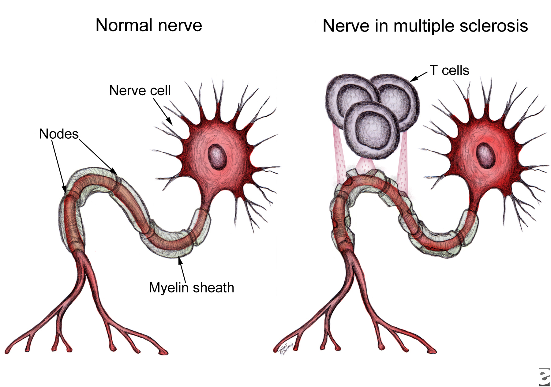
Multiple_sclerosis_T_cellsNeurological damage in multiple sclerosis (MS) is caused by autoreactive immune cells, which attack myelin sheathing on axons of the brain and spinal cord, leading to inflammation and myelin loss.  The MS drug Copaxone is a copolymer of glutamic acid, lysine, alanine, and tyrosine (YEAK) that is thought to impair myelin attack by inhibiting MBP self antigen presentation to autoreactive T cells; a related copolymer in which phenylalanine replaces glutamic acid (YFAK) has been developed based on MBP binding to class II MHC.  However, little is known about these drugs’ molecular targets or mechanisms of actionIn vitro studies suggest IL-10 secretion by B cells or regulatory and Th2-CD4+ T cells is involved.  Recent data demonstrate YEAK and YFAK also have MHC-independent effects on macrophages and dendritic cells, although the receptor which mediates these effects is unknown.  In a recent article in The Journal of Immunology, Koenig and colleagues isolated YEAK- and YFAK-interacting proteins from macrophage lysates and identified structures required for copolymer interaction with cells.

Following incubation with RAW264.7 macrophage lysate, biotinylated copolymers were recovered using avidin-coated beads and the associated cellular proteins were identified using mass spectroscopy.  One high-frequency hit with known surface expression and involvement in immune signaling was gp96.  Cell surface gp96 directly activates innate immune cell cytokine production, acts as a class I MHC antigen chaperone, and has been proposed as a Th2-specific co-stimulatory molecule.  CD91 has been implicated in gp96 stimulation of antigen-presenting cells (APC) and is involved in signaling and endocytosis of several ligands.  App was also identified as a surface protein that interacts with YEAK and YFAK.  A β-amyloid species precursor in Alzheimer’s disease, its function on myeloid cells is not well understood.

Macrophages secrete CCL22, a chemoattractant for regulatory and Th2 T cells, in response to YEAK or YFAK.  Koenig et al. studied this response in wild-type versus gp96-, CD91-, and App-deficient cells and found no impairment in any of the knock-out cell lines, indicating that despite their interactions with YEAK and YFAK, neither gp96, CD91, nor App are involved in cell signaling by these copolymers.

Lysine confers a positive charge on these copolymers, leading Koenig et al. to propose that cellular binding may be mediated by electrostatic interaction rather than conformation.  Indeed, increasing salt concentration reduced protein interactions with biotinylated copolymer.  Importantly, using cell lines lacking specific sulfation enzymes, the authors demonstrated that YEAK and YFAK bind to negatively charged heparan sulfate proteoglycans (HSPG).  This interaction is functional: RAW264.7 cells stimulated with YFAK in the presence of heparin sulfate, a structurally similar competitor of HSPG, did not produce CCL22.

HSPG are glycoproteins which contain one or more covalently attached heparin sulfate (HS) chains.  Membrane HSPG are known to act as co-receptors for many growth factors and could therefore play a role in the cellular effects of YEAK by activating cell signaling through an associated receptor or preventing signaling by that receptor’s natural ligand.  For example, YEAK binding to HS, a co-receptor for gp96 binding to CD91, may alter cellular uptake of gp96-peptide complexes via CD91, affecting self antigen cross-presentation and T cell activation by APC.

Alternatively, HSPG also function as receptors for constitutive as well as ligand-induced endocytosis.  YEAK interaction with HSPG may promote its fluid-phase uptake and delivery to intracellular target(s) in a manner similar to that of cationic cell-penetrating peptides.  In fact, gene ontology term enrichment analysis highlights “RNA binding” as a molecular function of YEAK- and YFAK-interacting proteins identified in this study, supporting a potential cytosolic or nuclear site of action.

Significant work remains to define the receptors and molecular mechanisms of action for these copolymers and aid rational design of future immune-modulating drugs.  The authors’ list of 222 copolymer-interacting proteins and characterization of sulfated glycosaminoglycans as the moieties responsible for functional interaction of these copolymers with innate immune cells serve as a solid foundation for further research in this area.

Further Reading:

Amino acid copolymers that alleviate experimental autoimmune encephalomyelitis in vivo interact with heparan sulfates and glycoprotein 96 in APCs.  Koenig PA, Spooner E, Kawamoto N, Strominger JL, Ploegh HL.  J Immunol.  2013 Jul 1; 191(1):XXX.  Epub ahead of print 2013 June 5.

Heparan sulphate proteoglycans fine-tune mammalian physiology.  Bishop JR, Schuksz M, Esko JD.  Nature. 2007 Apr 26; 446(7139):1030-7.

Interactions between heparan sulfate and proteins – design and functional implications.  Lindahl U, Li JP.  Int Rev Cell Mol Biol. 2009; 276:105-59.

Cell surface heparan sulfate proteoglycans influence MHC class II-restricted antigen presentation.  Léonetti M, Gadzinski A, Moine G.  J Immunol. 2010 Oct 1; 185(7):3847-56.


INTESTINAL BACTERIA LINKED TO LYMPHOMA

The human gut harbors approximately one thousand different bacterial species (intestinal microbiota). Intestinal microbiota number 100 trillion cells; over 90 percent of the cells in the body are bacteria. The composition of each person’s microbiome — the body’s bacterial make-up — is very different, due to the types of bacteria people ingest in their early lives, as well as the effects of diet and lifestyle.

Several studies implicated intestinal bacteria in various cancers. Gram-negative Helicobacter species were found to be associated with liver cancer, colon cancer, and breast cancer. A recent study published in the peer reviewed journal Nature by Yoshimoto et al. (2013) reported that gut bacteria of obese mice unleash high levels of an acid that promotes liver cancer. In rodents, intestinal bacteria influence obesity, intestinal inflammation and certain types of epithelial cancers. However, in human, little is known about the identity of the bacterial species that promote the growth or protect the body from cancer. Therefore, studies are warranted to determine whether differences in peoples’ microbiomes affect their risk for cancer, and whether changing the bacteria can reduce this risk. A clinical trial at the National Cancer Institute (NCI) is currently evaluating the relationship between intestinal bacteria and breast cancer risk (Clinical Trials.gov number: NCT01461070).

intestinal  bacteria

For the first time, a recent study by Yamamoto et al. (2013) demonstrated a relationship between intestinal microbiota and onset of lymphoma (a type of blood cancer of B or T lymphocytes). Yamamoto and colleagues studied mice with ataxia-telangiectasia (A-T), a genetic disease that in humans and mice is associated with a high rate of B-cell lymphoma. These investigators discovered that of mice with A-T, those with certain microbial species lived much longer than those with other bacteria before developing lymphoma, and had less of the gene damage (genotoxicity) that causes lymphoma. A high-throughput sequence analysis of rRNA genes identified the bacteria Lactobacillus johnsonnii in abundance in more cancer-resistant mouse colonies compared to cancer-prone mouse colonies.This study by Yamamoto et al. also created a detailed catalog of bacteria types with promoting or protective effects on genotoxicity (a chemical or other agent that damages cellular DNA, resulting in mutations or cancer) and lymphoma, which could be used in the future to formulate combination therapies that kill the bacteria that promote cancer (such as antibiotics) and increase the presence of the bacteria that protect from cancer (like probiotics).

References:

1.   Ward, J.M., et al., Chronic active hepatitis in mice caused by Helicobacter hepaticus. Am J Pathol, 1994. 145(4): p. 959-68.

2.   Yoshimoto, S., et al., Obesity-induced gut microbial metabolite promotes liver cancer through senescence secretome. Nature, 2013. 499(7456): p. 97-101.

3.   Yamamoto, M.L., et al., Intestinal Bacteria Modify Lymphoma Incidence and Latency by Affecting Systemic Inflammatory State, Oxidative Stress, and Leukocyte Genotoxicity. Cancer Res, 2013. 73(14): p. 4222-4232.


Remodeling of the Tumor Extracellular Matrix Activates YAP in Fibroblasts to Produce Cancer Associated Fibroblasts

When cells undergo transformation and initiate the formation of a solid tumor mass, they cause profound changes on the phenotypes of the cells that surround them1. However, in addition to the changes in cellular phenotype, there is a change in the extracellular matrix that coincides with tumor formation1. It has been demonstrated that the majority of solid tumors have increased stiffness in their extracellular matrix (ECM), which may lead to increased activation of pro-tumor signaling pathways, such as Src, FAK, and RhoA2-4. Recently, it was discovered that increased matrix stiffness may also lead to increased activity of the oncogenic YAP/TAZ complex, which is connected to the Hippo signaling pathways, transcriptional regulators that increase cellular proliferation, decreased cellular contact inhibition, increased cancer stem cell phenotype, and increased metastasis5. However, in a fibroblastrecent edition of Nature Cell Biology Calvo et al. demonstrated that 6.  Not only do the authors demonstrate that YAP/TAZ is active in CAFs, but YAP/TAZ is necessary for CAF development6. They show that CAF activation leads to matrix remodeling towards increased stiffness, via myosin light chain 2 (MYL9/MLC) expression, establishing a feed-forward loop where the ECM plays a vital role6.

The authors first isolated fibroblasts in different stages towards becoming a CAF and saw that both mechanical-responsive signaling machinery (SMA, FN1, Paxillin, MYL9, MYH10, DIAH1 & F-actin) and mechanical tension were increased in populations containing CAFs. Moreover, tumor cell invasion, and angiogenesis of the tumor microenvironment (shown via endomucin and second-harmonic microscopy) were increased in samples that contained more tumor-associated-like fibroblasts (indicated by vimentin)6.

Because of the role of cell-cell and cell-ECM contact in the Hippo signaling pathway, the authors sought to understand whether this pathway is activated in CAFs. They found that YAP, and its co-factor, TAZ to be only upregulated and co-localized in the nucleus of transformed fibroblasts; the target of the activated YAP-TAZ complex6.  Furthermore, upon depletion of YAP, the ability for CAFs to cause matrix stiffness by contraction lessened as well as CAFs ability to form collagen networks and facilitate angiogenesis. Interestingly, when TAZ was inhibited, there was no change in functionality, which may lead to a TAZ-independent function for YAP.

Upon microarray analysis of CAFs treated with siRNA that targets YAP, Calvo et al. found that the expression of many of the genes involved in mechanosensing and motility to be diminished6. Furthermore, when these individual genes were silenced, there was an overall decrease in the amount of cellular invasion of tumors. Many of the YAP-mediated genes, such as ANLN and DIAPH3, were involved in matrix remodeling and cellular invasion. Interestingly, modification of only one protein overexpression resulted in high amounts of matrix-remodeling and invasion: myosin regulatory light polypeptide 9 (MYL9). While not transcriptionally controlled by the YAP/TAZ complex, the authors demonstrate that YAP/TAZ is able to control MYL9 by post-translational modifications, placing YAP as a critical factor in regulating matrix-remodeling and invasion through MYL96.

Calvo et al. next posited that YAP/TAZ activation may not be exclusive to CAFs, but may also occur in normal fibroblasts when placed in a cancerous environment6. They found that fibroblasts placed in culture with tumor conditioned media had higher nuclear translocation of YAP, and higher gel contraction (akin to matrix stiffening) comparable to known promoters of pro-contractile function: L-alpha-lysophosphatidic acid (LPA) and transforming growth factor-beta (TGFβ). However, actomyosin inhibition (by blebbistatin) could not be rescued with LPA and TGFβ. Therefore, while soluble factors may activate matrix contraction, a functional cytoskeleton is essential for matrix contraction. Because of the necessary role of the cytoskeleton, the authors tested whether inhibition of RhoA kinase (ROCK), a kinase involved in regulating translocation and structure of the cell by the cytoskeleton, would affect the nuclear localization of YAP6. Inhibition of ROCK decreased YAP nuclearization and decreased the matrix stiffness. Of note, like ROCK inhibition, inhibition of Src also affected the nuclear localization of YAP as well as complex formation with TEAD1 and TEAD4. However, Src modulation of YAP is downstream of cytoskeletal changes in tension since Src inhibition did not affect stress fibers6.

Since activation of YAP in CAFs  is connected to actomycin-mediated matrix stiffness, and this activation of YAP expresses MYL9, and expression of MYL9 results in matrix-remodeling towards stiffness, the authors posit that this pathway forms a feed-forward loop6. This loop could lead to constitutive activation of YAP pathway in CAFs, causing a robust response and stabilizing the CAF phenotype. However, it is not known what other mechanisms, as well as regulatory mechanisms of YAP, are involved in this process as well as whether the YAP-ECM tension pathway may play a regulatory role in normal fibroblasts.

References:

1. Boudreau, A., van’t Veer, L. J. & Bissell, M. J. An “elite hacker”: breast tumors exploit the normal microenvironment program to instruct their progression and biological diversity. Cell adhesion & migration 6, 236-248, doi:10.4161/cam.20880 (2012).

2. Levental, K. R. et al. Matrix crosslinking forces tumor progression by enhancing integrin signaling. Cell 139, 891-906, doi:10.1016/j.cell.2009.10.027 (2009).

3. Guilluy, C. et al. The Rho GEFs LARG and GEF-H1 regulate the mechanical response to force on integrins. Nature cell biology 13, 722-727, doi:10.1038/ncb2254 (2011).

4. Sawada, Y. et al. Force sensing by mechanical extension of the Src family kinase substrate p130Cas. Cell 127, 1015-1026, doi:10.1016/j.cell.2006.09.044 (2006).

5. Harvey, K. F., Zhang, X. & Thomas, D. M. The Hippo pathway and human cancer. Nature reviews. Cancer 13, 246-257, doi:10.1038/nrc3458 (2013).

6. Calvo, F. et al. Mechanotransduction and YAP-dependent matrix remodelling is required for the generation and maintenance of cancer-associated fibroblasts. Nature cell biology 15, 637-646, doi:10.1038/ncb2756 (2013).


Upcoming Immunology Conferences: October – December, 2013

I previously posted on Immunology Conferences: July – September, 2013 and 2013 Conferences in Tumor Immunology and Cancer ImmunotherapyThis post lists upcoming Immunology-related conferences from October – December, 2013.

conferencesOctober
Aegean Conference: 6th International Conference on Autoimmunity: Mechanisms & Novel Treatments

October 2 – 7, 2013.

Corfu, Greece

Early registration deadline: July 25, 2013.

Abstract submission deadline: July 25, 2013.

Travel award application deadline: June 30, 2013.


IL-1-mediated inflammation and diabetes: From basic science to clinical applications

October 10 – 11, 2013.

Nijmegen, The Netherlands

Abstract submission deadline: August 1, 2013.


European Macrophage & Dendritic Cell Society Meeting: Myeloid Cells: Microenvironment, Microorganisms & Metabolism From Basic Science to Clinical Applications

October 10 – 12, 2013.

Erlangen, Germany


13th International Workshop on Langerhans Cells

October 10-13, 2013

Royal Tropical Institute, Amsterdam, The Netherlands

Early Registration deadline: August 1, 2013.

Abstract submission deadline: August 19, 2013.


The International Symposium on Immunotherapy

October 11-12, 2013

London, UK

Early Registration deadline: August 9, 2013.

Abstract submission deadline: August 9, 2013.


46th Annual Meeting of the Society for Leukocyte Biology

October 20-22, 2013

Newport Marriott, Newport, RI, USA

Late Breaking Abstract submission deadline: August 6, 2013.

Online Registration deadline: October 7, 2013.


16th Annual New York State Immunology Conference

October 20-23, 2013

Sagamore Resort and Conference Center, Bolton Landing, NY, USA

Abstract submission deadline: July 31, 2013.

Registration deadline: September 6, 2013.


Cold Spring Harbor Asia Conference: Tumour Immunology and Immunotherapy

October 28 – November 1, 2013.

Suzhou, China

Abstract submission deadline: August 16, 2013.

Early Registration deadline: August 16, 2013.


Keystone Symposium: Advancing Vaccines in the Genomics Era

October 31 – November 4, 2013.

Rio de Janeiro, Brazil

Abstract Deadline: July 30, 2013.

Early Registration Deadline: August 29, 2013.


November

The Lancet and Cell: What Will it Take to Achieve an AIDS-free World?

November 3–5, 2013.

San Francisco, California, USA.

Abstract submission deadline: July 26, 2013.

Early Registration Deadline: September 13, 2013.


International Primary Immunodeficiencies Congress

November 7–8, 2013.

Lisbon, Portugal

Early Registration deadline: July 19, 2013.


Asia Pacific Congress of Allergy, Asthma and Clinical Immunology

November 14–17, 2013.

Taipei City, Taiwan

Abstract Submission Deadline: July 31, 2013.

Early Registration: August 30, 2013.

Registration Deadline: October 25, 2013.


Cold Spring Harbor Asia Conference: Bacterial Infection and Host Defence

November 18–22, 2013.

Suzhou, China

Abstract submission deadline: September 6, 2013.

Early Registration Deadline: September 6, 2013.


6th Autoimmunity Congress Asia

November 20–22, 2013.

Hong Kong

Abstract Submission Deadline: July 20, 2013.

Early Registration deadline: August 6, 2013.


Harnessing Immunity to Prevent and Treat Disease

November 20–23, 2013.

Cold Spring Harbor Laboratory, New York, USA

Abstract submission deadline: September 6, 2013.


11th Annual UC Irvine Immunology Fair
November 21–22, 2013.

Irvine, California, USA

Abstract submission deadline: October 19, 2013.

Poster contest deadline: November 2, 2013.


December

EMBO Workshop: Complex Systems in Immunology

December 2–4, 2013.

Biopolis, Singapore

Abstract submission deadline: September 1, 2013.

Registration deadline: September 1, 2013.


British Society for Immunology Congress

December 2–5, 2013.

Liverpool, UK

Abstract submission deadline: September 6, 2013.

Early Registration deadline: September 30, 2013.


Annual Scientific Meeting of the Australasian Society for Immunology

December 2–5, 2013.

Wellington, New Zealand

Abstract submission deadline: September 1, 2013.

Early Registration deadline: September 1, 2013.


UK Primary Immunodeficiency Network Forum

December 6–7, 2013.

Liverpool, UK

Abstract submission deadline: September 6, 2013.

Early Registration deadline: September 30, 2013.


2013 American Society for Cell Biology Annual Meeting

December 14-18, 2013

New Orleans, LA, USA

Abstract submission deadline (Minisymposium Talk, ePoster Talk, and/or Poster Presentation): July 30, 2013.

Abstract submission deadline (Poster Presentation Only): September 4, 2013.

Early Registration deadline: October 10, 2013.


Websites that list upcoming Conferences & Events in Immunology, Tumor Immunology, and Cancer Immunotherapy:

The American Association of Immunologists (AAI) Meetings and Events Calendar

Nature Reviews Immunology’s list of conferences

Cancer Immunity Journal’s List of Conferences

FASEB Scientific Research Conferences Calendar

Highlight: A silver bullet against bacteria?

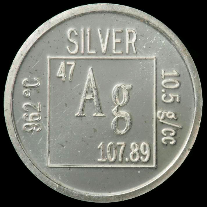
The development of multi-resistant bacterial strains is a major medical problem, especially in hospitals. New antibiotics are constantly under development, but the success rate seems to have slowed down in recent years. However, two recent publications suggest that instead of finding novel antibiotics, an alternative strategy could be to increase the sensitivity of the bacteria to the antibiotics already in use.s12sIn the first report [1], the authors demonstrated that silver ions interfered with several metabolic pathways and increased the permeability of the cell membrane, both made the bacteria much more susceptible to antibiotics. The use of silver to combat bacteria is not unprecedented and actually has been used at least since Grecian times to treat wounds and to preserve water. However, with the advent of potent antibiotics in the 1940s their use fell out of favor.

The authors uncovered two independent mechanisms of how silver is beating up bacteria. They studied E. coli a Gram-negative bacteria that is especially difficult to treat with many drugs due to its thick cell wall.

First, when exposed to silver ions the bacteria produced more reactive oxygen species (ROS). ROS are chemically highly reactive molecules that can bind unspecifically to closeby proteins and DNA and thereby irreversibly alter or damage those structures. Small amounts of ROS are constantly produced during several chemical reactions of the normal metabolism, but cell stress in any of its forms can greatly increase their production. The widespread damage that ROS can inflict weakens the cell and if the damage is too severe it can ultimately lead to cell death.

The second mechanism of silver is its ability to affect two metabolic pathways of the bacterial cells. On the one hand, the ability of the bacteria to maintain their iron level was disturbed. On the other hand, the formation of disulfide bonds, which are crucial for the structural integrity and function of many proteins, was affected in the presence of the silver ions. Both of these actions can be understood as severe forms of cell stress.

As a result of the ROS production and the metabolic impairment, the bacterial cell wall became more permeable. This is important as Gram-negative bacteria have a thick extra cell coating that prevents large molecules to enter. Many antibiotics are too big to enter through this bacterial cell wall, but the silver treatment allowed the antibiotic to enter the cells. By gaining access to the cell, the gram-negative bacteria became sensitive to large molecule antibiotics that usually work only with Gram-positive bacteria that lack such a thick cell coating. This finding greatly expands the arsenal of antibiotics that can be used against Gram-negative bacteria.

Importantly, bacteria that were weakened and made permeable by the silver ions became highly susceptible to even low amounts of antibiotics. The authors tested this in vivo with a mouse model of urinary tract infection. When the antibiotic treatment of the infected mice was supplemented with small amounts of silver ions, the silver greatly augmented the efficiency of the antibiotic: 10 fold to up to 1,000 fold. In one experiment, only 10% of the infected mice that were treated with the antibiotic alone survived, but when treated additional with the silver ions 90% survived!

This silver sensitization was also effective with two types of infections that are particular difficult to treat: dormant bacteria that remain inactive during the antibiotic treatment and rebound afterwards, and bacteria that produce slime layers, called biofilms. Biofilms can be visualized as huge amounts of extra coating produced by the bacteria that make them stick to surfaces (e.g. catheters in the clinic) and provides them with an extra shielding against antibiotics.

However, before somebody now starts grinding his silver spoons into his food, the caveat has to be noted that silver has some side effects: it can accumulate in your body, e.g. in the skin and when it is then exposed to sun can turn you into a smurf, quite literally, as the skin turns irreversible blue-grayish. The medical term is ‘Argyria’ and one stunning example is Paul Karason. Although the concentrations of silver used by Morones-Ramirez et al. were much lower, it still shows that the use of silver will likely be very limited in humans.

In a similar vein to the report by Morones-Ramirez et al., another recent study [2] showed that very high doses of vitamin C also could trigger the production of above-mentioned ROS in the bacterium Mycobacterium tuberculosis, the causative agent of tuberculosis. Thereby, vitamin C was able to kill the bacteria, either directly or in concert with antibiotics. Similar to the case above, the efficiency of the antibiotic was greatly increased when applied together with the vitamin C. However, starting to eat now vitamin C in bucket loads might be a bit premature too.

Vitamin C structure
Vitamin C structure

Nonetheless, both reports can be viewed as proof-of-principle studies. In both studies agents that by themselves are rather harmless to bacteria could massively increase their sensitivity towards antibiotics! Having established such potential it is likely that other substances will be described in the near future that are safer and still have this prominent potential to boost the efficiency of antibiotics.

References:

[1] Morones-Ramirez, J. R., Winkler, J. A., Spina, C. S. & Collins, J. J. Silver Enhances Antibiotic Activity Against Gram-Negative Bacteria. Science Translational Medicine 5, 190ra81–190ra81 (2013).

[2] Vilchèze, C., Hartman, T., Weinrick, B. & Jacobs, W. R. Mycobacterium tuberculosis is extraordinarily sensitive to killing by a vitamin C-induced Fenton reaction. Nat Commun 4, 1881 (2013).