FDA APPROVED NEW DRUG TO TREAT ADVANCED PROSTATE CANCER

Prostate cancer (PCa) is the second most common cause of male cancer-related death.  According to the National Cancer Institute, in the United States in 2013 the estimated new cases of prostate cancer would be 238,590 and deaths would be 29,720.

Bone metastases are a serious problem in men with advanced PCa. Bone metastases increase the risk of skeletal-related events (SREs) which include pathological fractures, spinal cord compression, bone pain. Both bone metastases and SREs are associated with an unfavorable prognosis and greatly affect quality of life.

Numerous studies have shown the importance of androgens (steroid hormones) in the development of PCa (although the exact role of androgen in PCa development is yet to be determined). Therefore, continuous androgen deprivation has been the standard therapy for metastatic hormone-sensitive disease. Despite a high response rate, resistance to androgen-deprivation therapy occurs in most patients, resulting in a median survival of 2.5 to 3 years. Thus, resulting in the development of castration resistant (CRPC) or hormone-refractory (HRPC) stage. Standard chemotherapy has not proven to be very effective in cases of metastatic CRPC, with a 10–20% response rate and approximately one-year median survival. In the United States docetaxel and cabazitaxel are the only Food and Drug Administration (FDA)-approved chemotherapies for the treatment of metastatic CRPC. Even though these drugs palliate symptoms, the overall survival benefit is moderate. In addition, a cellular immunotherapeutic agent sipuleucel-T (Provenge; Dendreon Corp) has been shown to increase overall survival period by 4.1 months on average but not progression-free survival time for patients with metastatic CRPC.

On May 15th, 2013, the U.S. FDA approved Xofigoradium Ra 223 dichloride (Xofigo®; Bayer HealthCare Pharmaceuticals) to treat men with metastatic castration-resistant prostate cancer with bone metastases after receiving medical or surgical treatment. The efficacy of Xofigo® was evaluated in a single clinical trial (Phase 3 ALSYMPCA trial) of 809 men with metastatic castration-resistant prostate cancer. Compared to the controls (patients received placebo plus standard care) with median survival of 11.2 months, patients who received Xofigo® lived a median of 14 months. The side effects noted during the clinical trials among patients treated with Xofigo® were nausea, diarrhea, vomiting, and swelling of the leg, foot, or ankle.

The alpha particle-emitting pharmaceutical Xofigo® is a radio-therapeutic drug. It mimics calcium and forms complexes with the bone mineral hydroxyapatite at areas of increased bone turnover, such as bone metastases. This drug is administered as an intravenous injection.

Overall, Xofigo® was found to extend the survival of men with metastatic prostate cancer and expected to be available in the clinic within a few weeks.


References:

1.         Yagoda A, Petrylak D: Cytotoxic chemotherapy for advanced hormone-resistant prostate cancer. Cancer 1993, 71(3 Suppl):1098-1109.

2.         Harrison MR, Wong TZ, Armstrong AJ, George DJ: Radium-223 chloride: a potential new treatment for castration-resistant prostate cancer patients with metastatic bone disease. Cancer Manag Res 2013, 5:1-14.

3.         Karantanos T, Corn PG, Thompson TC: Prostate cancer progression after androgen deprivation therapy: mechanisms of castrate resistance and novel therapeutic approaches. Oncogene 2013.

ISSCR 2013 Meeting Updates: Is a Cure in Sight for Type 1 Diabetes?

Last week, the International Society for Stem Cell Research (ISSCR) held its 11th Annual Meeting in Boston, MA.  Over 3,000 stem cell researchers from around the world gathered to hear the leading experts share their research and perspectives in this fast-paced field.  One interesting topic was recent advances in cell-replacement therapy for treatment of type 1 diabetes.

Diabetes mellitus is a metabolic disease that results from a failure in glucose regulation, often leading to severe hyperglycemia and tissue/organ damage.  Pancreatic β-cells respond to high blood glucose levels by secreting insulin, which acts on other tissues to promote glucose uptake from the blood.  Type 1 diabetes (T1D) results from autoimmune destruction of insulin-producing β-cells of the pancreas.  The lack of insulin leads to increased blood and urine glucose.  T1D is often fatal unless treated with exogenous administration of insulin daily and regular blood glucose monitoring for the patient’s entire life.  However, this treatment does not match the effect of having endogenous β-cells.  Thus, scientists in the field of regenerative medicine have focused on strategies for generation of β-cells for cell-replacement therapy.

Douglas Melton presented his lab’s describe the imagecurrent progress on generation of functional β-cells from human embryonic stem cells (hESCs).  Since 2006, D’Amour et al. demonstrated that they had developed a robust differentiation protocol to produce hESC-derived pancreatic endocrine cells capable of synthesizing insulin 1.  However, the derived cells failed to secrete insulin appropriately in response to the addition of glucose, a required function of true β-cells.  Thus, one focus of Melton’s group has been to identify the signals to generate functional β-cells.  Pancreatic islets are complex structures consisting of multiple cell types, including the insulin-producing β-cells as well as endothelial cells in the surrounding blood vessels.  Thus, endothelial signals are known to promote pancreatic development 2.  Melton’s group found that co-culture of β-cells with endothelial cells promotes functional maturation of the hESC-derived β-cells.  In addition, they recently identified another hormone, betatrophin, which is secreted by the liver and functions in promoting β-cell replication 3.  Thus, increasing the levels of this hormone may generate more β-cells.  Although various groups have demonstrated that large amounts of glucose-responsive, insulin-secreting β-cells can be generated in vitro, one concern that remains to be addressed is how the cells will be protected from an autoimmune attack once delivered to the patient.  One potential strategy involves encapsulation of the β-cells into an immunoprotective device prior to delivery.

Other mature cells have also iPSDerivationbeen proposed as a source of new β-cells.  Sarah Ferber presented her lab’s work on inducing liver cells to transdifferentiate into β-cells for autologous cell-replacement therapy.  Transdifferentiation is the process by which one type of adult cell is directly converted into another type of cell.  They used the transcription factor, pancreatic and duodenal homeobox gene 1 (PDX-1), and soluble factors to induce the developmental shift of adult human liver cells into functional insulin-producing cells 4.  Not only did the transdifferentiated liver cells produce insulin, but they also released in a glucose-regulated manner.  When transplanted into diabetic, immunodeficient mice, the cells ameliorated hyperglycemia over a 60-day period.  Thus, PDX-1-induced transdifferentiated liver cells offer the potential to replace β-cells’ function in vivo.  Furthermore, transplantation of autologous β-cells would circumvent a host versus graft immune response, as well as allow the patient to be the donor of his or her own insulin-producing cells.

In summary, Melton’s and Ferber’s presentations demonstrated the tremendous progress that has been made in the last decade to generate functional β-cells for use in treatment of T1D.

References

1          D’Amour, K. A. et al. Production of pancreatic hormone-expressing endocrine cells from human embryonic stem cells. Nat Biotechnol 24, 1392-1401, doi:10.1038/nbt1259 (2006).

2          Nikolova, G. et al. The vascular basement membrane: a niche for insulin gene expression and Beta cell proliferation. Dev Cell 10, 397-405, doi:10.1016/j.devcel.2006.01.015 (2006).

3          Yi, P., Park, J. S. & Melton, D. A. Betatrophin: A Hormone that Controls Pancreatic beta Cell Proliferation. Cell 153, 747-758, doi:10.1016/j.cell.2013.04.008 (2013).

4          Sapir, T. et al. Cell-replacement therapy for diabetes: Generating functional insulin-producing tissue from adult human liver cells. Proc Natl Acad Sci U S A 102, 7964-7969, doi:10.1073/pnas.0405277102 (2005).

Highlight: Is too much salt bad for your guts?

salt

Whether eating too much table salt in our diet is bad for our health has long been debated. Links have been proposed to several cardiovascular diseases. But, a recent expert committee for the Institute of Medicine concluded that the data do not support such a link [1], keeping the discussion going. Two recent publications in Nature, however, suggest that too much dietary salt might impact our immune system instead and potentially increase the likelihood of autoimmune diseases.

CD4 T lymphocytes can differentiate in specialized subsets that promote or help diverse immune responses. Called T helper (Th) cells, particular subsets are named after prominent cytokines they produce, e.g. IL-17 in the case of Th17 T cells. Th17 cells are important for protection of the body against many bacterial and fungal infections and they are prevalent in the intestinal tissue were they are believed to aid the barrier function of the gut to keep the intestinal bacteria were they belong [2]. However, the “too much of a good thing” proverb applies to lymphocytes too and in the case of Th17 T cells this is exemplified by their pathogenic involvement in several autoimmune diseases. Therefore, the control of cell number and function of Th17 cells requires a delicate balance.

It was known that there is a cross talk between the gut lumen and the Th17 cell response. For example, a few years back it was shown that the frequency of a common bacterium within the gut microbiota could influence the prevalence of Th17 cells in the intestinal tissue [3]. The two new studies demonstrate that table salt (sodium chloride, NaCl) is a surprising new factor on the list to influence the frequency and function of Th17 cells [4-6].Lymphocyte activation

Adding 40 mM NaCl – a level found in the intestinal tissue of animals after feeding of a high salt diet – to in vitro cultures augmented the differentiation of Th17 cells [4, 5]. Similar to in vitro, feeding mice with a high salt diet increased the frequency of Th17 cell in the intestinal tissue, but not in the lymph nodes or the spleen. In both settings (in vitro and in vivo) the resulting Th17 cells were capable of producing large amounts of pro-inflammatory cytokines. By analysis of the mRNA expression, both reports characterized the MAP-kinase p38, NFAT5 (nuclear factor of activated T cells 5) and SGK1 (serum glucocorticoid-regulated kinase-1) as critical molecular players in sensing NaCl and mediating its effect. The elimination of any of these factors from the T cells, either by genetic ablation or by impeding the expression by means of RNA-interference (shRNA), blocked the increased Th17 cell differentiation in the presence of NaCl. Although all three proteins are part of the same pathway, SGK1 appeared to be central in the regulation of the NaCl induced effect. Although this finding is surprising, the results are in line with the known function of SGK1 in sodium transport and homeostasis [7]. SGK1 expression was not only induced by increased NaCl concentrations, but also by the cytokine IL-23, which has a critical role in stabilizing and reinforcing the TH17 phenotype [2]. As NaCl also increased the expression of the IL-23 receptor this established a positive feedback loop that strengthened the Th17 cell differentiation. Importantly, both groups also showed that raising the levels of dietary salt could augment the severity of EAE (experimental autoimmune encephalomyelitis), a mouse model for the autoimmune disease multiple sclerosis [4-6].

In summary, these reports demonstrate that high levels of salt in the diet could make mice susceptible to a form of autoimmune disease that involves pathogenic Th17 T cells. The data suggest that high concentration of NaCl might be an environmental risk factor for autoimmune diseases. However, it should be pointed out that high concentration of NaCl did not induce autoimmune responses by itself, as the EAE animal model requires the immunization with a know self-antigen. Autoimmunity is a complex interplay of numerous genetic pre-disposing and environmental factors. In this regard these new reports [4, 5] suggest that high dietary salt concentrations might tilt the balance a bit towards autoimmunity in genetically predisposed individuals.

However, the reality will likely be more complicated – as it usually is. For example, it will be critical to show that the correlation between dietary NaCl and Th17 cells is valid also in humans. Furthermore, with this knew knowledge other factors might come to light soon. For example, SGK1 expression is also stimulated by several hormones including endogenous steroids like stress hormones [7], suggesting that the induction of Th17 cell might be augmented by stress as well. Therefore, these intriguing new reports [4, 5] will surely spur now the required research to clarify these points. Till then, going slow on sodium-laden junk food might be generally a justified suggestion.                                 

References:

[1] Strom, Brian (2013). Sodium Intake in Populations: Assessment of Evidence. Washington, DC: The National Academies Press: The Institute of Medicine.

[2] Weaver, C. T., Elson, C. O., Fouser, L. A. & Kolls, J. K. The Th17 pathway and inflammatory diseases of the intestines, lungs, and skin. Annu Rev Pathol 8, 477–512 (2013).

[3] Ivanov, I. I. et al. Induction of Intestinal Th17 Cells by Segmented Filamentous Bacteria. Cell 139, 485–498 (2009).

[4] Kleinewietfeld, M. et al. Sodium chloride drives autoimmune disease by the induction of pathogenic TH17 cells. Nature (2013). doi:10.1038/nature11868.

[5] Wu, C. et al. Induction of pathogenic TH17 cells by inducible salt-sensing kinase SGK1. Nature (2013). doi:10.1038/nature11984.

[6] O’Shea, J. J. & Jones, R. G. Autoimmunity: Rubbing salt in the wound. Nature 496, 437–439 (2013).

[7] Lang, F. & Shumilina, E. Regulation of ion channels by the serum- and glucocorticoid-inducible kinase SGK1. The FASEB Journal 27, 3–12 (2013).

Positive Selection vs Negative Selection for Cell Isolation

In a previous post, I covered the current options for isolating pure cell populations. One immediate question you will have to ask yourself is whether you would prefer positive selection or negative selection (depletion) for the isolation of your cell type of interest.

Positive selection involves the isolation of a target cell population by using an antibody that specifically binds that population. As an example, a positive selection kit for T cells would use an antibody specific for the CD3 molecule on T cells. Negative selection, however, involves the depletion of all cell types except your cell type of interest. With our T cell isolation example, our negative selection kit would likely involve antibodies specific for B cells (CD19), monocytes (CD14), NK cells (CD56), and so on. With the depletion of these cell types we would only be left with our cells of interest, in this case T cells (CD3).

The Advantages of Positive Selection

Positive selection and negative selection each havepositiveSelection their advantages. Positive selection offers greater purity due to the specificity of the reaction. You know in our example that positive selection of T cells will only yield a high purity of T cells due to the binding of selection antibodies to CD3 molecules. Negative selection, however, is inherently leakier since it is impossible to design a perfect depletion cocktail to target all cells that do not carry CD3 molecules. It is important to point out though that all of the popular cell isolation companies have made quite excellent kits that yield good purity levels when done properly. The difference in purity between positive selection and negative selection is roughly 99% to 95% pure, both of which are more than serviceable.

Another advantage of positive selection is that it offers the ability for a follow-up selection, or sequential isolations. Since negative selection works by binding all cells except the target cells with bead-bound antibodies, there is no way to do further isolations with the negative population. However, the negative flow through population from positive isolation will not have bead-bound antibodies and therefore is available for either another positive selection or a negative selection of your choice.

The Advantages of Negative Selection

The disadvantage of positive selection of course isnegativeSelection that your isolated cells will carry bead-bound antibodies. Not surprisingly, the kit manufacturers will tell you that this is not a concern, but it is something you need to keep and mind and use at your discretion. While neither the antibodies nor the beads should activate your isolated cells, it may in some way affect your downstream experiments. If you feel this could be an issue and you would prefer ‘untouched’ cells, then negative selection may be the right choice for you. First, however, be sure the negative selection kit actually depletes all necessary cells in order to achieve a pure target population. Often these kits are designed for common target tissues, such as peripheral bloods, lymph nodes, and spleens. Unfortunately negative selection kits may not work well for other target tissues. For example, my own work involves isolation of T cells from tumor samples. Since stock negative selection kits do not contain depletion antibodies for tumor cells, negative selection is not an option for our assays, and as a result we are forced to use positive selection.

It is important to choose an optimal cell isolation strategy specific to your assay, your target cells, and your tissue source. In my next post I will offer some tips for sorting through the various kits and technologies many companies offer for cell isolation.

RESISTANCE TO ANTI-EGFR THERAPIES IN COLORECTAL CANCER

Colorectal cancer (CRC) originates in the tissues of the colon (the longest part of the large intestine), rectum and appendix, and is also known as colon cancer. Most CRCs are adenocarcinomas (cancers that begin in cells that make and release mucus and other fluids). According to the National Cancer Institute (NCI) the estimated new cases of colorectal cancer in the United States in 2013 will be 102,480.

Based on the genetics and etiology of the disease, CRC is usually classified into three specific types: sporadic, inherited, or familial.

Sporadic colorectal carcinomas: describe the imageAccount for approximately 70% of CRC. Sporadic carcinomas are devoid of any familial or inherited predisposition and are common in persons over 50 years of age.

Inherited colorectal carcinomas: This group of CRC includes those in which colonic polyps (an extra piece of tissue that grow in the colon) are a major manifestation of disease and those in which they are not. The nonpolyposis predominant syndromes include hereditary nonpolyposis CRC (HNPCC) (Lynch syndrome I) and the cancer family syndrome (Lynch syndrome II).

Familial colorectal carcinomas: This is the least understood pattern of CRC. In affected families, CRC develops too frequently to be considered sporadic but not in a pattern consistent with an inherited syndrome. Up to 25% of all cases of CRC may fall into this category.

Several studies suggested that like many other types of cancers accumulation of genetic changes were also associated with the development of CRC. Each of these event confers selective growth advantage, ultimately results in uninhibited cell growth, proliferation, and clonal tumor development. Two major mechanisms of genomic alterations that have been implicated in CRC development and progression are chromosomal instability and microsatellite instability. In addition, genes which have been implicated in the tumorigenesis of CRC include p53, p16, p14, APC, β-catenin, E-cadherin, Transforming Growth Factor (TGF)β, SMADs, MLH1, MSH2, MSH6, PMS2, AXIN, STK11, PTEN, DCC, and KRAS. Among these, oncogenic mutation of KRAS is considered a standard molecular biomarker that predicts the clinical benefit for targeted inhibition with epidermal growth factor receptor (EGFR) inhibitors. The EGFR-targeted monoclonal antibodies cetuximab and panitumumab are effective only in a subset of metastatic CRC, and 50% patients who initially respond to cetuximab or panitumumab develop resistance through KRAS mutations. Emergence of secondary resistance to anti-EGFR antibodies has also been implicated through expression of EGFR ligands, HER2 amplification, and deregulation of EGFR recycling process. Altogether, these account for 70-80% of the cases of resistance to anti-EGFR antibodies. This suggests that there might be additional mechanisms of resistance to these agents in CRC.

A recent study by Bardelli et al. (Cancer Discovery, June 6, 2013), the authors addressed the molecular basis of resistance to anti-EGFR therapy in CRC patients who did not develop KRAS mutations. In their study, Bardelli and colleagues identified amplification of the MET proto-oncogene responsible for acquired resistance. Presence of the MET amplicon was detected 3 months after therapy initiation in circulating cell-free DNA of CRC patients. The role of MET amplification in limiting the efficacy of anti-EGFR antibodies was further verified in preclinical CRC models and in patient-derived colorectal cancer xenografts. Marked tumor regression was observed in these models when tumors were treated with a MET inhibitor JNJ-38877605 combined with cetuximab. Therefore, collectively this study suggests that a CRC patient population developing resistance through MET amplification could benefit from combined treatment of MET inhibitor with anti-EGFR monoclonal antibody.

REFERENCES:

Bardelli A, Corso S, Bertotti A, Hobor S, Valtorta E, Siravegna G, Sartore-Bianchi A, Scala E, Cassingena A, Zecchin D, Apicella M, Migliardi G, Galimi F, Lauricella C, Zanon C, Perera T, Veronese S, Corti G, Amatu A, Gambacorta M, Diaz LA, Jr., Sausen M, Velculescu VE, Comoglio P, Trusolino L, Di Nicolantonio F, Giordano S, Siena S (2013) Amplification of the MET Receptor Drives Resistance to Anti-EGFR Therapies in Colorectal Cancer. Cancer Discov 3: 658-673.

Center MM, Jemal A, Smith RA, Ward E (2009) Worldwide variations in colorectal cancer. CA Cancer J Clin 59: 366-378.

Misale S, Yaeger R, Hobor S, Scala E, Janakiraman M, Liska D, Valtorta E, Schiavo R, Buscarino M, Siravegna G, Bencardino K, Cercek A, Chen CT, Veronese S, Zanon C, Sartore-Bianchi A, Gambacorta M, Gallicchio M, Vakiani E, Boscaro V, Medico E, Weiser M, Siena S, Di Nicolantonio F, Solit D, Bardelli A (2012) Emergence of KRAS mutations and acquired resistance to anti-EGFR therapy in colorectal cancer. Nature 486: 532-536.

Sameer AS (2013) Colorectal cancer: molecular mutations and polymorphisms. Front Oncol 3: 114.

Tumor Immunotherapies Combine Big for Synergy in Phase I Trials

There are currently two immune cell targeting cancer_immunotherapyimmunotherapeutic agents that have received FDA approval for treatment of various malignancies.  The first approved in 2010 was the autologous dendritic cell vaccine Provenge (Sipuleucel-T) by Dendreon Corporation, for hormone refractory metastatic prostate cancer.  A second immunotherapeutic gaining FDA approval in 2011 for late stage melanoma, was an antibody called Ipilimumab, which inhibits CTLA-4, a major negative regulator of T cell activation.  Antagonists to PD-1 such as Nivolumab and/or PD-L1, another receptor/ligand pair of T cell negative regulators, are expected to join this crowd by 2015.  However, early results from combinatorial immunotherapeutics trials have demonstrated that significant synergy may be achieved by combining several immunotherapeutic modalities. A report in the June edition of The New England Journal of Medicine by Wolchok et al., demonstrates impressive synergistic results with combination therapy of Ipilimumab and Nivolumab in achieving deep and durable tumor regression in patients with advanced metastatic melanoma.

CTLA-4 and PD-1 are negative regulatory immune checkpoint inhibitors expressed on activated T cells and share homology with the TCR co-stimulatory receptor CD28.  CTLA-4 strongly competes with CD28 for binding to CD80 (B7-1) and CD86 (B7-2) on antigen presenting cells thereby limiting CD28-activation signals, and furthermore recruits inhibitory molecules into the TCR signaling synapse.  PD-1 interacts with ligands homologous with the B7 family, PD-L1 (B7H1) and PD-L2 (B7-DC), which are upregulated on tumor and stromal cells and activated antigen presenting cells.  PD-1 interaction with its ligands leads to recruitment of SHP1 and SHP2 phosphatases to the immune synapse, resulting in inhibition of TCR-mediated signaling.  PD-1 and CTLA-4 are considered non-redundant in their functions, and thus blocking both of these has been proposed to have a synergistic effect on T cell function in cancer, which was previously demonstrated in murine tumor models.

In previous clinical trials, Bristol-Myers Squibb’s Ipilimumab (MDX-010, Yervoy, IgG1) with or without a gp100 peptide vaccine extended overall survival of previously treated metastatic melanoma patients by almost 4 months versus gp100 peptide alone.  In a second study, Ipilimumab plus darcarbazine versus darcarbazine alone was tested in previously untreated metastatic melanoma patients.  The addition of Ipilimumab to the darcarbazine regimen extended overall survival by approximately two months, and lent to significantly higher survival rates at 1, 2, and 3 years later.  Bristol-Myers Squibb’s PD-1 blocking antibody (BMS-936558, IgG4) showed significant clinical efficiency in metastatic or advanced non–small-cell lung cancer, melanoma, and renal-cell cancer.

In this dose-escalation phase I trial reported on by Wolchok et al., 53 patients with advanced melanoma were concurrently treated with Ipilimumab and Nivolumab, and 33 patients received sequenced treatment.  Although the primary goal of a phase I trial is to evaluate safety, the clinical responses of the concurrently treated patients were exciting enough to garner much attention.  In the concurrent regimen, 40% of patients had an objective response (modified WHO criteria), and 16 patients had a reduction in their tumor burden by 80% or more at 12 weeks, 5 being complete responses. Not only were responses faster and more pronounced than the previous clinical trials evaluating these inhibitors alone, but in the responding patients, the reduction in tumor burden was quite durable over the course of the study.  Thus, results from phase III clinical trials comparing the combination to the inhibitors alone will be eagerly awaited.  As an interesting note, tumor expression of PD-L1 has been proposed to be an indication of efficacy for Nivolumab.  However, even in patients with PD-L1-negative tumors, responses to this combination regimen were observed.

Despite the strong promise of these inhibitors in combination, adverse events were notably higher than the inhibitors had exhibited alone in previous trials.  Although no treatment-related deaths were reported, 72% of patients exhibited grade 3 or 4 adverse events, 53% of patients exhibited treatment-related grade 3 or 4 adverse events, and 21% of patients discontinued therapy due to treatment-related adverse events.  Adverse events were however manageable with either immunosuppressant or hormone-replacement therapies.

Many different cancer immunotherapeutics are now being tested in clinical trials, including a number of therapies combining immunotherapeutic modalities in the hopes to achieve synergy.  The results from the current trial indicate that anti-tumor T cells are not only present in tumor-bearing patients, but when uninhibited, can lend significantly to tumor-killing.  This is truly an exciting time for cancer immunology.

Further Reading:

Nivolumab plus Ipilimumab in Advanced Melanoma.  Wolchok JD, Kluger H, Callahan MK, Postow MA, Rizvi NA, Lesokhin AM, Segal NH, Ariyan CE, Gordon RA, Reed K, Burke MM, Caldwell A, Kronenberg SA, Agunwamba BU, Zhang X, Lowy I, Inzunza HD, Feely W, Horak CE, Hong Q, Korman AJ, Wigginton JM, Gupta A, Sznol M. N Engl J Med. 2013 Jun 2.

Safety, Activity, and Immune Correlates of Anti–PD-1 Antibody in Cancer. Topalian, S.L. et al. N. Engl. J. Med. 366, 2443–2454 (2012).

Improved Survival with Ipilimumab in Patients with Metastatic Melanoma.  Hodi, F.S. et al. N. Engl. J. Med. 363, 711–723 (2010).

Ipilimumab plus dacarbazine for previously untreated metastatic melanoma.  Robert C, Thomas L, Bondarenko I, O’Day S, M D JW, Garbe C, Lebbe C, Baurain JF, Testori A, Grob JJ, Davidson N, Richards J, Maio M, Hauschild A, Miller WH Jr, Gascon P, Lotem M, Harmankaya K, Ibrahim R, Francis S, Chen TT, Humphrey R, Hoos A, Wolchok JD. N Engl J Med. 2011 Jun 30;364(26):2517-26.

Ipilimumab: an anti-CTLA-4 antibody for metastatic melanoma.  Lipson EJ, Drake CG. Clin Cancer Res. 2011 Nov 15;17(22):6958-62.

A unique set of cell surface markers for induced T regulatory cells

Transplantation of antigen-specific T cells during cancer immunotherapy has generated many notable results in the fight against diverse cancers. In these scenarios, the transplanted T cell populations act as a sort of highly mobile army that can track and kill tumor cells wherever they hide. In contrast, the goal of therapy during autoimmunity is to suppress immune activity, not increase its potency. Would transplantation of a “peacekeeper” immune cell-type be able to specifically quell autoimmune reactions? T regulatory cells (Tregs) are attractive candidates for the peacekeeper role based on their ability to dominantly suppress auto-reactive cell populations. As opposed to studies of cancer immunotherapy, clinical trials for Treg adoptive transplant are hampered by the lack of specific cell surface markers for these populations that enable their purification1. The defining characteristics of Treg populations (eg. FoxP3, IL-10, TGF-beta) are intracellular proteins whose analysis and quantification requires permeabilization and, hence, destruction of the cells. Positive selection for markers such as CD4 and CD25 which are expressed on the surface of certain types of Tregs also enriches for effector T cell populations whose functions upon transplantation may serve to further stimulate immune activity in an autoreactive host.Adoptive t cell transfer

In this month’s issue of Nature Medicine, Gagliani et al. sought to address the need for Treg-specific cell surface markers2. The authors focused on a particular type of inducible Treg called Type 1 regulatory T cells (Tr1 cells). These are a highly suppressive population of CD4+ T cells that are thought to control immune reactions both through IL-10 secretion and direct, Granzyme B-mediated destruction of myeloid antigen presenting cells. Galiani et al. were able to isolate Tr1 clones from the peripheral blood of healthy donors using a limiting-dilution assay: CD4+ T cells were plated in wells at a density of 1 cell/well, grown in conditions known to be suitable for Tr1 development, and then assessed for high levels of IL-10 secretion. The isolated Tr1 clones were stimulated in vitro and their gene expression profiles were measured at different time points and compared to that of naïve CD4+ T cells (Th0 cells). Under these conditions, the authors found that Tr1 cells uniquely expressed genes for two cell surface markers, CD49b and LAG-3. Used independently, these markers would enrich for multiple T cell types. But when used in combination, CD49b and LAG-3 allowed the investigators to isolate Tr1 cells from human peripheral blood which expressed high-levels of IL-10 and were able to suppress T cell proliferation in vitro. The authors went on to show that this cell-surface marker combination could also be used to isolate Tr1 cells from well-defined mouse models of Treg function. Finally, authors showed that CD49b and LAG-3 could effectively enrich for Tr1 cells from a highly-expanded, in vitro-polarized bulk population. This raises the possibility of generating large numbers of highly pure IL-10 secreting Tr1 cells for adoptive transplantation during autoimmunity.

Gagliani et al. have effectively used gene profiling of a target cell type to identify cell-surface markers for a previously difficult-to-analyze population. These new markers should facilitate further clinical study of adoptive transplant of Treg populations for autoimmunity. Now that it is possible to identify Tr1 cells from blood, it will be interesting to see how numbers of these cells correlate to different disease states and how they change in response to immune modulatory treatments. Furthermore, coupling the polarization and enrichment of Tr1 cells to tetramer-based identification of antigen-specific T cells may allow for highly-selective targeting of autoimmune reactions.

References:

1. Human T regulatory cell therapy: take a billion or so and call me in the morning. Riley JL, June CH, Blazar BR. Immunity. 2009 May;30(5):656-65. doi: 10.1016/j.immuni.2009.04.006.

2. Coexpression of CD49b and LAG-3 identifies human and mouse T regulatory type 1 cells. Gagliani N, Magnani CF, Huber S, Gianolini ME, Pala M, Licona-Limon P, Guo B, Herbert DR, Bulfone A, Trentini F, Di Serio C, Bacchetta R, Andreani M, Brockmann L, Gregori S, Flavell RA, Roncarolo MG. Nat Med. 2013 Jun;19(6):739-46. doi: 10.1038/nm.3179. Epub 2013 Apr 28.

Phase 1 Trial: Tolerance To MS Autoantigens Using Peptide-Coupled PBMCs

Multiple sclerosis (MS) is a degenerative inflammatory disease of the brain and the spinal cord, with its onset of symptoms occurring between the ages of 20 and 40. MS is categorized into two major forms: the most common form which accounts for 85%–90% of MS cases is relapsing-remitting MS (RRMS) whose victims usually develop secondary progressive MS (SPMS) over time. The second category, termed primary progressive MS (PPMS), accounts for approximately 10%–15% of MS cases that present with disability from the onset of the disease, progressing steadily with very little to no remissions in symptoms. It is not clear which factors are responsible for differentiating these different courses. In fact, up to date, there is little known about the underlying factors responsible for the complex heterogeneity, such as variation in immune abnormalities, observed among MS patients.Multiple sclerosis T cells

Although the etiology of MS remains unclear, it is predominantly considered to be driven by CD4+ T-cells autoreactivity to self-antigens expressed in the central nervous system (CNS), particularly to the myelin antigens. Three myelin sheath proteins that have been recognized as key autoantigens in MS include myelin basic protein (MBP), myelin oligodendrocyte protein (MOG), and proteolipid protein (PLP). Previous studies suggest epitope spreading may occur during the immune response to these three antigens in relapsing-remitting MS models. This notion is further supported by the existence of different target myelin epitopes in MS patients, which may be indicative of changes in the specificity of T-cell pathogenic response over time. These observations suggest the involvement of epitope spreading in MS, while providing a viable cause for the unfavorable efficacy reported from the several MS clinical trials that utilized a single antigen/peptidic-epitope in their therapeutic approach. In other words, previous clinical trials targeted pathogenic T-cells that are reactive against a single target antigen/epitope, which do not take into account the change of specificity in the pathogenic response overtime.

Antigen-coupled cell tolerance is a therapeutic approach aimed at antigen-specific T-cell tolerance via coupling target peptide(s) to carrier agents. In a recent study published in Science Translational Medicine, Lutterotti’s group report promising outcomes from their first-in-man MS clinical trial, demonstrating antigen-specific tolerance by autologous myelin peptide–coupled cells that utilizes a single infusion of autologous peripheral blood mononuclear cells (PBMCs) as the carrier cells. Seven myelin peptides which are believed to be key targets of autoreactive CD4+ T-cells in MS peptides (MOG1–20, MOG35–55, MBP13–32, MBP83–99, MBP111–129, MBP146–170, and PLP139–154), were chemically bound to the surface of patient-isolated PBMCs in the presence of the chemical cross-linker 1-ethyl-3-(3-dimethylaminopropyl)-carbodiimide (EDC), followed by reinfusion back to the patient. Here, Lutterotti’s group validated the safety and feasibility of this antigen-coupled cell tolerization therapeutic approach in nine MS patients. Furthermore, they reported promising tolerability resulting from their approach, since patients’ immune autoreactivity to myelin peptides were reduced by 50 to 75 percent.  While all nine patients in this study displayed T-cell reactivity to at least one of the seven targeted myelin-peptides, seven were RRMS patients and two were SPMS patients.


describe the image
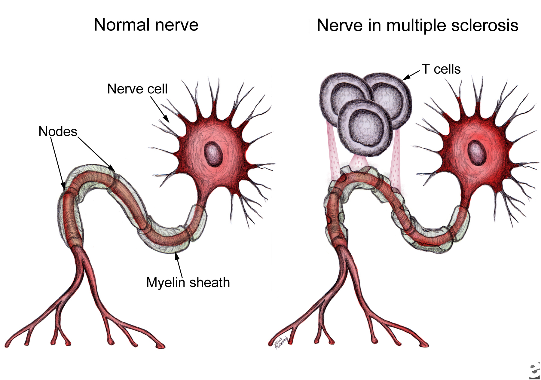
These results support the epitope-spreading hypothesis, which indicates that MS patients make antibodies against one or a few myelin proteins, but as the disease progresses, the autoimmune response spreads to other myelin sheath epitopes. In their recent publication, Lutterotti et al. provide sufficient evidence necessitating emphasis not only on the specific target antigens, but also on the facility to inhibit epitope spreading, preferably prior to diversification of the CD4+ T-helper cell autoreactivity.


Lutterotti’s study is a significant step towards finding an effective strategy not only for treatment of MS, but also other T-cell-driven autoimmune disorders. Nonetheless, this therapeutic method must be tested on a much larger sample and geographically distinct population to demonstrate whether the efficacy reported here would be consistent in most if not all MS subtypes. The phase II of this clinical trial is planned to take place in the near future, during which more will be known about the long-term safety and efficacy of this technique. Regardless, the data presented by this study, at the very least, have set the requirement for future antigen-specific therapies to include the ability to target not only the previously activated autoreactive T-cells, but also the naïve autoreactive T-cells specific for several myelin epitopes.

Further Reading:

Antigen-Specific Tolerance by Autologous Myelin Peptide–Coupled Cells: A Phase 1 Trial in Multiple Sclerosis

Identification of New Potential Drug Targets for Treatment of Lupus

autoantibodies Systemic lupus erythematosus (SLE) is a complex autoimmune disease that afflicts tens of millions of people worldwide.  The most prominent feature is generation of “autoantibodies” to self-proteins and nucleic acids, resulting in immune complex (IC) formation and organ inflammation.  Affected patients may demonstrate rashes, joint pain, anemia, or kidney damage, and untreated complications can often be fatal.  In addition, most SLE patients demonstrate continuously elevated levels of interferon (IFN) α, which is naturally produced by activated plasmacytoid dendritic cells (pDCs) 1.  pDCs are a rare subset of DCs found in the blood and peripheral lymphoid organs that function in host defense by secreting proinflammatory cytokines to initiate the innate immune response.  pDCs are activated following engagement of Toll-like receptors (TLRs), which recognize molecular signatures of bacteria and viruses.  Studies have shown that the frequency of circulating pDCs is significantly reduced in SLE patients, due to increased migration to inflammatory sites in affected organs 2.  Although pDCs have been implicated in contributing to autoimmunity via continuous type I IFN production, their exact role in lupus pathogenesis has not been clearly elucidated.

Recently, in PNAS, Baccala et al. provided direct evidence that in the absence of pDCs, the disease manifestations of Lupus were significantly decreased 3.  Since IRF8 is a hematopoietic cell-specific transcription factor known to be essential for pDC development 4, the authors knocked out IRF8 in NZB mice, a widely used mouse model for SLE.  Appropriately, pDCs were absent in IRF8-deficientNZB mice, and type I IFNs were undetectable even after injection with CpG DNA, a standard method of inducing the interferon pathway.  Interestingly, autoantibody production was almost completely abrogated and kidney disease was drastically improved compared to wild-type NZB mice.  Taken together, their results suggest that without pDCs, SLE disease manifestations are significantly reduced.

Next, the authors sought to examine specifically how pDCs promote systemic autoimmunity.  They used another mouse model with a mutation in Slc15a4, which is characterized by normal development of pDCs but an absence of type I IFN production by pDCs.  It is still unclear how a mutation in Slc15a4 leads to a disruption in proinflammatory cytokine production in pDCs, but since Slc15a4 is a peptide/histidine transporter, others hypothesize that it transports free histidine from the endosome to the cytosol to enable cathepsin-mediated cleavage of endosomal TLRs required for subsequent signaling 5.  Similar to the IRF8-deficient NZB mice, Slc15a4 mice had significantly reduced autoantibodies, decreased kidney disease, and extended survival.  This finding rules out the possibility that pDCs contribute to disease through other functions outside of type I IFN production.

In summary, Baccala et al. provide direct evidence that pDCs contribute to the abnormal manifestations of SLE via hyperproduction of type I IFNs.  Thus, IRF8 and Slc15a4 serve as new potential drug targets for treatment of SLE.  Current therapies involve broad immunosuppressive drugs, which suppress multiple arms of the immune system, increasing a patient’s risk for various infections and cancer.  Specific pharmacologic inhibition of IRF8 or Slc15a4 could prevent Lupus-specific flare-ups, as well as manifestations of other autoimmune diseases.

References

1          Gilliet, M., Cao, W. & Liu, Y. J. Plasmacytoid dendritic cells: sensing nucleic acids in viral infection and autoimmune diseases. Nat Rev Immunol 8, 594-606, doi:10.1038/nri2358 (2008).

2          Ronnblom, L. The type I interferon system in the etiopathogenesis of autoimmune diseases. Ups J Med Sci 116, 227-237, doi:10.3109/03009734.2011.624649 (2011).

3          Baccala, R. et al. Essential requirement for IRF8 and SLC15A4 implicates plasmacytoid dendritic cells in the pathogenesis of lupus. Proc Natl Acad Sci U S A 110, 2940-2945, doi:10.1073/pnas.1222798110 (2013).

4          Tsujimura, H., Tamura, T. & Ozato, K. Cutting edge: IFN consensus sequence binding protein/IFN regulatory factor 8 drives the development of type I IFN-producing plasmacytoid dendritic cells. J Immunol 170, 1131-1135 (2003).

5          Park, B. et al. Proteolytic cleavage in an endolysosomal compartment is required for activation of Toll-like receptor 9. Nat Immunol 9, 1407-1414, doi:10.1038/ni.1669 (2008).

RESISTANCE TO LUNG CANCER THERAPY: A CASE REPORT

A study recently published in The New England Journal of Medicine (Jun 1st, 2013) identified an acquired mutation in the ROS1 kinase domain resulting in resistance to crizotinib in a woman with metastatic lung adenocarcinoma.

Crizotinib is an oral ATP-competitive selective Non Small cell Lung Cancer resized 600inhibitor of the anaplastic lymphoma kinase (ALK) and MET tyrosine kinase that inhibits tyrosine phosphorylation of activated ALK at nanomolar concentrations. In 2011, crizotinib was approved by the U.S. Food and Drug Administration (FDA) for treatment of patients with locally advanced or metastatic non-small-cell lung cancer (NSCLC) that are ALK-positive. Activating mutations or translocations of the ALK gene have been discovered in various types of cancer, including anaplastic large-cell lymphoma, neuroblastoma, inflammatory myofibroblastic tumor, and non–small-cell lung cancer. Because of its role in lung cancer, ALK receptor tyrosine represents a potential therapeutic target.

In addition to ALK mutations or translocations, chromosomal rearrangements in another tyrosine kinase receptor, ROS1, was identified in a molecular subset of NSCLC with distinct clinical characteristics that are similar to those observed in patients with ALK-rearranged NSCLC. Crizotinib was found highly sensitive in lung cancer patients who harbor rearrangements in ALK or ROS1. However, resistance to crizotinib was reported in lung cancer due to secondary mutations in ALK. To overcome this problem a new compound CH5424802 has been identified and is currently in clinical trials (ClinicalTrials.gov number, NCT01588028) for ALK-positive NSCLC.

A 48-year-old woman with metastatic lung cancer and a distant history of light smoking was initially treated with first line of chemotherapy with carboplatin and pemetrexed. Genetic analysis with patient’s cancer cells showed no mutation in oncogenic KRAS or EGFR and no ALK translocations. Additional molecular testing revealed ROS1 rearrangement lead to expression of a fusion protein CD74-ROS1. After three cycles of chemotherapy, marked disease progression was noted and patient’s condition deteriorated. The patient was then enrolled in a clinical trial evaluating the safety and efficacy of crizotinib in cancer patients with ROS1 translocations (ClinicalTrials.gov number, NCT00585195). Computed tomographic scan (CT) obtained two months after treatment noted dramatic response to treatment. However, one month later, while the patient was still taking crizotinib, disease progression was observed and unfortunately the patient expired. Molecular analysis of tumor samples from all sites of disease detected a mutation glycine to arginine Gly2032Arg (G2032R) spanning CD74-ROS1 fusion area that had not been observed in pretreated samples. No other mutation of ROS1 kinase was identified by deep sequencing. Thus this suggested that appearance of G2032R mutation was an early event in crizotinib-resistant tumor cells.

To identify role of G2032R mutation in crizotinib resistance, 293T cells were transfected with either mutated or nonmutated G2032R CD74-ROS1 and subsequently treated with tyrosine kinase inhibitors crizotinib and TAE648. Cells transfected with a mutated form of ROS1 exhibited a half-maximal inhibitory concentration (IC50) value greater than 1000 nM while for nonmutated cells it was approximately 30 nM for crizotinib and 50 nM for TAE648. Crystal structure analysis of ROS1 revealed an arginine at position 2032 resulted in steric interference of crizotinib binding. Collectively, this study reported a mechanism of acquired resistance to crizotinib in a cancer driven by oncogenic ROS1 fusion. Therefore, in the context of these observations, it may be necessary to identify novel compounds that specifically target the G2032R ROS1 mutant to overcome the development of crizotinib resistance in cancers driven by ROS1.


References:

1. Awad MM, Katayama R, McTigue M, et al. Acquired Resistance to Crizotinib from a Mutation in CD74-ROS1. N Engl J Med 2013.

2. Bergethon K, Shaw AT, Ou SH, et al. ROS1 rearrangements define a unique molecular class of lung cancers. J Clin Oncol 2012;30:863-70.

3. Sakamoto H, Tsukaguchi T, Hiroshima S, et al. CH5424802, a selective ALK inhibitor capable of blocking the resistant gatekeeper mutant. Cancer Cell 2011;19:679-90.


describe the image

Arup Chakraborty is postdoctoral research fellow at the National Cancer Institute, Bethesda, MD. He earned a doctoral degree from Texas Tech University, and his primary research interest is in the field of clinical cancer mainly in mechanisms of resistance to molecularly targeted therapies