ISSCR 2013 Meeting Updates: Can Alzheimer’s Disease be modeled in a dish?

Human pluripotent stem cells (hPSCs) can differentiate into all cell types of the body, and can thereby serve as great models to examine the pathological mechanisms of various human diseases.  At the International Society for Stem Cell Research (ISSCR) 11th Annual Meeting, various stem cell experts highlighted the current human stem cell models for Alzheimer’s disease, and discussed the potential future directions of the field.

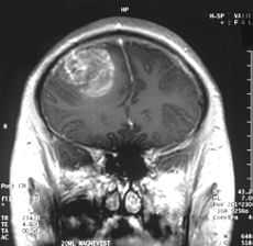
Alzheimer’s disease (AD) is the most common neurodegenerative dementia, affecting ~30 million people worldwide.  AD occurs in two main forms:  early-onset, familial AD (FAD) and late-onset, sporadic AD (SAD).  Both 40004_webare characterized by extensive neuronal loss and the aggregation of two proteins in the brain: amyloid β peptide (Aβ) and tau.  Aβ peptide is derived from the amyloid precursor protein (APP) via cleavage by two proteases, β-secretase and γ-secretase.  According to the amyloid cascade hypothesis, elevated levels of Aβ are necessary and sufficient to trigger disease 1.  Tau is synthesized in neurons and normally functions in binding to tubulin and stabilization of microtubules.  However, in AD, tau is hyper-phosphorylated, resulting in dissociation from microtubules, aggregation, and formation of neurofibrillary tangles (NFTs).  Although the pathological hallmarks of AD consist of these amyloid plaques and NFTs, how the two are related to each other and how they contribute to clinical onset and progression of AD is still under investigation.  By the time a patient manifests symptoms of a mild dementia, there is already significant neuronal loss and substantial accumulation of plaques and tangles.  One major limitation to our understanding of AD has been the lack of live, patient-specific neurons to examine disease progression.

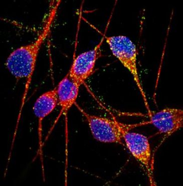
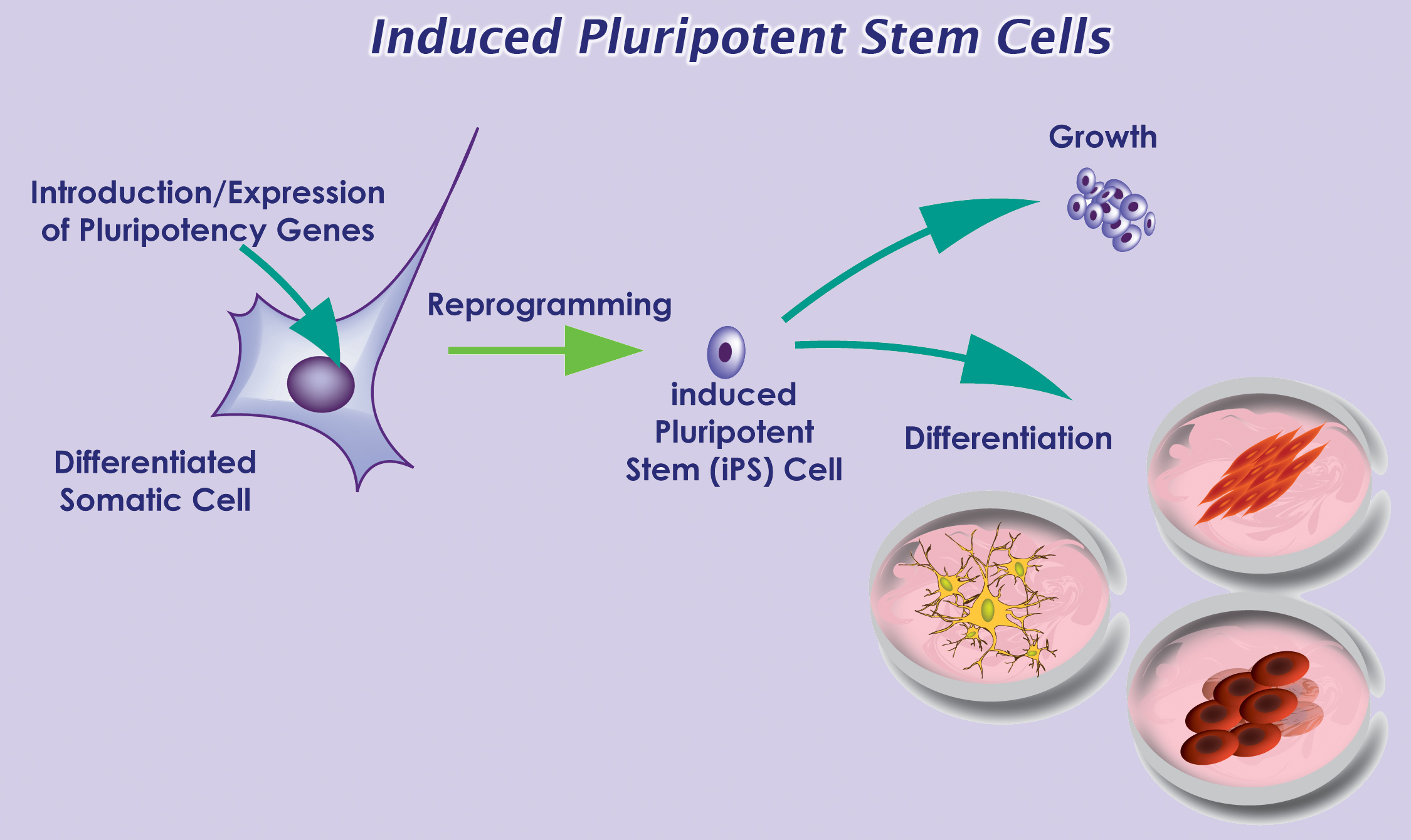
With recent advances in reprogramming technology, scientists can now generate induced pluripotent stem cells (iPSCs), and thereby use live, patient-specific models to examine disease phenotypes in a dish.  At the ISSCR meeting, Larry Goldstein presented his lab’s recent work on using hiPSC models to study AD.  They generated iPSCs from two patients with FAD caused by a duplication of the APP gene, two patients with SAD, and two control individuals.  Next, neurons were generated from the iPSC lines by directed differentiation and fluorescence-activated cell sorting (FACS) purification 2.  Neurons from one SAD and two FAD patients demonstrated significantly higher levels of secreted Aβ and phosphorylated tau (p-tau) 3.  To determine whether there is an association between APP processing and elevated p-tau levels, they treated iPSC-derived neurons with γ-secretase and β-secretase inhibitors.  Interestingly, pharmacologic inhibition of β-secretase resulted in a significant reduction in the levels of Aβ and p-tau.  Treatment with the γ-secretase inhibitor only reduced Aβ levels, but not p-tau levels.  This suggests that products of APP processing other than Aβ might contribute to elevated p-tau levels, highlighting a potential weakness with the amyloid cascade hypothesis.

Other groups have proposed alternative hypotheses to explain AD pathogenesis.  Haruhisa Inoue presented his group’s work on using human iPSC models to examine how intracellular Aβ oligomers contribute to AD.  They generated iPSCs from one patient with FAD caused by the APP-E693Δ mutation, two patients with SAD, and three control individuals.  Corticol neurons were derived using small molecule inhibitors of bone morphogenic protein (BMP) and activin/nodal signaling as previously described 4.  Aβ oligomers accumulated in neurons derived from the FAD patient and one SAD patient, but not in the control neurons 5.  Specifically, the Aβ oligomers accumulated in the endoplasmic reticulum (ER), and triggered ER and oxidative stress in the neurons.  In addition, treatment with docosahexaenoic acid (DHA) alleviated the stress responses.  Although the drug has previously failed in some clinical trials of AD treatment, Inoue’s work suggests that DHA might be effective for a subset of patients.

In summary, Goldstein and Inoue presented convincing evidence that human iPSC models can be used to study early AD pathogenesis and patient-specific drug responses.  Although it can take decades for symptoms to manifest in patients, disease phenotypes can be observed using iPSC models.  However, the fact that only one out of two SAD patients generated a disease phenotype highlights the need of future iPSC studies to examine larger numbers of patients to account for the observed heterogeneity in AD pathogenesis.

References:

1          Hardy, J. & Selkoe, D. J. The amyloid hypothesis of Alzheimer’s disease: progress and problems on the road to therapeutics. Science 297, 353-356, doi:10.1126/science.1072994 (2002).

2          Yuan, S. H. et al. Cell-surface marker signatures for the isolation of neural stem cells, glia and neurons derived from human pluripotent stem cells. PLoS One 6, e17540, doi:10.1371/journal.pone.0017540 (2011).

3          Israel, M. A. et al. Probing sporadic and familial Alzheimer’s disease using induced pluripotent stem cells. Nature 482, 216-220, doi:10.1038/nature10821 (2012).

4          Morizane, A., Doi, D., Kikuchi, T., Nishimura, K. & Takahashi, J. Small-molecule inhibitors of bone morphogenic protein and activin/nodal signals promote highly efficient neural induction from human pluripotent stem cells. J Neurosci Res 89, 117-126, doi:10.1002/jnr.22547 (2011).

5          Kondo, T. et al. Modeling Alzheimer’s disease with iPSCs reveals stress phenotypes associated with intracellular Abeta and differential drug responsiveness. Cell Stem Cell 12, 487-496, doi:10.1016/j.stem.2013.01.009 (2013).


New Findings in Cell Based Therapy for GBM

Glioblastoma multiforme (GBM) is the most common and lethalGlioblastoma type of malignant primary brain tumors that account for over 70% of all intracranial cancers.  The current course of GBM treatment consists of surgical resection of the main tumor mass, followed by administration of radiation and chemotherapy. Surgical resection of the primary tumor leads to injury to the surrounding normal tissue, while chemotherapy and radiotherapy cause toxicity to the healthy tissue in the brain.  These undesirable secondary effects of glioma treatments have a major impact on patients’ physical, cognitive and emotional functioning. Nonetheless, despite this aggressive treatment regimen and its harmful side effects, GBM remains virtually incurable, with post-diagnosis median survival persisting less than 14 months.  This dismal prognosis is due to a combination of unique anatomical features of the central nervous system (CNS), in addition to GBMs’ (glioma cells’) exceptional invasive capacity. Glioma cells infiltrate the brain’s highly dense parenchyma, migrating along the corpus callosum and creating new masses within the hemisphere contralateral to the initial tumor mass homing.

Thus, recurrence in postoperative GBM patients is in essence inevitable. Furthermore, GBMs are not only heterogeneous among individual patients, but they are also highly heterogeneous within a single tumor mass. Recent studies have shown compelling evidence of a therapeutically resistant subpopulation of malignant glioma cells that exhibit stem-cell like characteristics, such as multipotency, the ability to self-renew and invade/migrate; these tumor-initiating cells are referred to as glioma stem cells (GSCs) and are believed to be responsible for tumor initiation and recurrence in GBM patients. Furthermore, GSCs have been observed to ensconce within similar niches to neural stem cells (NSCs).

NSCs and mesenchymal stem cells (MSCs) have been shown to have an exceptional migratory ability within brain’s parenchyma and possess a notable inherent tumor tropism.  Thus, several cell-based therapy (CBT) studies and clinical models of malignant tumors have incorporated autonomous tracking of tumor cells by employing NSCs and MSCs to deliver multiple therapeutic genes to specific tumor loci. This target specific drug-delivery model has NSCs or MSCs transduced to express one pro-drug-activating enzyme, which catalyzes the conversion of a particular pro-drug into an active toxic agent, which results in the localization of the chemotherapeutics specifically at the tumor sites.  In addition to its ability to deliver effective cytotoxic damage to the tumor without causing damage to the healthy surrounding tissue, the resulting bystander effect of this system causes cell death not only to the drug-delivery-vehicle cells, but also to the surrounding glioma cells. Although a number of different enzyme/prodrug systems have been utilized in relevant studies, HSV-thymidine kinase (HSV-tk)/ deoxyguanosine analog ganciclovir (GCV) has been the most commonly tested in animal and in vitro models. HSV-tk phosphorylates GCV and produces deoxyguanosine triphosphate which is a polymerase-I inhibitor and DNA chain terminator; cell death occurs upon incorporation of the nucleotide analog within DNA chains.

In recent study published in the journal of Molecular Therapy, Blanco’s group report their findings regarding the interaction between human MSCs (hMSCs) and gliomas and the underlying mechanism for the effectiveness of hAMSC based therapy in GBMs.  In their previous work Blanco’s group showed that the administration of hAMSCs expressing HSV thymidine kinase in glioma tumors significantly promotes tumor growth, whereas induction of cytotoxicity by administration of the prodrug GCV demonstrated a significant antitumor response.

According to this new study, hMSCs differentiate to endothelial lineage (supported by the expression of CD31 marker) within tumors, and integrate in the tumor vascular system where they adopt an endothelial phenotype. Further, Blanco proposed the notion of hMSCs’ ability to home to privileged vascular structures where GSCs also reside, is the underlying characteristic that leads to the effectiveness of cytotoxic hMSCs in regulating the bystander killing of tumor cells.

Although Blanco’s study provides invaluable insights into the GSCs’ niche and its role in malignant brain tumor CBT, hMSCs’ tumor growth promotion is a quality, which makes these cells less than ideal for utilization in human clinical trials in the near future. Future studies on this mechanism using primary patient tumor cells rather than the U87 glioma cell line and human NSCs are needed to further confirm these observations.

Further reading:

Juli R. Bagó, Maria Alieva, Carolina Soler, Núria Rubio, Jerónimo Blanco. Endothelial differentiation of adipose tissue-derived mesenchymal stromal cells in glioma tumors: implications for cell based therapy. Molecular Therapy.

ISSCR 2013 Meeting Updates: Is a Cure in Sight for Type 1 Diabetes?

Last week, the International Society for Stem Cell Research (ISSCR) held its 11th Annual Meeting in Boston, MA.  Over 3,000 stem cell researchers from around the world gathered to hear the leading experts share their research and perspectives in this fast-paced field.  One interesting topic was recent advances in cell-replacement therapy for treatment of type 1 diabetes.

Diabetes mellitus is a metabolic disease that results from a failure in glucose regulation, often leading to severe hyperglycemia and tissue/organ damage.  Pancreatic β-cells respond to high blood glucose levels by secreting insulin, which acts on other tissues to promote glucose uptake from the blood.  Type 1 diabetes (T1D) results from autoimmune destruction of insulin-producing β-cells of the pancreas.  The lack of insulin leads to increased blood and urine glucose.  T1D is often fatal unless treated with exogenous administration of insulin daily and regular blood glucose monitoring for the patient’s entire life.  However, this treatment does not match the effect of having endogenous β-cells.  Thus, scientists in the field of regenerative medicine have focused on strategies for generation of β-cells for cell-replacement therapy.

Douglas Melton presented his lab’s describe the imagecurrent progress on generation of functional β-cells from human embryonic stem cells (hESCs).  Since 2006, D’Amour et al. demonstrated that they had developed a robust differentiation protocol to produce hESC-derived pancreatic endocrine cells capable of synthesizing insulin 1.  However, the derived cells failed to secrete insulin appropriately in response to the addition of glucose, a required function of true β-cells.  Thus, one focus of Melton’s group has been to identify the signals to generate functional β-cells.  Pancreatic islets are complex structures consisting of multiple cell types, including the insulin-producing β-cells as well as endothelial cells in the surrounding blood vessels.  Thus, endothelial signals are known to promote pancreatic development 2.  Melton’s group found that co-culture of β-cells with endothelial cells promotes functional maturation of the hESC-derived β-cells.  In addition, they recently identified another hormone, betatrophin, which is secreted by the liver and functions in promoting β-cell replication 3.  Thus, increasing the levels of this hormone may generate more β-cells.  Although various groups have demonstrated that large amounts of glucose-responsive, insulin-secreting β-cells can be generated in vitro, one concern that remains to be addressed is how the cells will be protected from an autoimmune attack once delivered to the patient.  One potential strategy involves encapsulation of the β-cells into an immunoprotective device prior to delivery.

Other mature cells have also iPSDerivationbeen proposed as a source of new β-cells.  Sarah Ferber presented her lab’s work on inducing liver cells to transdifferentiate into β-cells for autologous cell-replacement therapy.  Transdifferentiation is the process by which one type of adult cell is directly converted into another type of cell.  They used the transcription factor, pancreatic and duodenal homeobox gene 1 (PDX-1), and soluble factors to induce the developmental shift of adult human liver cells into functional insulin-producing cells 4.  Not only did the transdifferentiated liver cells produce insulin, but they also released in a glucose-regulated manner.  When transplanted into diabetic, immunodeficient mice, the cells ameliorated hyperglycemia over a 60-day period.  Thus, PDX-1-induced transdifferentiated liver cells offer the potential to replace β-cells’ function in vivo.  Furthermore, transplantation of autologous β-cells would circumvent a host versus graft immune response, as well as allow the patient to be the donor of his or her own insulin-producing cells.

In summary, Melton’s and Ferber’s presentations demonstrated the tremendous progress that has been made in the last decade to generate functional β-cells for use in treatment of T1D.

References

1          D’Amour, K. A. et al. Production of pancreatic hormone-expressing endocrine cells from human embryonic stem cells. Nat Biotechnol 24, 1392-1401, doi:10.1038/nbt1259 (2006).

2          Nikolova, G. et al. The vascular basement membrane: a niche for insulin gene expression and Beta cell proliferation. Dev Cell 10, 397-405, doi:10.1016/j.devcel.2006.01.015 (2006).

3          Yi, P., Park, J. S. & Melton, D. A. Betatrophin: A Hormone that Controls Pancreatic beta Cell Proliferation. Cell 153, 747-758, doi:10.1016/j.cell.2013.04.008 (2013).

4          Sapir, T. et al. Cell-replacement therapy for diabetes: Generating functional insulin-producing tissue from adult human liver cells. Proc Natl Acad Sci U S A 102, 7964-7969, doi:10.1073/pnas.0405277102 (2005).

Identification of a new HSC viral transduction enhancer, Vectofusin-1

HSC gene therapy is an emerging therapeutic option for several disorders of the blood and immune system.  Ex vivo cell therapies are based on the ability to isolate CD34+ cells from a patient or a normal donor, expansion ex vivo with genetic modification, and systemic administration into the patient following myeloablative treatment.  An efficient method for gene transfer into HSCs is required for successful gene therapy.  Lentiviral vectors (LVs) have emerged as a robust and versatile tool for ex vivo and in vivo gene delivery into multiple cell types, including HSCs.  LVs can either be pseudotyped with viral envelope glycoproteins that confer a broad tropism, such as the vesicular stomatitis virus G (VSV-G) protein, or those that confer a specific HSC tropism, including gibbon ape leukemia virus (GALVTR), feline endogenous retrovirus RD114 (RD114TR), or amphotropic murine leukemia virus (MLV-A) proteins.  However, viral envelopes vary in transduction efficiency.  Thus, transduction protocols often involve the addition of factors to enhance viral entry, including cationic polymers (polybrene) 1 or fibronectin fragments (Retronectin) 2.

gene therapy

Recently, in Molecular Therapy-Nucleic Acids, Fenard et al identified another viral entry enhancer, Vectofusin-1 3.  Vectofusin-1 is a synthetic, histidine-rich cationic amphipathic peptide derived from the LAH4 peptide family.  LAH4 peptides and their derivatives are known to be efficient DNA transfection agents 4.  In this study, the authors examined whether Vectofusin-1 would also enhance gene transfer of LVs into CD34+ cells derived from human umbilical cord blood.  Indeed, Vectofusin-1 significantly increased the transduction efficiency of LVs pseudotyped with various envelopes (VSV-G, GALVTR, RD114TR, MLV), with transduction levels ranging from 50-80% compared to undetectable transduction levels in its absence.  In addition, the increased transduction efficiency was not cytotoxic.  Addition of Vectofusin-1 during transduction of CD34+ cells did not negatively affect subsequent myeloerythroid differentiation in colony-forming cell (CFC) assays in vitro, or hematopoietic reconstitution in immunodeficient BALB-Rag/γC mice in vivo.  The mechanism for the increased transduction efficiency was attributed to insertion of the peptide in the viral and cellular membranes, resulting in an enhancement in both adhesion and fusion of the viral particles with the cell’s plasma membrane.

In short, the authors demonstrated that Vectofusin-1 is a promising LV entry enhancer that can be potentially used in ex vivo transduction of HSCs for subsequent use in clinical applications.  Addition of Vectofusin-1 to the transduction medium had similar effects as the commonly used Retronectin, although the latter is used to coat plates, suggesting a different mechanism of action.  Future experiments will determine whether Vectofusin-1 and Retronectin can be used together to synergistically enhance HSC transduction.


References:

1          Davis, H. E., Morgan, J. R. & Yarmush, M. L. Polybrene increases retrovirus gene transfer efficiency by enhancing receptor-independent virus adsorption on target cell membranes. Biophys Chem 97, 159-172 (2002).

2          Pollok, K. E. & Williams, D. A. Facilitation of retrovirus-mediated gene transfer into hematopoietic stem and progenitor cells and peripheral blood T-lymphocytes utilizing recombinant fibronectin fragments. Curr Opin Mol Ther 1, 595-604 (1999).

3          Fenard, D. et al. Vectofusin-1, a new viral entry enhancer, strongly promotes lentiviral transduction of human hematopoietic stem cells. Mol Ther Nucleic Acids 2, e90, doi:10.1038/mtna.2013.17 (2013).

4          Kichler, A., Leborgne, C., Marz, J., Danos, O. & Bechinger, B. Histidine-rich amphipathic peptide antibiotics promote efficient delivery of DNA into mammalian cells. Proc Natl Acad Sci U S A 100, 1564-1568, doi:10.1073/pnas.0337677100 (2003).

Reprogramming of old HSCs reverses functional defects associated with aging

HSCs must continuously self-renew to replenish the pool of mature blood cells throughout the life an adult.  One requirement for extensive self-renewal is high telomerase activity to prevent telomere shortening.  HSCs isolated from adult bone marrow have shorter telomeres than cells from fetal liver or umbilical cord blood 1, suggesting that proliferative potential may decrease with age.  Also, HSC aging is associated with decreased lymphoid potential, as well as an up-regulation of genes involved in leukemic transformation 2.  Consequently, “aging” HSCs may have functional defects that might be detrimental for therapeutic strategies involving genetic manipulation and transplantation of HSCs for the treatment of various blood disorders.

describe the image

Previous studies have demonstrated that during reprogramming, developing iPSCs express epigenetic features of ESCs, and lose those of the starting somatic cell 3.  In addition, a previous study reported a significant elongation of telomeres in derived iPSCs compared to the starting somatic cell 4.  Thus, it may be possible that reprogramming reverses functional defects associated with HSC aging.

Recently, in Blood, Wahlestedt et al examined whether characteristics of aging HSCs are reversible 5.  First, they derived iPSCs from young and aged murine HSCs.  To examine their differentiation potential, they injected the derived iPSCs into murine blastocysts and analyzed the engraftment of the donor cells in the developing chimeric embryos.  Overall, iPSCs derived from aged HSCs demonstrated similar differentiation potential compared to that of younger HSCs.  The engraftment of bone marrow mononuclear cells from primary chimeric mice in a competitive transfer experiment was comparable to that of young HSCs.  Aged HSCs, on the other hand, demonstrated a significant reduction in repopulation capacity.  Interestingly, aged iPSC-derived HSCs also generated naïve T cells at similar levels as young HSCs.

Next, the authors examined telomere length following re-differentiation of the young and aged iPSCs.  Telomeres in aged HSCs were ~11% shorter compared to young HSCs.  However, telomeres of the HSCs derived from the aged iPSCs demonstrated a 2-fold elongation compared to blastocyst control HSCs.  This 2-fold elongation was maintained even after transplantation.  Overall, these results indicate that iPSC induction from HSCs results in elongation of telomeres.

In short, Wahlestedt et al demonstrated that reprogramming does indeed reverse some of the functional defects associated with chronologically aged HSCs, including decreased differentiation potential and shortened telomeres.  However, the study did not address whether the iPSCs derived from aged HSCs had an increased DNA mutation frequency, since HSC aging is also associated with a higher mutation rate.  It would also be interesting to determine whether the above phenomena are also observed in reprogramming of aged human HSCs.  If iPSC induction does indeed result in an “epigenetic reset,” then HSCs derived from iPSCs may have unique characteristics favorable for use in clinical settings.


References

1          Vaziri, H. et al. Evidence for a mitotic clock in human hematopoietic stem cells: loss of telomeric DNA with age. Proc Natl Acad Sci U S A 91, 9857-9860 (1994).

2          Rossi, D. J. et al. Cell intrinsic alterations underlie hematopoietic stem cell aging. Proc Natl Acad Sci U S A 102, 9194-9199, doi:10.1073/pnas.0503280102 (2005).

3          Maherali, N. et al. Directly reprogrammed fibroblasts show global epigenetic remodeling and widespread tissue contribution. Cell Stem Cell 1, 55-70, doi:10.1016/j.stem.2007.05.014 (2007).

4          Marion, R. M. et al. Telomeres acquire embryonic stem cell characteristics in induced pluripotent stem cells. Cell Stem Cell 4, 141-154, doi:10.1016/j.stem.2008.12.010 (2009).

5          Wahlestedt, M. et al. An epigenetic component of hematopoietic stem cell aging amenable to reprogramming into a young state. Blood, doi:10.1182/blood-2012-11-469080 (2013).

Mechanical Properties of 3D Cell Culture Affect Stem Cell Phenotype

The field of regenerative medicine holds great promise as we gain greater understanding of how stem cells differentiate into the many cell types found in our bodies.  However, the clinical applications of these stem cells have been hampered by the challenges in replicating the in vitro capabilities of these cells once transplanted into the body.  This is partially due to the fact that most stem cell work is based on culturing cells in a flat two-dimensional (2D) format.

Cell culture in 2D has been routinely used in laboratories for over 40 years.  Cells are grown on flat dishes made of coated or uncoated polystyrene plastic or glass that are very stiff and arguably primitive.  These cells attach and spread on the surface to form unnatural cell attachments to other cells and to deposited proteins that are denatured on this synthetic surface.  Thus, the culturing of cells in 2D does not accurately reproduce the extracellular matrix (ECM) found in native tissue resulting in an alteration of many complex biological responses.  The native microenvironment provides mechanical signals, soluble factors, communication between neighboring cells and communication between the cell and its matrix.  This spatial and temporal organization affects normal cell fate including division, proliferation, migration, differentiation and apoptosis.

To overcome these challenges scientists have developed several three-dimensional (3D) culture methods such as cell spheroids, micro-carrier cultures, scaffolds, or tissue-engineered models.  Cell spheroids, self-assembled spherical clusters of cell colonies, are simple, reproducible and similar to physiological tissues compared to other methods involving ECM scaffolds and hydrogel systems (water-swollen polymer networks).  They are created from single culture or co-culture techniques such as hanging drop, rotating culture, non-adhesive surfaces or concave plate methods.  A similar method is the development of epithelial tissues to form polarized sheets.  However, as the size and complexity of the 3D model increases, so does the requirement for a scaffold which will ideally produce features naturally found within the ECM required for native cell function.

In this 3D cell culture environment, cells synthesize and secrete a flexible and pliable extracellular matrix in their native configuration.  Gap junctions are increased in 3D culture allowing cells to communicate with each other via exchange of ions, small molecules and electrical currents.  Surface adhesion molecules and receptors critical for cell function are also maximized.  The various effects of 3D culture versus 2D culture on differentiation, drug metabolism, expression, cell function, morphology, proliferation, viability, response to stimuli and in vivo relevance are vast and ever expanding (JUST THE FACTS: Specific effects of 3D vs. 2D cell culture).

In a recent study published April 11, 2013 online in Advanced Functional Materials, researchers at Case Western University describe how micropatterning technology influences stem cell fate decisions.  Micropatterning technology is the use of a technique to influence the network pattern of microgels aka intelligent hydrogel systems.  While this technology has recently become an important tool for spatially controlling stem cell microenvironment, very little is known about the effect of the size of the micropatterned regions, which influences hydrogel stiffness and transport properties, on stem cell behavior.  The researchers developed a 3D micropatterned hydrogel system that was either single-crosslinked or dual-crosslinked and evaluated human adipose-derived stem cell (hASC) behavior.  The cells grew into clusters in the singly-crosslinked regions where the size of the hASC clusters depended on the micropattern size (increased cell-cell interactions may have promoted cell proliferation), while hASCs encapsulated in the dual-crosslinked regions remained mostly isolated and had lower proliferation rates.  Interestingly, osteogenic (bone) and chondrogenic (cartilage) differentiation of the hASCs increased as the micropattern size increased but there was no effect on adipogenic (fat cells) differentiation.  The researchers believe that controlling local biomaterial properties may allow them to guide the formation of complex tissues.

Another study published March 24, 2013 in Nature Materials describes how mechanotransduction (how cells take information about its physical environment and t3D Traction image
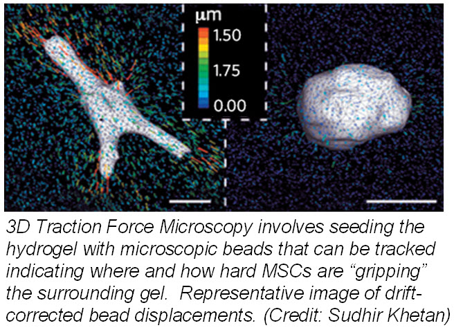
ranslate that into chemical signals) can influence stem cell fate.  Researchers from University of Pennsylvania showed that cell fate is regulated by cell-generated tension that is enabled through cell-mediated degradation of the covalently crosslinked matrix.  When cultured on “softer” 2D covalently crosslinked gels (RGD-modified methacrylated hyaluronic acid hydrogels), mesenchymal stem cells (MSCs) differentiated into adipocytes when cultured in bipotential adipogenic/ osteogenic media.  In contrast, MSCs cultured on “harder” 2D alginate gels differentiated into chondrocytes.  This phenomenon was not present in 3D hydrogels and was attributed to the inability of cells to degrade the covalent cross-linked bonds resulting in MSCs differentiating into adipocytes.  Introduction of proteolytically cleavable crosslinks and utilizing 3D traction force microscopy, revealed that MSC differentiation into bone cells was dependent on the cells to better anchor themselves into the environment and degradation signals.

These two studies provide insight into how the microenvironment can affect the fate of stem cells.  Understanding how the microenvironment influences stem cell behavior is important for tissue engineering approaches.  Cell-based assays have the potential to provide reliable data for regenerative medicine but scientists need to bridge the in vitro and in vivo gap by growing cells within a microenvironment that establishes physiological cell-cell and cell-substrate interactions that regulate proliferation and differentiation.  Hence, 3D models will provide more reliable and meaningful therapeutic results compared to 2D tests.


Further reading:

Haycock JW.  3D cell culture: a review of current approaches and techniques.  Methods Mol Biol. 2011; 695:1-15

Oju Jeon, Eben Alsberg.  Regulation of Stem Cell Fate in a Three-Dimensional Micropatterned Dual-Crosslinked Hydrogel System.  Advanced Functional Materials.  Article first published online: 11 April 2013

Wei Song, Naoki Kawazoe, and Guoping Chen.  Dependence of Spreading and Differentiation of Mesenchymal Stem Cells on Micropatterned Surface Area.  Journal of Nanomaterials.  Volume 2011 (2011), 9 pages

Sudhir Khetan, Murat Guvendiren, Wesley R. Legant, Daniel M. Cohen, Christopher S. Chen and Jason A. Burdick.  Degradation-mediated cellular traction directs stem cell fate in covalently crosslinked three-dimensional hydrogels. Nature Materials. Published online 24 March 2013


Efficient generation of iPSCs from human cord blood and peripheral blood

The initial finding that pluripotency could be induced in human somatic cells revolutionized the field of regenerative medicine, since patient-specific stem cells can now be generated to further examine the causes and mechanisms of various human diseases.  Since the discovery of human iPSCs in 2007 1, various studies have focused on improving the reprogramming methods in order to increase the induction efficiency, as well as to further simplify the protocol.  iPSCs are commonly generated from dermal fibroblasts.  However, skin biopsies are required to isolate fibroblast cells, highlighting the necessity to identify an alternative source of cells for reprogramming that would involve less invasive surgical procedures for isolation.

Cord blood cells and peripheral blood mononuclear cells (PMNCs) are attractive sources for the generation of iPSCs due to the low invasiveness of their collection, as well as the abundance of blood banks for potential donors.  iPSCs were first derived from human peripheral blood in 2009 2.  CD34+ cells were mobilized from peripheral blood and subsequently transduced with retroviruses delivering OCT4, SOX2, KLF4, and MYC vectors.   Although the reprogramming was successful, use of retroviral vectors requires genomic integration of transgenes that may increase the risk of tumor formation during clinical applications.  Thus, recent studies have focused on generation of “integration-free” iPSCs.  Yet, development of integration-free methods often means compromising the reprogramming efficiency.  iPSC induction in CD34+ cells using non-integrating episomal plasmids resulted in ~0.03% reprogramming efficiency 3.

hESC H9p40 resized 600 resized 600
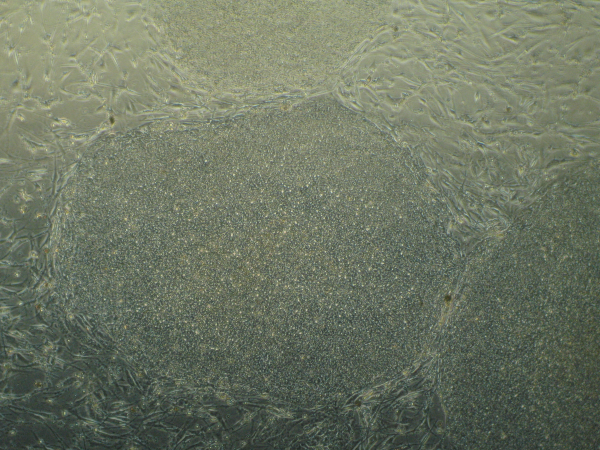
Recently, in Stem Cells, Yamanaka’s group reported a protocol that increased the efficiency of iPSC induction from CD34+ cord blood and peripheral blood 4.  They previously identified an efficient combination of episomal plasmids for reprogramming of adult fibroblasts, termed the “Y4” combination, consisting of plasmids encoding OCT3/4, SOX2, KLF4, L-MYC, LIN28, and an shRNA for TP53 5.  Transfection of CD34+ cells from human cord blood with the Y4 combination resulted in up to 0.1% reprogramming efficiency across two donors.  iPSC induction efficiency of PMNCs isolated from peripheral blood with the Y4 mixture, on the other hand, was inconsistent across donors.  To further increase the reproducibility of iPSC induction from multiple donors, the authors added a vector encoding EBNA1, which is required for episomal plasmid replication and should thereby increase expression of the episomal plasmids.  Addition of the EBNA1 vector to the Y4 mixture resulted in 0.1% reprogramming efficiency in PMNCs across seven donors.  Both, the CD34+– and PMNC-derived iPSCs were molecularly and functionally identical to hESCs.

In summary, Okita et al identified a new protocol allowing efficient generation of integration-free iPSCs from blood.  Previous studies reported ~0.02%- 0.03% induction efficiency from peripheral blood 2 and isolated CD34+ cells 3.  Here, the authors reported a reprogramming efficiency of ~0.06%, with a maximum of 0.1%.  In addition, the new protocol could induce iPSCsfrom frozen PMNCs as efficiently as from freshly isolated cells.  Thus, with the increasing number of potential donors available at cord blood banks, iPSCs can be now efficiently generated for use in autologous or allogeneic stem cell therapy.


References

1          Takahashi, K. et al. Induction of pluripotent stem cells from adult human fibroblasts by defined factors. Cell 131, 861-872, doi:10.1016/j.cell.2007.11.019 (2007).

2          Loh, Y. H. et al. Generation of induced pluripotent stem cells from human blood. Blood 113, 5476-5479, doi:10.1182/blood-2009-02-204800 (2009).

3          Mack, A. A., Kroboth, S., Rajesh, D. & Wang, W. B. Generation of induced pluripotent stem cells from CD34+ cells across blood drawn from multiple donors with non-integrating episomal vectors. PLoS One 6, e27956, doi:10.1371/journal.pone.0027956 (2011).

4          Okita, K. et al. An efficient nonviral method to generate integration-free human-induced pluripotent stem cells from cord blood and peripheral blood cells. Stem Cells 31, 458-466, doi:10.1002/stem.1293 (2013).

5          Okita, K. et al. A more efficient method to generate integration-free human iPS cells. Nat Methods 8, 409-412, doi:10.1038/nmeth.1591 (2011).

How do HSCs deal with aging?

hematopoietic stem cells resized 600
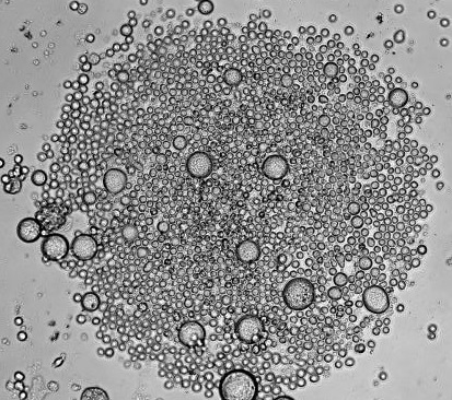
Mature blood cells are relatively short-lived, and require replenishment from multipotent HSCs. Thus, HSCs must self-renew to generate an adequate pool of HSCs, as well as differentiate to give rise to more mature blood cells.  A balance between self-renewal and differentiation ensures that the hematopoietic system can be functionally sustained throughout the lifetime of an adult body.  However, as HSCs age, they accumulate DNA damage, often compromising their functionality.  DNA damage can be further propagated both to daughter stem cells and downstream lineages, and may increase the risk of developing blood disorders 1.

Depending on the nature of the damage, cells use two major response pathways to combat cellular stress.  If the damage is excessive and functionality is compromised, cells usually undergo apoptosis for self-elimination.  In contrast, autophagy allows cells a window of survival.  Autophagy is a process of self-degradation in which organelles or portions of the cytoplasm are sequestered within double-membrane vesicles, known as autophagosomes, and then delivered to lysosomes for degradation 2.  The resulting breakdown products are released through permeases and recycled in the cytosol.  Thus, autophagy can be used to generate high-energy compounds during conditions of metabolic stress.

Recently, in Nature, Warr et al found that metabolic stress and old age induce autophagy in HSCs 3.  The authors isolated HSCs and myeloid progenitors from the bone marrow of transgenic mice systemically expressing GFP fused to LC3, an autophagosome marker 4.  They used cytokine withdrawal to induce metabolic stress and measured autophagy induction by examining the formation and turnover of LC3-GFP.  Myeloid progenitors expressed LC3-GFP in the presence and absence of cytokines.  In contrast, HSCs did not express LC3-GFP in the presence of cytokines, but demonstrated autophagosome formation following cytokine withdrawal.  Furthermore, when the mice were starved in vivo, autophagy flux increased in HSCs.

The authors speculated that autophagy “protects” HSCs from starvation-induced apoptosis, and indeed, hematopoietic-specific deletion of an essential autophagy machinery component, ATG12, resulted in a significant increase in caspase activation in starved HSCs in vivo.  FOXO3A was identified as the specific transcriptional regulator that maintains pro-autophagy gene expression, and was expressed higher in HSCs compared to progenitors.  Interestingly, HSCs isolated from old mice retained their autophagic potential, and was found to be required for their survival.

In summary, Warr et al demonstrated that long-lived HSCs mount a protective survival autophagy response to combat metabolic stress, whereas short-lived progenitors do not.  Previous studies suggested that impaired autophagy might contribute to the aging phenotype 5.  However, this study directly showed that the pro-autophagy gene expression program is still intact in old HSCs and is essential for continued survival of these cells.  Future studies will address whether autophagy increases the incidence of age-related blood disorders since it protects damaged, old HSCs from elimination by apoptosis.


References 

1          Rossi, D. J., Jamieson, C. H. & Weissman, I. L. Stems cells and the pathways to aging and cancer. Cell 132, 681-696, doi:10.1016/j.cell.2008.01.036 (2008).

2          He, C. & Klionsky, D. J. Regulation mechanisms and signaling pathways of autophagy. Annu Rev Genet 43, 67-93, doi:10.1146/annurev-genet-102808-114910 (2009).

3          Warr, M. R. et al. FOXO3A directs a protective autophagy program in haematopoietic stem cells. Nature 494, 323-327, doi:10.1038/nature11895 (2013).

4          Mizushima, N., Yamamoto, A., Matsui, M., Yoshimori, T. & Ohsumi, Y. In vivo analysis of autophagy in response to nutrient starvation using transgenic mice expressing a fluorescent autophagosome marker. Mol Biol Cell 15, 1101-1111, doi:10.1091/mbc.E03-09-0704 (2004).

5          Rubinsztein, D. C., Marino, G. & Kroemer, G. Autophagy and aging. Cell 146, 682-695, doi:10.1016/j.cell.2011.07.030 (2011).


Generation of Red Blood Cells from Human Pluripotent Stem Cells

After the brief review of the in vitro systems for hematopoietic differentiation of pluripotent stem cells (PSCs), I would now like to take a closer look at the functional properties of PSCs-derived blood cells and discuss their potential for clinical application.

182028 303876673057855 753085483 n

Erythrocytes or red blood cells (RBCs) are the most abundant cell population comprising of ~45% of the total blood volume, whose main function is to deliver oxygen to the body tissues. Red blood cells lack a nucleus and most organelles to provide maximum space for hemoglobin – a complex metalloprotein containing heme groups whose iron atoms temporarily bind to oxygen molecules and release them throughout the body.

Mammalian erythroid progenitors originate from a megakaryocyte-erythroid progenitor (MEP) and undergo the gradual process toward terminal differentiation. Two globin gene switches occur during development: the embryonic to fetal globin switch, which coincides with the transition from embryonic (yolk sac) to definitive (fetal liver) hematopoiesis; and fetal to adult switch, which occurs during the perinatal period. During erythroblast differentiation, the chromatin condenses while the hemoglobin concentration increases. Chromatin condensation involves histone deacetylation and unknown signals that activate the Rac-GTPases-mDia2 pathway, which is required for the formation of a contractile actin ring and subsequent enucleation, the process in which the nucleus is rapidly squeezed out of the cell.

In vivo, erythroid precursors proliferate, differentiate, and enucleate within specialized niches called erythroblastic islands. These hematopoietic compartments are composed of erythroblasts surrounding a central macrophage. The central macrophage communicates with erythroblasts through a number of signaling molecules and phagocytizes their nuclei after enucleation.

EnuclErNitch

In 1977, the American biochemist Eugene Goldwasser isolated the human protein erythropoietin (EPO), which stimulates red blood cell production. EPO became a blockbuster product that changed the lives of millions of patients suffering from anemia. Although EPO with other specific additives allow red blood cells to mature in vitro without a supportive role of macrophages, the resulting proliferation and enucleation efficiency of red blood cells is lower than their capacities in vivo, suggesting the importance of a niche microenvironment.

Blood transfusions are a common treatment for severe anemia and massive blood loss due to trauma. A type O-negative red blood cell can be transfused to patients of all blood types and is always in great demand. Thus, the derivation of (O)Rh-negative RBCs from PSCs could be an effective way to overcome shortages in donated red blood cells.

Red blood cells can be produced from human pluripotent stem cells (hESCs and hiPSCs) through various differentiation systems, such as an embryoid body (EB) formation and coculturing hPSCs on top of stromal feeder cells. In general, the existing methods are sufficient for a large-scale production of hPSC-derived red blood cells, whose in vitro expansion capacity is greater than the expansion potential of the bone marrow, peripheral blood, or even cord blood-derived erythroid progenitors. Despite the large amounts of RBCs obtained in many studies, the majority of the resulting RBCs expresses embryonic ε– and fetal γ-globins with low levels of detectable adult β-globin. Although no differences were observed between hiPSC and hESC lines in terms of erythroid commitment and expression of erythroid markers, iPSC-derived red blood cells have lower proliferation activity and produce less enucleated cells.

Robert Lanza’s group suggested the idea of developing an early hemato-endothelial progenitor, a hemangioblast, which can be expanded and cryopreserved.This study, published in Nature Methods in 2007, demonstrated the regenerative properties of blast cells that differentiate into multiple hematopoietic lineages as well as into endothelial cells. The extended coculture of these cells on OP9 feeders facilitated enucleation in up to 65% of cells and the expression of β-globin in up to 15% of the cells.

Lapillonne and colleagues employed a feeder free, two-step differentiating system to produce mature blood cells from hESCs and  iPSCs. In the first step, researchers initiated erythropoiesis by conditioning embryoid bodies in the presence of cytokines. To obtain mature erythrocytes, they further cultured cells in the presence of EPO, SCF, IL3 and 10% of human plasma for another 25 days. The resulting population contained up to 10% of enucleated cultured RBC from hiPSC, and 66% of enucleated RBC from hESC. The vast majority (~93%) of PSCs-derived red blood cells expressed the tetrameric form of fetal hemoglobin HbF (α2γ2). The CO-rebinding kinetics of hemoglobin from hESC- and hiPSC-derived erythroid cells was almost identical to those of cord blood cells suggesting that the HbF in these erythrocytes is functional.

Several studies have shown a time-dependent increase in β-globin expression, the oxygen dissociation curve and G6PD activities similar to normal RBCs. Nevertheless, significant progress is needed in the production of terminally differentiated/enucleated erythrocytes. Thus, at least two major steps are required for future therapeutic use of in vitro generated RBCs: (i) finding a cost-effective method for generating fully maturated, enucleated erythrocytes, and (ii) the evaluation of their biophysical parameters such as membrane surface potential, pliability, half-life in vivo, hemoglobin packing, gas exchange properties, and immunogenicity.


Further Reading:

1. Peng Ji, Maki Murata-Hori, Harvey F. Lodish Formation of mammalian erythrocytes: Chromatin condensation and enucleation Trends Cell Biol. 2011 July; 21(7): 409–415.

2. Joel Anne Chasis, Narla Mohandas Erythroblastic islands: niches for erythropoiesis Blood. 2008 August 1; 112(3): 470–478.

3. Lu SJ, Feng Q, Caballero S, Chen Y, Moore MA, Grant MB, Lanza R. Generation of functional hemangioblasts from human embryonic stem cells. Nat Methods. 2007 Jun;4(6):501-9.

4. Hélène Lapillonne, Ladan Kobari, Christelle Mazurier et al. Red blood cell generation from human induced pluripotent stem cells: perspectives for transfusion medicine Haematologica. 2010 October; 95(10): 1651–1659.

5. Dias J, Gumenyuk M, Kang H, Vodyanik M, Yu J, Thomson JA, Slukvin II. Generation of red blood cells from human induced pluripotent stem cells. Stem Cells Dev. 2011 Sep;20(9):1639-47.

6. Chang KH, Bonig H, Papayannopoulou T. Generation and characterization of erythroid cells from human embryonic stem cells and induced pluripotent stem cells: an overview. Stem Cells Int. 2011;2011:791604.


Pictures:

1. Scanning electron microscope (SEM) image of a single red blood cell on the tip of a needle: http://www.rsc.org/chemistryworld/regulars

2. Confocal immunofluorescence image of an island reconstituted from freshly harvested mouse bone marrow cells stained with erythroid-specific marker (red), macrophage marker (green) and DNA probe (blue). Central macrophage is indicated by an arrow and a multilobulated reticulocyte by an arrowhead. Joel Anne Chasis, Narla Mohandas Erythroblastic islands: niches for erythropoiesis Blood. 2008 August 1; 112(3): 470–478.

How Cardiosphere-derived Cells Regenerate the Injured Heart

According to the Centers for Disease Control, approximately one million heart attacks (Myocardial Infarction, or MI) occur per year in the U.S. [1].  Loss of functioning heart muscle due to an MI can result in congestive heart failure which is the leading cause of death and disability inAmerica.  It is estimated that about half of those with congestive heart failure die within five years of diagnosis [1].  New therapies are desperately needed to treat and prevent the clinical complications that follow a heart attack. Unfortunately, the heart muscles (cardiomyocytes) do not ordinarily regenerate once damage has occurred by MI and thus many researchers are looking into stem cell therapies for heart failure and disease.

The widely accepted view that the heart was not capable of regeneration was established in 1925 by Karsner et al. who demonstrated that the heart grew larger due to cardiac hypertrophy (increase in cell size) as opposed to hyperplasia (increase in cell number) [2].  However, in 2009, Bergmann et al. demonstrated that the human heart was capable of self-renewal by measuring the age of cardiomyocytes in individuals exposed to carbon-14 generated by nuclear bomb tests during the Cold War [3].  The work of Hsieh et al. showed that adult heart regeneration was due to an actual subset of progenitor cells as opposed to increased proliferation of resident cardiomyocytes [4].  This group used an α-myosin heavy chain Cre-Lox transgenic mouse model (Mer-CreMer-ZEG mouse), where 100% of the cardiomyocytes are b-galactosidase (b-gal)  positive but activation of Cre recombinase by tamoxifen results in 80% EGFP (Enhanced Green Fluorescent Protein) positive cardiomyocytes and 20% b-gal positive cardiomyocytes.  After myocardial infarction there was a 15% increase in b-gal positive cardiomyocytes indicating that cardiomyocytes were being generated from stem/ progenitor cells, which were never exposed to Cre recombinase. Thus, while there is insufficient capacity of mammalian adult heart tissue to undergo self-repair, injured myocardium can increase cardiomyocyte generation from non-myocyte precursors.

While protocols aimed to mobilize endogenous stem/ progenitor cells to sites of injury or disease would be greatly beneficial to cardiac patients, transplantation of autologous or allogeneic cells is also a promising alternative.  Cell therapies may result in phenotypic replacement of heart cells, reduce scar formation, increase survival and function of endogenous cardiac cells by increasing in vivo bioavailability of growth factors, and exert immunomodulation by releasing soluble molecules and express immune-relevant receptors (chemokine receptors and CAMs (cellular adhesion markers)).  The first clinical trial using a stem cell therapy for cardiac disease was performed over a decade ago using autologous bone marrow cells (BMCs) in patients after acute MI which reported improved heart function and myocardial perfusion [5].  Since then, numerous adult stem cell therapies targeting cardiac disease have been launched with the majority of therapies involving BMCs [6].

describe the imageRecently, a phase I clinical trial based on administration of autologous cardiosphere-derived cells (CDCs) to assess safety in patients with left ventricular dysfunction after MI was completed by Dr. Eduardo Marban atCedars-SinaiMedicalCenter[7].  The formation of cardiospheres was first described by Messina’s group who showed that human and mouse heart explants generated a layer of fibroblast-like cells over which small, phase-bright cells migrated, and once these phase-bright cells are transferred to non-adherent plates, they generate three-dimensional spherical structures [3].  Marban’s group modified Messina’s protocol by placing the cardiospheres in adherent plates where the cells begin to grow in monolayer, hence cardiosphere-derived cells, allowing for easier and faster expansion.  Promising preclinical data which showed a reduction in infarct size and improved cardiac function after transplantation of CDCs in a porcine animal model prompted the phase I clinical trial [8].

This trial, known as CADUCEUS (CArdiosphere-Derived aUtologous stem CElls to reverse ventricUlar dySfunction), enrolled patients 2-4 weeks after MI (with left ventricular ejection of 25-45%) who received either standard of care (no intervention, usual medical management after MI), low cell dose (12.5 million) or high cell dose (25 million) of autologous CDCs (ClinicalTrials.gov NCT00893360).  Percutaneous endomyocardial biopsies were used to obtain the heart tissue necessary for generation of the predetermined dose of autologous CDCs (usually within 36 days of sampling) and cells were delivered through an angioplasty catheter in the infarct-related artery. Results from the trial showed that the CDCs were relatively safe: no patients died, developed cardiac tumors or experienced a major adverse cardiac event (MACE).  Preliminary efficacy suggested that patients treated with CDCs had reductions in scar mass and increases in viable heart mass as measured by magnetic resonance imaging (MRI), as well as increases in regional contractility and systolic wall thickening at 12 months compared to controls [7].  However, the trial did not confirm whether there was true increased myocardial mass, whether the CDCs themselves could possibly distort the myocardial architecture and/or how the CDCs potentially could regenerate the injured heart.

Recently Marban’s group has shed light on the potential mechanism of how the CDC therapy reduces scarring and regenerates healthy tissue after an MI, published in EMBO Molecular Medicine [9].  They applied lineage tracing systems using the Mer-CreMer-ZEG mouse with additional labeling tools to investigate the cellular origins of regeneration in an adult mouse after surgical induction of MI followed by intramyocardial injection of mouse CDCs.  Impressive results revealed that after MI new cardiomyocytes arise from both pre-existing cardiomyocytes and undifferentiated progenitors and that transplanted CDCs further up-regulate host cardiomyocyte proliferation and recruitment of endogenous progenitors to the infarct site.  The increase in cardiomyocyte proliferation and progenitor recruitment was accompanied by structural and functional changes in the infracted heart, specifically decreased scar size, increased infracted wall thickness and myocardium (cardiomyocyte hypertrophy was excluded), and increased cardiac function.  Interestingly, these effects occurred despite robust CDC engraftment indicating that CDCs do not contribute to phenotypic replacement but promote heart regeneration by indirect means.  These promising findings indicate that stem cell therapies may stimulate dormant surviving cells after injury and boost natural repair mechanisms.

However, there still remain many challenges for developing cell therapies for cardiac disease.  First, the technology is still immature.  With autologous approaches, patient-to-patient variability can arise such as purity and potency of the cells.  Harvesting the source material, obtaining a sufficient quantity of cells, delivery of the end product and the optimal site of delivery will need to be established.  In the future, Marban’s group plans on using allogeneic CDCs obtained from donor organs, which allows for an off-the-shelf-therapy, to treat patients following MI.  A thorough cell biological characterization of the CDCs will be required to understand the molecular identity and mechanism(s) of action of these cells.  Second, the underlying mechanism(s) of cardiac repair and/ or regeneration after MI remain elusive.  Additionally, treating chronic or congestive heart failure with the aim of generating new contracting heart muscle is more complex.  Third, it is difficult to make conclusive statements about the results obtained from many of the clinical trials since they have small patient size and therefore limited statistical power.  It is unclear how the findings will generalize to a larger population of patients.  Fourth, most of the clinical trials use surrogate endpoints which are measures of an effect of a certain treatment expected to predict clinical benefit (or harm, or lack of benefit or harm) such as measuring cardiac function.  Even though surrogate endpoints may correlate with a real clinical endpoint, they do not have a guaranteed relationship.  Therefore, clinically meaningful endpoints such as improvement in overall survival and prevention of future heart failure are needed.  In the CADUCEUS trial there was only enhanced regional structure/ function but no significant effect on global functional endpoints such as ejection fraction.  A phase II double-blinded placebo-controlled clinical trial will need to be performed to assess true efficacy.  It will be several more years before we have a clear understanding of the true potential of cell therapy in cardiac disease.  In the meantime, studies to understand the cellular sources and underlying cellular mechanisms involved in cardiac regeneration are still needed.


1.         CDC. Heart Disease Facts.  updated October 16, 2012; Available from: http://www.cdc.gov/heartdisease/facts.htm.

2.         Karsner, H.T., O. Saphir, and T.W. Todd, The State of the Cardiac Muscle in Hypertrophy and Atrophy. Am J Pathol, 1925. 1(4): p. 351-372 1.

3.         Carvalho, A.B., B.K. Fleischmann, and A.C. Campos de Carvalho, Cardiac Stem Cells, in Resident Stem Cells and Regenerative Therapy, R.C.d.S. Goldenberg, Editor. 2012, Academic Press: Waltham, MA. p. 141-155.

4.         Hsieh, P.C., et al., Evidence from a genetic fate-mapping study that stem cells refresh adult mammalian cardiomyocytes after injury. Nat Med, 2007. 13(8): p. 970-4.

5.         Strauer, B.E., et al., Repair of infarcted myocardium by autologous intracoronary mononuclear bone marrow cell transplantation in humans. Circulation, 2002. 106(15): p. 1913-8.

6.         Sheridan, C., Cardiac stem cell therapies inch toward clinical litmus test. Nat Biotechnol, 2013. 31(1): p. 5-6.

7.         Makkar, R.R., et al., Intracoronary cardiosphere-derived cells for heart regeneration after myocardial infarction (CADUCEUS): a prospective, randomised phase 1 trial. Lancet, 2012. 379(9819): p. 895-904.

8.         Johnston, P.V., et al., Engraftment, differentiation, and functional benefits of autologous cardiosphere-derived cells in porcine ischemic cardiomyopathy. Circulation, 2009. 120(12): p. 1075-83, 7 p following 1083.

9.         Malliaras, K., et al., Cardiomyocyte proliferation and progenitor cell recruitment underlie therapeutic regeneration after myocardial infarction in the adult mouse heart. EMBO Mol Med, 2013. 5(2): p. 191-209.