INTESTINAL BACTERIA LINKED TO LYMPHOMA

The human gut harbors approximately one thousand different bacterial species (intestinal microbiota). Intestinal microbiota number 100 trillion cells; over 90 percent of the cells in the body are bacteria. The composition of each person’s microbiome — the body’s bacterial make-up — is very different, due to the types of bacteria people ingest in their early lives, as well as the effects of diet and lifestyle.

Several studies implicated intestinal bacteria in various cancers. Gram-negative Helicobacter species were found to be associated with liver cancer, colon cancer, and breast cancer. A recent study published in the peer reviewed journal Nature by Yoshimoto et al. (2013) reported that gut bacteria of obese mice unleash high levels of an acid that promotes liver cancer. In rodents, intestinal bacteria influence obesity, intestinal inflammation and certain types of epithelial cancers. However, in human, little is known about the identity of the bacterial species that promote the growth or protect the body from cancer. Therefore, studies are warranted to determine whether differences in peoples’ microbiomes affect their risk for cancer, and whether changing the bacteria can reduce this risk. A clinical trial at the National Cancer Institute (NCI) is currently evaluating the relationship between intestinal bacteria and breast cancer risk (Clinical Trials.gov number: NCT01461070).

intestinal  bacteria

For the first time, a recent study by Yamamoto et al. (2013) demonstrated a relationship between intestinal microbiota and onset of lymphoma (a type of blood cancer of B or T lymphocytes). Yamamoto and colleagues studied mice with ataxia-telangiectasia (A-T), a genetic disease that in humans and mice is associated with a high rate of B-cell lymphoma. These investigators discovered that of mice with A-T, those with certain microbial species lived much longer than those with other bacteria before developing lymphoma, and had less of the gene damage (genotoxicity) that causes lymphoma. A high-throughput sequence analysis of rRNA genes identified the bacteria Lactobacillus johnsonnii in abundance in more cancer-resistant mouse colonies compared to cancer-prone mouse colonies.This study by Yamamoto et al. also created a detailed catalog of bacteria types with promoting or protective effects on genotoxicity (a chemical or other agent that damages cellular DNA, resulting in mutations or cancer) and lymphoma, which could be used in the future to formulate combination therapies that kill the bacteria that promote cancer (such as antibiotics) and increase the presence of the bacteria that protect from cancer (like probiotics).

References:

1.   Ward, J.M., et al., Chronic active hepatitis in mice caused by Helicobacter hepaticus. Am J Pathol, 1994. 145(4): p. 959-68.

2.   Yoshimoto, S., et al., Obesity-induced gut microbial metabolite promotes liver cancer through senescence secretome. Nature, 2013. 499(7456): p. 97-101.

3.   Yamamoto, M.L., et al., Intestinal Bacteria Modify Lymphoma Incidence and Latency by Affecting Systemic Inflammatory State, Oxidative Stress, and Leukocyte Genotoxicity. Cancer Res, 2013. 73(14): p. 4222-4232.


Remodeling of the Tumor Extracellular Matrix Activates YAP in Fibroblasts to Produce Cancer Associated Fibroblasts

When cells undergo transformation and initiate the formation of a solid tumor mass, they cause profound changes on the phenotypes of the cells that surround them1. However, in addition to the changes in cellular phenotype, there is a change in the extracellular matrix that coincides with tumor formation1. It has been demonstrated that the majority of solid tumors have increased stiffness in their extracellular matrix (ECM), which may lead to increased activation of pro-tumor signaling pathways, such as Src, FAK, and RhoA2-4. Recently, it was discovered that increased matrix stiffness may also lead to increased activity of the oncogenic YAP/TAZ complex, which is connected to the Hippo signaling pathways, transcriptional regulators that increase cellular proliferation, decreased cellular contact inhibition, increased cancer stem cell phenotype, and increased metastasis5. However, in a fibroblastrecent edition of Nature Cell Biology Calvo et al. demonstrated that 6.  Not only do the authors demonstrate that YAP/TAZ is active in CAFs, but YAP/TAZ is necessary for CAF development6. They show that CAF activation leads to matrix remodeling towards increased stiffness, via myosin light chain 2 (MYL9/MLC) expression, establishing a feed-forward loop where the ECM plays a vital role6.

The authors first isolated fibroblasts in different stages towards becoming a CAF and saw that both mechanical-responsive signaling machinery (SMA, FN1, Paxillin, MYL9, MYH10, DIAH1 & F-actin) and mechanical tension were increased in populations containing CAFs. Moreover, tumor cell invasion, and angiogenesis of the tumor microenvironment (shown via endomucin and second-harmonic microscopy) were increased in samples that contained more tumor-associated-like fibroblasts (indicated by vimentin)6.

Because of the role of cell-cell and cell-ECM contact in the Hippo signaling pathway, the authors sought to understand whether this pathway is activated in CAFs. They found that YAP, and its co-factor, TAZ to be only upregulated and co-localized in the nucleus of transformed fibroblasts; the target of the activated YAP-TAZ complex6.  Furthermore, upon depletion of YAP, the ability for CAFs to cause matrix stiffness by contraction lessened as well as CAFs ability to form collagen networks and facilitate angiogenesis. Interestingly, when TAZ was inhibited, there was no change in functionality, which may lead to a TAZ-independent function for YAP.

Upon microarray analysis of CAFs treated with siRNA that targets YAP, Calvo et al. found that the expression of many of the genes involved in mechanosensing and motility to be diminished6. Furthermore, when these individual genes were silenced, there was an overall decrease in the amount of cellular invasion of tumors. Many of the YAP-mediated genes, such as ANLN and DIAPH3, were involved in matrix remodeling and cellular invasion. Interestingly, modification of only one protein overexpression resulted in high amounts of matrix-remodeling and invasion: myosin regulatory light polypeptide 9 (MYL9). While not transcriptionally controlled by the YAP/TAZ complex, the authors demonstrate that YAP/TAZ is able to control MYL9 by post-translational modifications, placing YAP as a critical factor in regulating matrix-remodeling and invasion through MYL96.

Calvo et al. next posited that YAP/TAZ activation may not be exclusive to CAFs, but may also occur in normal fibroblasts when placed in a cancerous environment6. They found that fibroblasts placed in culture with tumor conditioned media had higher nuclear translocation of YAP, and higher gel contraction (akin to matrix stiffening) comparable to known promoters of pro-contractile function: L-alpha-lysophosphatidic acid (LPA) and transforming growth factor-beta (TGFβ). However, actomyosin inhibition (by blebbistatin) could not be rescued with LPA and TGFβ. Therefore, while soluble factors may activate matrix contraction, a functional cytoskeleton is essential for matrix contraction. Because of the necessary role of the cytoskeleton, the authors tested whether inhibition of RhoA kinase (ROCK), a kinase involved in regulating translocation and structure of the cell by the cytoskeleton, would affect the nuclear localization of YAP6. Inhibition of ROCK decreased YAP nuclearization and decreased the matrix stiffness. Of note, like ROCK inhibition, inhibition of Src also affected the nuclear localization of YAP as well as complex formation with TEAD1 and TEAD4. However, Src modulation of YAP is downstream of cytoskeletal changes in tension since Src inhibition did not affect stress fibers6.

Since activation of YAP in CAFs  is connected to actomycin-mediated matrix stiffness, and this activation of YAP expresses MYL9, and expression of MYL9 results in matrix-remodeling towards stiffness, the authors posit that this pathway forms a feed-forward loop6. This loop could lead to constitutive activation of YAP pathway in CAFs, causing a robust response and stabilizing the CAF phenotype. However, it is not known what other mechanisms, as well as regulatory mechanisms of YAP, are involved in this process as well as whether the YAP-ECM tension pathway may play a regulatory role in normal fibroblasts.

References:

1. Boudreau, A., van’t Veer, L. J. & Bissell, M. J. An “elite hacker”: breast tumors exploit the normal microenvironment program to instruct their progression and biological diversity. Cell adhesion & migration 6, 236-248, doi:10.4161/cam.20880 (2012).

2. Levental, K. R. et al. Matrix crosslinking forces tumor progression by enhancing integrin signaling. Cell 139, 891-906, doi:10.1016/j.cell.2009.10.027 (2009).

3. Guilluy, C. et al. The Rho GEFs LARG and GEF-H1 regulate the mechanical response to force on integrins. Nature cell biology 13, 722-727, doi:10.1038/ncb2254 (2011).

4. Sawada, Y. et al. Force sensing by mechanical extension of the Src family kinase substrate p130Cas. Cell 127, 1015-1026, doi:10.1016/j.cell.2006.09.044 (2006).

5. Harvey, K. F., Zhang, X. & Thomas, D. M. The Hippo pathway and human cancer. Nature reviews. Cancer 13, 246-257, doi:10.1038/nrc3458 (2013).

6. Calvo, F. et al. Mechanotransduction and YAP-dependent matrix remodelling is required for the generation and maintenance of cancer-associated fibroblasts. Nature cell biology 15, 637-646, doi:10.1038/ncb2756 (2013).


FIRST TARGETED THERAPEUTIC APPROACH FOR CHRONIC LYMPHOCYTIC LEUKEMIA

Chronic lymphocytic leukemia (CLL) is a slow-growing cancer in which a large number of immature lymphocytes (white blood cells) are found mostly in the blood and bone marrow. It is the most common leukemia in the Western world with incidence rates as high as ~4 per 100,000 individuals in the USA. According to the National Cancer Institute (NCI) it is estimated that in 2013, approximately 15,680 people (9,720 men and 5,960 women) will be diagnosed with CLL and 4,580 men and women will die of CLL. Even though few durable remissions were noted following treatment with chemotherapeutic agents such as chlorambucil, cyclophosphamide, and fludarabine, in the majority of cases these agents are effective for palliation but do not improve survival. An alternative treatment option using chemoimmunotherapy (combination of a chemotherapeutic agent with an anti-CD20 antibody rituximab) was found to have limited efficacy and increased toxicity. In addition, treatment options for CLL are further limited by lack of common genetic target. Nonetheless, many studies reported the association of B-cell receptor (BCR) signaling in the survival of CLL tumor-cells. A downstream component of BCR signaling, a receptor tyrosine kinase, Burton’s tyrosine kinase (BTK) was noted for activation of the Akt, ERK, NF-κB pathways associated with CLL-cell survival.

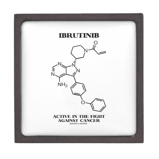
Bruton’s tyrosine kinase is essential for B-cell development and function. BTK deficiency in man or mice results in the B-cell specific immunodeficiencies X-linked agammaglobulinemia (XLA) or x-linked immune deficiency (xid), respectively. It is also implicated in the pathogenesis of B-cell cancers. Studies suggest that the levels of BTK represent a rate-limiting step in BCR signaling and thereby B-cell activation and survival. Therefore, inhibition of BTK in CLL could serve as an effective treatment strategy. In vitro studies reported that following inhibition of BTK with selective inhibitor CLL cells lose their resistance to apoptosis. Preclinical studies also demonstrated that that BTK-deficiency completely abrogated CLL development in mice.IBRUTINIBWith the accumulating evidence of the role of BCR pathway involving BTK in CLL, first targeted therapeutic approach for CLL was tested clinically with BTK inhibitors. A study published recently in The New England Journal of Medicine (July 4, 2013) by Byrd et al. reported a high frequency of durable remissions in patients with relapsed or refractory CLL with a BTK inhibitor, ibrutinib. A phase I study of ibrutinib (previously known as PCI-32765) showed mild-to-moderate toxicity and clinical antitumor activity in patients with relapsed or refractory B-cell cancers; 11 of the 16 patients in the study had CLL or small lymphocytic lymphoma. These preliminary results prompted the initiation of a phase Ib–II study of ibrutinib in CLL; this study involved two different therapeutic doses in patients with relapsed or refractory disease.

In the phase Ib-II multicenter study of ibrutinib, Byrd et al. (2013) assessed the safety, efficacy, and pharmacokinetics of this inhibitor in patients with CLL or small lymphocytic lymphoma (ClinicalTrials.gov number NCT01105247). Among 85 patients enrolled in this study, 51 received 420 mg and 34 received 840 mg ibrutinib orally once daily. In both cohorts the overall response rate was 71%. This treatment resulted in durable response were the 26-month estimated progression-free survival was 75% and the rate of overall survival was 83%. The pharmacodynamic study showed that ibrutinib was able to successfully inhibit BTK. However, disease progression was noted in 13% patients during follow-up. The most common toxicities observed during ibrutinib treatment were diarrhea, fatigue, and upper respiratory tract infection.

As compared to other single agent therapies for relapsed CLL this targeted therapy of BTK inhibition exhibited more durable responses. The durable remissions observed in this study suggest that many patients may be treated successfully with ibrutinib.

References:

1. Gribben, J.G. and S. O’Brien, Update on therapy of chronic lymphocytic leukemia. J Clin Oncol, 2011. 29(5): p. 544-50.

2.  Advani, R.H., et al., Bruton tyrosine kinase inhibitor ibrutinib (PCI-32765) has significant activity in patients with relapsed/refractory B-cell malignancies. J Clin Oncol, 2013. 31(1): p. 88-94.

3. Byrd, J.C., et al., Targeting BTK with ibrutinib in relapsed chronic lymphocytic leukemia. N Engl J Med, 2013. 369(1): p. 32-42.


New Findings in Cell Based Therapy for GBM

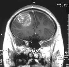
Glioblastoma multiforme (GBM) is the most common and lethalGlioblastoma type of malignant primary brain tumors that account for over 70% of all intracranial cancers.  The current course of GBM treatment consists of surgical resection of the main tumor mass, followed by administration of radiation and chemotherapy. Surgical resection of the primary tumor leads to injury to the surrounding normal tissue, while chemotherapy and radiotherapy cause toxicity to the healthy tissue in the brain.  These undesirable secondary effects of glioma treatments have a major impact on patients’ physical, cognitive and emotional functioning. Nonetheless, despite this aggressive treatment regimen and its harmful side effects, GBM remains virtually incurable, with post-diagnosis median survival persisting less than 14 months.  This dismal prognosis is due to a combination of unique anatomical features of the central nervous system (CNS), in addition to GBMs’ (glioma cells’) exceptional invasive capacity. Glioma cells infiltrate the brain’s highly dense parenchyma, migrating along the corpus callosum and creating new masses within the hemisphere contralateral to the initial tumor mass homing.

Thus, recurrence in postoperative GBM patients is in essence inevitable. Furthermore, GBMs are not only heterogeneous among individual patients, but they are also highly heterogeneous within a single tumor mass. Recent studies have shown compelling evidence of a therapeutically resistant subpopulation of malignant glioma cells that exhibit stem-cell like characteristics, such as multipotency, the ability to self-renew and invade/migrate; these tumor-initiating cells are referred to as glioma stem cells (GSCs) and are believed to be responsible for tumor initiation and recurrence in GBM patients. Furthermore, GSCs have been observed to ensconce within similar niches to neural stem cells (NSCs).

NSCs and mesenchymal stem cells (MSCs) have been shown to have an exceptional migratory ability within brain’s parenchyma and possess a notable inherent tumor tropism.  Thus, several cell-based therapy (CBT) studies and clinical models of malignant tumors have incorporated autonomous tracking of tumor cells by employing NSCs and MSCs to deliver multiple therapeutic genes to specific tumor loci. This target specific drug-delivery model has NSCs or MSCs transduced to express one pro-drug-activating enzyme, which catalyzes the conversion of a particular pro-drug into an active toxic agent, which results in the localization of the chemotherapeutics specifically at the tumor sites.  In addition to its ability to deliver effective cytotoxic damage to the tumor without causing damage to the healthy surrounding tissue, the resulting bystander effect of this system causes cell death not only to the drug-delivery-vehicle cells, but also to the surrounding glioma cells. Although a number of different enzyme/prodrug systems have been utilized in relevant studies, HSV-thymidine kinase (HSV-tk)/ deoxyguanosine analog ganciclovir (GCV) has been the most commonly tested in animal and in vitro models. HSV-tk phosphorylates GCV and produces deoxyguanosine triphosphate which is a polymerase-I inhibitor and DNA chain terminator; cell death occurs upon incorporation of the nucleotide analog within DNA chains.

In recent study published in the journal of Molecular Therapy, Blanco’s group report their findings regarding the interaction between human MSCs (hMSCs) and gliomas and the underlying mechanism for the effectiveness of hAMSC based therapy in GBMs.  In their previous work Blanco’s group showed that the administration of hAMSCs expressing HSV thymidine kinase in glioma tumors significantly promotes tumor growth, whereas induction of cytotoxicity by administration of the prodrug GCV demonstrated a significant antitumor response.

According to this new study, hMSCs differentiate to endothelial lineage (supported by the expression of CD31 marker) within tumors, and integrate in the tumor vascular system where they adopt an endothelial phenotype. Further, Blanco proposed the notion of hMSCs’ ability to home to privileged vascular structures where GSCs also reside, is the underlying characteristic that leads to the effectiveness of cytotoxic hMSCs in regulating the bystander killing of tumor cells.

Although Blanco’s study provides invaluable insights into the GSCs’ niche and its role in malignant brain tumor CBT, hMSCs’ tumor growth promotion is a quality, which makes these cells less than ideal for utilization in human clinical trials in the near future. Future studies on this mechanism using primary patient tumor cells rather than the U87 glioma cell line and human NSCs are needed to further confirm these observations.

Further reading:

Juli R. Bagó, Maria Alieva, Carolina Soler, Núria Rubio, Jerónimo Blanco. Endothelial differentiation of adipose tissue-derived mesenchymal stromal cells in glioma tumors: implications for cell based therapy. Molecular Therapy.