BLOCKADE OF CTLA-4 AND PD-1 ENHANCED TUMOR REGRESSION IN MELANOMA

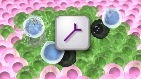
One of the primary roles of the immune system is the specific identification and elimination of tumor cells on the basis of their expression of tumor-specific antigens or molecules induced by cellular stress. This process is referred to as tumor immune surveillance. In this process the immune system recognizes malignant and/or pre-malignant cells and removes them. However, tumor cells do escape from tumor immune surveillance, and therefore, therapies targeted to enhance antitumor immunity is currently in development.

Blockade of immune checkpoints  is the most promising approach to activate therapeutic antitumour immunity. Immune checkpoints refer to a group of inhibitory pathways connected into the immune system that are important for maintaining self-tolerance. In peripheral tissues immune surveillance also modulates the duration and amplitude of physiological immune responses in order to minimize collateral tissue damage. Studies have suggested that tumor cells adopt many immune-checkpoint pathways as a major mechanism of immune resistance. Immune checkpoint receptors cytotoxic T-lymphocyte-associated antigen 4 (CTLA-4, also known as CD152) and programmed death 1 (PD-1) receptor appear to play important roles in antitumor immunity and have been most actively studied in the context of clinical cancer immunotherapy.

monoclonal3CTLA-4 is expressed on T cells and down modulates the amplitude of T cell activation. Several preclinical studies demonstrated significant antitumor responses following blockade of CTL4-A with limited immune toxicities. This led to the development of two fully humanized  CTLA-4 antibodies ipilimumab and tremelimumab. In clinical trials, ipilimumab demonstrated survival benefits for patients with metastatic melanoma, and was approved by the US Food and Drug Administration (FDA) for the treatment of advanced melanoma in 2010.

On the other hand, PD-1limits T cell effector functions within tissues. Tumor  cells block antitumor immune responses in the tumor microenvironment by upregulating ligands (PDL1 and PDL2) for PD1. Several studies detected increased PD1 expression by tumor infiltrating lymphocytes and the increased expression of PD1 ligands in melanoma, ovarian, lung, renal-cell cancers and in lymphomas. This provided an important rationale to target PD1 in order to enhance antitumor immunity. The fully human antibody nivolumab was found to produce durable objective responses in patients with melanoma, renal-cell cancer, and non-small-cell lung cancer.

Even though individual blocking of CTLA-4 and PD-1 have shown substantial clinical antitumor activity, studies suggest that blocking a single inhibitory receptor only leads to up-regulation of the unblocked pathway. Therefore, in order  to enhance antitumor immunity within the tumor microenvironment it appears to require simultaneous blockade of multiple negative co-stimulatory receptors. In preclinical studies, concurrent inhibition of CTLA-4 and PD-1 resulted in more pronounced antitumor activity than blockade of either pathway alone. On the basis of these observations, a phase I study was conducted to investigate the safety and efficacy of combined inhibition of CTLA-4 and PD-1in advanced melanoma patients and published recently in The New England Journal of Medicine (July 11, 2013). In their study, Wolchok and collagues (2013) treated 53 patients concurrently, and 33 patients sequentially with nivolumab and ipilimumab. Rapid responses were observed in concurrent-regimen cohorts as compared with sequential-regimen cohorts. The objective response rate in the concurrent-regimen cohorts was 40% along with 53% patients exhibited tumor regression of 80% or more. The objective response rate in the sequenced-regimen cohorts was 20% and 13% patients had tumor regression of 80% or more. In both groups, treatment related adverse events were managed with the use of immunosuppressants.

Collectively this study suggested that combined blockade of CTLA-4 and PD-1 would be more effective to enhance antitumor immunity compared to single inhibition of either CTLA-4 or PD-1.

References:

1.  Swann, J.B. and M.J. Smyth, Immune surveillance of tumors. J Clin Invest, 2007. 117(5): p. 1137-46.

2.   Pardoll, D.M., The blockade of immune checkpoints in cancer immunotherapy. Nat Rev Cancer, 2012. 12(4): p. 252-64.

3.   Topalian, S.L., et al., Safety, activity, and immune correlates of anti-PD-1 antibody in cancer. N Engl J Med, 2012. 366(26): p. 2443-54.

4.   Wolchok, J.D., et al., Nivolumab plus ipilimumab in advanced melanoma. N Engl J Med, 2013. 369(2): p. 122-33.

PD-1 re-expression is differentially regulated by IL-12 vs IFNα in CD8 T cells

TUMOR_immunotherapyDownregulation of immune functions following responses to pathogen infections is critical for limiting damage to the host by the immune system.  T cell activity is known to be downregulated by a variety of negative regulatory mechanisms including negative checkpoint regulatory proteins, a family of CD28-related molecules.  PD-1 is one such molecule that is transiently expressed on activated T cells.   The ligands for PD-1 are PD-L1 and PD-L2, members of the B7 family of molecules which are upregulated on antigen presenting cells and tumor cells.  Interaction of PD-1 with its ligand leads to inhibition of TCR-mediated signaling via recruitment of SHP1 and SHP2 phosphatases to the TCR synapse.  In the August 2013 edition of The Journal of Immunology, Gerner et al., demonstrate that CD8 T cells initially activated in the presence of IL-12 and IFNα differentially re-express PD-1 upon antigen restimulation.

Cytokines play roles in regulation of nearly every aspect of immune responses.  The cytokine milieu present during T cell activation directs differentiation into the different functional classes of CD4 T helper or CD8 T cells.  This study sought to determine the differences in anti-tumor CD8 T cell effector functions mediated when T cells are activated in the presence of various cytokines.  IL-12 and IFNα activate both overlapping and distinct gene programs and promote cytotoxic CD8 T cell responses.  Thus, these cytokines were chosen for comparison in this study.

In this system, CD8+ OT-1 cells were activated ex-vivo in the presence of either IL-12 or IFNα, and transferred into B16-OVA tumor-bearing mice.  T cells activated in the presence of IL-12 were found to mediate tumor-growth inhibition significantly better than if they had been activated in the presence of IFNα.  Over time in tumor-bearing mice, transferred IFNα-matured OT-1 cells were observed to decline in number and lost the ability to produce IFNγ ex vivo upon restimulation, indicating these cells may be exhausted.

Because PD-1 is known to be a marker and mediator of T cell exhaustion, PD-1 expression was examined.  Initial induction levels of PD-1 were comparable on OT-1 cells following ex vivo activation with IFNα or IL-12.  Following transfer into tumor-bearing mice, PD-1 levels declined over time on both types of cells isolated from the spleen and on IL-12 matured cells isolated from the tumor.  However, PD-1 expression was high on transferred IFNα-matured cells when isolated from the tumor.  Similar results were seen when cells were transferred into mice that subsequently received an injection of the OVA peptide.  Thus, CD8+ T cells matured in the presence of IFNα appear to re-express significantly higher levels of PD-1 upon antigen restimulation than IL-12 matured T cells.

PD-1 and PD-L1 targeting with inhibitory antibodies have emerged as promising avenues in tumor immunotherapy.  In this study, anti-PD-1 antibody administration had no additional anti-tumor effect in mice that received IL-12-matured T cells, while in mice that received IFNα-matured T cells, anti-PD-1 antibodies led to inhibition of tumor-growth to a level similar to that in mice that had received IL-12-matured T cells.  Thus, the relatively poor ability of IFNα-matured T cells to efficiently inhibit tumor growth appears to be largely due to PD-1 upregulation.  Finally, when T cells were matured with both IL-12 and IFNα, the effect of IL-12 was dominant.

Many questions remain regarding the mechanisms mediating PD-1 re-expression in IFNα vs. IL-12 matured T cells.  However, since IL-12 activity was dominant over IFNα on regulating PD-1 expression, IL-12 administration during immunotherapy regimens may enhance anti-tumor T cell responses by blocking the mechanisms by which IFNα enhances PD-1 re-expression.

Further Reading:

Cutting Edge: IL-12 and Type I IFN Differentially Program CD8 T Cells for Programmed Death 1 Re-expression Levels and Tumor Control.  Gerner MY, Heltemes-Harris LM, Fife BT, Mescher MF. J Immunol. 2013 Aug 1;191(3):1011-5. doi: 10.4049/jimmunol.1300652. Epub 2013 Jun 26.

INTESTINAL BACTERIA LINKED TO LYMPHOMA

The human gut harbors approximately one thousand different bacterial species (intestinal microbiota). Intestinal microbiota number 100 trillion cells; over 90 percent of the cells in the body are bacteria. The composition of each person’s microbiome — the body’s bacterial make-up — is very different, due to the types of bacteria people ingest in their early lives, as well as the effects of diet and lifestyle.

Several studies implicated intestinal bacteria in various cancers. Gram-negative Helicobacter species were found to be associated with liver cancer, colon cancer, and breast cancer. A recent study published in the peer reviewed journal Nature by Yoshimoto et al. (2013) reported that gut bacteria of obese mice unleash high levels of an acid that promotes liver cancer. In rodents, intestinal bacteria influence obesity, intestinal inflammation and certain types of epithelial cancers. However, in human, little is known about the identity of the bacterial species that promote the growth or protect the body from cancer. Therefore, studies are warranted to determine whether differences in peoples’ microbiomes affect their risk for cancer, and whether changing the bacteria can reduce this risk. A clinical trial at the National Cancer Institute (NCI) is currently evaluating the relationship between intestinal bacteria and breast cancer risk (Clinical Trials.gov number: NCT01461070).

intestinal  bacteria

For the first time, a recent study by Yamamoto et al. (2013) demonstrated a relationship between intestinal microbiota and onset of lymphoma (a type of blood cancer of B or T lymphocytes). Yamamoto and colleagues studied mice with ataxia-telangiectasia (A-T), a genetic disease that in humans and mice is associated with a high rate of B-cell lymphoma. These investigators discovered that of mice with A-T, those with certain microbial species lived much longer than those with other bacteria before developing lymphoma, and had less of the gene damage (genotoxicity) that causes lymphoma. A high-throughput sequence analysis of rRNA genes identified the bacteria Lactobacillus johnsonnii in abundance in more cancer-resistant mouse colonies compared to cancer-prone mouse colonies.This study by Yamamoto et al. also created a detailed catalog of bacteria types with promoting or protective effects on genotoxicity (a chemical or other agent that damages cellular DNA, resulting in mutations or cancer) and lymphoma, which could be used in the future to formulate combination therapies that kill the bacteria that promote cancer (such as antibiotics) and increase the presence of the bacteria that protect from cancer (like probiotics).

References:

1.   Ward, J.M., et al., Chronic active hepatitis in mice caused by Helicobacter hepaticus. Am J Pathol, 1994. 145(4): p. 959-68.

2.   Yoshimoto, S., et al., Obesity-induced gut microbial metabolite promotes liver cancer through senescence secretome. Nature, 2013. 499(7456): p. 97-101.

3.   Yamamoto, M.L., et al., Intestinal Bacteria Modify Lymphoma Incidence and Latency by Affecting Systemic Inflammatory State, Oxidative Stress, and Leukocyte Genotoxicity. Cancer Res, 2013. 73(14): p. 4222-4232.


Remodeling of the Tumor Extracellular Matrix Activates YAP in Fibroblasts to Produce Cancer Associated Fibroblasts

When cells undergo transformation and initiate the formation of a solid tumor mass, they cause profound changes on the phenotypes of the cells that surround them1. However, in addition to the changes in cellular phenotype, there is a change in the extracellular matrix that coincides with tumor formation1. It has been demonstrated that the majority of solid tumors have increased stiffness in their extracellular matrix (ECM), which may lead to increased activation of pro-tumor signaling pathways, such as Src, FAK, and RhoA2-4. Recently, it was discovered that increased matrix stiffness may also lead to increased activity of the oncogenic YAP/TAZ complex, which is connected to the Hippo signaling pathways, transcriptional regulators that increase cellular proliferation, decreased cellular contact inhibition, increased cancer stem cell phenotype, and increased metastasis5. However, in a fibroblastrecent edition of Nature Cell Biology Calvo et al. demonstrated that 6.  Not only do the authors demonstrate that YAP/TAZ is active in CAFs, but YAP/TAZ is necessary for CAF development6. They show that CAF activation leads to matrix remodeling towards increased stiffness, via myosin light chain 2 (MYL9/MLC) expression, establishing a feed-forward loop where the ECM plays a vital role6.

The authors first isolated fibroblasts in different stages towards becoming a CAF and saw that both mechanical-responsive signaling machinery (SMA, FN1, Paxillin, MYL9, MYH10, DIAH1 & F-actin) and mechanical tension were increased in populations containing CAFs. Moreover, tumor cell invasion, and angiogenesis of the tumor microenvironment (shown via endomucin and second-harmonic microscopy) were increased in samples that contained more tumor-associated-like fibroblasts (indicated by vimentin)6.

Because of the role of cell-cell and cell-ECM contact in the Hippo signaling pathway, the authors sought to understand whether this pathway is activated in CAFs. They found that YAP, and its co-factor, TAZ to be only upregulated and co-localized in the nucleus of transformed fibroblasts; the target of the activated YAP-TAZ complex6.  Furthermore, upon depletion of YAP, the ability for CAFs to cause matrix stiffness by contraction lessened as well as CAFs ability to form collagen networks and facilitate angiogenesis. Interestingly, when TAZ was inhibited, there was no change in functionality, which may lead to a TAZ-independent function for YAP.

Upon microarray analysis of CAFs treated with siRNA that targets YAP, Calvo et al. found that the expression of many of the genes involved in mechanosensing and motility to be diminished6. Furthermore, when these individual genes were silenced, there was an overall decrease in the amount of cellular invasion of tumors. Many of the YAP-mediated genes, such as ANLN and DIAPH3, were involved in matrix remodeling and cellular invasion. Interestingly, modification of only one protein overexpression resulted in high amounts of matrix-remodeling and invasion: myosin regulatory light polypeptide 9 (MYL9). While not transcriptionally controlled by the YAP/TAZ complex, the authors demonstrate that YAP/TAZ is able to control MYL9 by post-translational modifications, placing YAP as a critical factor in regulating matrix-remodeling and invasion through MYL96.

Calvo et al. next posited that YAP/TAZ activation may not be exclusive to CAFs, but may also occur in normal fibroblasts when placed in a cancerous environment6. They found that fibroblasts placed in culture with tumor conditioned media had higher nuclear translocation of YAP, and higher gel contraction (akin to matrix stiffening) comparable to known promoters of pro-contractile function: L-alpha-lysophosphatidic acid (LPA) and transforming growth factor-beta (TGFβ). However, actomyosin inhibition (by blebbistatin) could not be rescued with LPA and TGFβ. Therefore, while soluble factors may activate matrix contraction, a functional cytoskeleton is essential for matrix contraction. Because of the necessary role of the cytoskeleton, the authors tested whether inhibition of RhoA kinase (ROCK), a kinase involved in regulating translocation and structure of the cell by the cytoskeleton, would affect the nuclear localization of YAP6. Inhibition of ROCK decreased YAP nuclearization and decreased the matrix stiffness. Of note, like ROCK inhibition, inhibition of Src also affected the nuclear localization of YAP as well as complex formation with TEAD1 and TEAD4. However, Src modulation of YAP is downstream of cytoskeletal changes in tension since Src inhibition did not affect stress fibers6.

Since activation of YAP in CAFs  is connected to actomycin-mediated matrix stiffness, and this activation of YAP expresses MYL9, and expression of MYL9 results in matrix-remodeling towards stiffness, the authors posit that this pathway forms a feed-forward loop6. This loop could lead to constitutive activation of YAP pathway in CAFs, causing a robust response and stabilizing the CAF phenotype. However, it is not known what other mechanisms, as well as regulatory mechanisms of YAP, are involved in this process as well as whether the YAP-ECM tension pathway may play a regulatory role in normal fibroblasts.

References:

1. Boudreau, A., van’t Veer, L. J. & Bissell, M. J. An “elite hacker”: breast tumors exploit the normal microenvironment program to instruct their progression and biological diversity. Cell adhesion & migration 6, 236-248, doi:10.4161/cam.20880 (2012).

2. Levental, K. R. et al. Matrix crosslinking forces tumor progression by enhancing integrin signaling. Cell 139, 891-906, doi:10.1016/j.cell.2009.10.027 (2009).

3. Guilluy, C. et al. The Rho GEFs LARG and GEF-H1 regulate the mechanical response to force on integrins. Nature cell biology 13, 722-727, doi:10.1038/ncb2254 (2011).

4. Sawada, Y. et al. Force sensing by mechanical extension of the Src family kinase substrate p130Cas. Cell 127, 1015-1026, doi:10.1016/j.cell.2006.09.044 (2006).

5. Harvey, K. F., Zhang, X. & Thomas, D. M. The Hippo pathway and human cancer. Nature reviews. Cancer 13, 246-257, doi:10.1038/nrc3458 (2013).

6. Calvo, F. et al. Mechanotransduction and YAP-dependent matrix remodelling is required for the generation and maintenance of cancer-associated fibroblasts. Nature cell biology 15, 637-646, doi:10.1038/ncb2756 (2013).


FIRST TARGETED THERAPEUTIC APPROACH FOR CHRONIC LYMPHOCYTIC LEUKEMIA

Chronic lymphocytic leukemia (CLL) is a slow-growing cancer in which a large number of immature lymphocytes (white blood cells) are found mostly in the blood and bone marrow. It is the most common leukemia in the Western world with incidence rates as high as ~4 per 100,000 individuals in the USA. According to the National Cancer Institute (NCI) it is estimated that in 2013, approximately 15,680 people (9,720 men and 5,960 women) will be diagnosed with CLL and 4,580 men and women will die of CLL. Even though few durable remissions were noted following treatment with chemotherapeutic agents such as chlorambucil, cyclophosphamide, and fludarabine, in the majority of cases these agents are effective for palliation but do not improve survival. An alternative treatment option using chemoimmunotherapy (combination of a chemotherapeutic agent with an anti-CD20 antibody rituximab) was found to have limited efficacy and increased toxicity. In addition, treatment options for CLL are further limited by lack of common genetic target. Nonetheless, many studies reported the association of B-cell receptor (BCR) signaling in the survival of CLL tumor-cells. A downstream component of BCR signaling, a receptor tyrosine kinase, Burton’s tyrosine kinase (BTK) was noted for activation of the Akt, ERK, NF-κB pathways associated with CLL-cell survival.

Bruton’s tyrosine kinase is essential for B-cell development and function. BTK deficiency in man or mice results in the B-cell specific immunodeficiencies X-linked agammaglobulinemia (XLA) or x-linked immune deficiency (xid), respectively. It is also implicated in the pathogenesis of B-cell cancers. Studies suggest that the levels of BTK represent a rate-limiting step in BCR signaling and thereby B-cell activation and survival. Therefore, inhibition of BTK in CLL could serve as an effective treatment strategy. In vitro studies reported that following inhibition of BTK with selective inhibitor CLL cells lose their resistance to apoptosis. Preclinical studies also demonstrated that that BTK-deficiency completely abrogated CLL development in mice.IBRUTINIBWith the accumulating evidence of the role of BCR pathway involving BTK in CLL, first targeted therapeutic approach for CLL was tested clinically with BTK inhibitors. A study published recently in The New England Journal of Medicine (July 4, 2013) by Byrd et al. reported a high frequency of durable remissions in patients with relapsed or refractory CLL with a BTK inhibitor, ibrutinib. A phase I study of ibrutinib (previously known as PCI-32765) showed mild-to-moderate toxicity and clinical antitumor activity in patients with relapsed or refractory B-cell cancers; 11 of the 16 patients in the study had CLL or small lymphocytic lymphoma. These preliminary results prompted the initiation of a phase Ib–II study of ibrutinib in CLL; this study involved two different therapeutic doses in patients with relapsed or refractory disease.

In the phase Ib-II multicenter study of ibrutinib, Byrd et al. (2013) assessed the safety, efficacy, and pharmacokinetics of this inhibitor in patients with CLL or small lymphocytic lymphoma (ClinicalTrials.gov number NCT01105247). Among 85 patients enrolled in this study, 51 received 420 mg and 34 received 840 mg ibrutinib orally once daily. In both cohorts the overall response rate was 71%. This treatment resulted in durable response were the 26-month estimated progression-free survival was 75% and the rate of overall survival was 83%. The pharmacodynamic study showed that ibrutinib was able to successfully inhibit BTK. However, disease progression was noted in 13% patients during follow-up. The most common toxicities observed during ibrutinib treatment were diarrhea, fatigue, and upper respiratory tract infection.

As compared to other single agent therapies for relapsed CLL this targeted therapy of BTK inhibition exhibited more durable responses. The durable remissions observed in this study suggest that many patients may be treated successfully with ibrutinib.

References:

1. Gribben, J.G. and S. O’Brien, Update on therapy of chronic lymphocytic leukemia. J Clin Oncol, 2011. 29(5): p. 544-50.

2.  Advani, R.H., et al., Bruton tyrosine kinase inhibitor ibrutinib (PCI-32765) has significant activity in patients with relapsed/refractory B-cell malignancies. J Clin Oncol, 2013. 31(1): p. 88-94.

3. Byrd, J.C., et al., Targeting BTK with ibrutinib in relapsed chronic lymphocytic leukemia. N Engl J Med, 2013. 369(1): p. 32-42.


Finding the Right Cancer Culprits Using Mutational Heterogeneity

Imagine this: you are a police officer on patrol and you receive a call that multiple 30-year-old Caucasian males were seen breaking and entering; stealing heirlooms from a nearby neighborhood. The suspects were last seen entering a convention center and, to your dismay, you arrive to find the entire convention center is an antique show containing several 30-year-old Caucasian males carrying heirlooms. What do you do to apprehend your perpetrators?  You could arrest everyone that fits the description and interrogate them. On the other hand, you could scan the crowd for clues that there is a group of people that do not belong, or also radio to the police station for more information to narrow down the crowd. Needless to say, without gaining more contextual information for prudent discernment of the situation, you may arrest the wrong men and let the criminals go free.genomes

This is where cancer genomics is today; the sophistication of sequencing techniques have allowed for datasets that can detect every genomic mutation within cancer cells. Unfortunately, mutation rates are not equal among all genes. While this may seem a non-issue, this could lead scientists to ascertain that a mutated gene is associated with cancer when, in fact, the gene that “matches the description” is more susceptible to mutation, but has no role in oncogenesis. This is exactly what occurred to researchers who found high mutation rates of olfactory genes within lung cancer1. Doubtful of the role of olfactory genes in lung tumorigenesis, these scientists ultimately concluded that the mutation of olfactory genes had no role in the transformation of the lung epithelial cells1.

In Nature, Lawrence et al. further explored this issue, showing that failure to correct for the variability of mutation rates across the genome could lead to false positives for cancer associated genes1. To illustrate the importance of incorporating heterogeneity into the methodologies of data analysis, the authors compared a datasets with similar mutation frequencies to datasets that had different average mutation frequencies and found, when failing to take into account variability of mutations, there was an increase false categorization of cancer associated genes. Furthermore, the authors demonstrate that an analysis of an increasing sample size, as seen in the “big data” datasets of  American Society of Clinical Oncology’s “CancerLinQ™”2 and the Cancer Genome Atlas3, without correcting mutation rates, may exacerbate  the amount of false positives for cancer associated genes by decreasing the threshold needed to reach statistical significance. Lawrence postulated that heterogeneity may affect the detection of appropriate cancers by failing to correct for three contextual events: heterogeneity in mutation rates amongst samples of the same cancer type (patient-specific context), heterogeneity in mutation rates based on nucleotides surrounding a sequence (sequence-specific context), and heterogeneity in mutation rates based on the time that the gene is replicated or transcribed (replication/transcription-specific context).  Using the mutated olfactory genes mentioned above, along 3083 tumor-control pairs spanning 27 different cancer types, the authors demonstrate the importance of these contextually-discerning mutation rates and construct an algorithm for further context-based analysis, called MutSigCV.

Lawrence et al. studied cancer samples of the same cancer type (3,083 tumor-normal pairs across 27 tumor types) with variable average mutation rate. The authors found that, among all pairs and tumor types, there was a 1,000-fold variance in median frequency of mutations within the sample size. In these samples, the lowest variances were amongst hematological and pediatric cancers while the highest were among tumors induced by environmental factors, such as smoking and radiation. Given the importance of having accurate knowledge of the variability of rate of mutation, this underscores the importance in treating different cancer types, as well as patients with the same cancer, with a context-specific treatment protocol.

However, correcting for mutational frequencies attributed to tissue types, and mutations caused by known carcinogens and differences in cancer types, the authors still found that there was high mutational variability within certain samples of the same cancer type. Since mutation variance cannot be wholly accounted for by carcinogens, Lawrence et al. postulated that nucleotide makeup of the gene sequence may play a role in the mutation rate variability. The authors tested mutational heterogeneity in multiple tumors by assaying for 96 possible mutations (taking into account flanking bases) that were simplified into a radial chart for analysis1. Lawrence et al found that certain tumor types clustered into certain mutated sequences with the same flanking nucleotides (for instance lung cancers had a really high C to A mutations) was predominate, but still varied, within a certain cancer type.

While both variance in median mutation rates, and predominance of a specific sequence mutation, within specific cancer types was significant, the most important aspect in mutational heterogeneity seems to be in regional areas across a whole genome of cancer types, attributing to an excess of fivefold differences in median mutation rates1. Lawrence et al. credited this to two factors: the amount a gene is transcribed for the time the DNA section is replicated. The authors discovered that mutation rates are highest in genes with low rates of transcription and late DNA replication events. Comparing falsely-implicated olfactory receptor genes to known cancer associated genes, Lawrence et al. demonstrate different transcription rates and different replication times, with olfactory genes being expressed at cells with lower rates and later replication times. In contrast, cancer associated genes have higher transcription rates and earlier replication times.  In other words, while normal and cancer associated genes are both gaining mutations, the events that lead to these mutations are different. Thus, without parsing out mutational rates compared to replication and transcription, one may falsely assume that similar mutation levels must determine a cancer associated genes.

In the end, the authors surmised that “the rich variation in mutational spectrum across tumours underscores the problems with using an overly simplistic model of the average mutational process for a tumour type and failing to account for heterogeneity within a tumor type.” They state that their new analysis algorithm, MutSigCV, takes into account these context dependent nuances, allowing for cancer genomic analysis of mutations that eliminates these false positives. Using MutSigCV, Lawrence et al. was able to take a list of 450 suspected cancer associated genes in lung carcinoma and narrow the list to 11 suspected genes; genes shown to be linked to cancer1. This underscores the importance of context-specific analysis of big data in terms of cancer genomics. Without such a process, the use of whole genome sequencing for mutation rates for novel drug targets may be inadequate, sending many pharmaceutical and biotech companies toward therapeutic targets that, while look like the right suspect, are just an innocent bystanders that “fit the description”.


References:

1          Lawrence, M. S. et al. Mutational heterogeneity in cancer and the search for new cancer-associated genes. Nature, doi:10.1038/nature12213 (2013).

2          DeMartino, J. K. & Larsen, J. K. Data Needs in Oncology: “Making Sense of The Big Data Soup”. Journal of the National Comprehensive Cancer Network 11, S-1-S-12 (2013).

3          Network, C. G. A. R. Comprehensive genomic characterization of squamous cell lung cancers. Nature 489, 519-525, doi:10.1038/nature11404 (2012).


FDA APPROVED NEW DRUG TO TREAT ADVANCED PROSTATE CANCER

Prostate cancer (PCa) is the second most common cause of male cancer-related death.  According to the National Cancer Institute, in the United States in 2013 the estimated new cases of prostate cancer would be 238,590 and deaths would be 29,720.

Bone metastases are a serious problem in men with advanced PCa. Bone metastases increase the risk of skeletal-related events (SREs) which include pathological fractures, spinal cord compression, bone pain. Both bone metastases and SREs are associated with an unfavorable prognosis and greatly affect quality of life.

Numerous studies have shown the importance of androgens (steroid hormones) in the development of PCa (although the exact role of androgen in PCa development is yet to be determined). Therefore, continuous androgen deprivation has been the standard therapy for metastatic hormone-sensitive disease. Despite a high response rate, resistance to androgen-deprivation therapy occurs in most patients, resulting in a median survival of 2.5 to 3 years. Thus, resulting in the development of castration resistant (CRPC) or hormone-refractory (HRPC) stage. Standard chemotherapy has not proven to be very effective in cases of metastatic CRPC, with a 10–20% response rate and approximately one-year median survival. In the United States docetaxel and cabazitaxel are the only Food and Drug Administration (FDA)-approved chemotherapies for the treatment of metastatic CRPC. Even though these drugs palliate symptoms, the overall survival benefit is moderate. In addition, a cellular immunotherapeutic agent sipuleucel-T (Provenge; Dendreon Corp) has been shown to increase overall survival period by 4.1 months on average but not progression-free survival time for patients with metastatic CRPC.

On May 15th, 2013, the U.S. FDA approved Xofigoradium Ra 223 dichloride (Xofigo®; Bayer HealthCare Pharmaceuticals) to treat men with metastatic castration-resistant prostate cancer with bone metastases after receiving medical or surgical treatment. The efficacy of Xofigo® was evaluated in a single clinical trial (Phase 3 ALSYMPCA trial) of 809 men with metastatic castration-resistant prostate cancer. Compared to the controls (patients received placebo plus standard care) with median survival of 11.2 months, patients who received Xofigo® lived a median of 14 months. The side effects noted during the clinical trials among patients treated with Xofigo® were nausea, diarrhea, vomiting, and swelling of the leg, foot, or ankle.

The alpha particle-emitting pharmaceutical Xofigo® is a radio-therapeutic drug. It mimics calcium and forms complexes with the bone mineral hydroxyapatite at areas of increased bone turnover, such as bone metastases. This drug is administered as an intravenous injection.

Overall, Xofigo® was found to extend the survival of men with metastatic prostate cancer and expected to be available in the clinic within a few weeks.


References:

1.         Yagoda A, Petrylak D: Cytotoxic chemotherapy for advanced hormone-resistant prostate cancer. Cancer 1993, 71(3 Suppl):1098-1109.

2.         Harrison MR, Wong TZ, Armstrong AJ, George DJ: Radium-223 chloride: a potential new treatment for castration-resistant prostate cancer patients with metastatic bone disease. Cancer Manag Res 2013, 5:1-14.

3.         Karantanos T, Corn PG, Thompson TC: Prostate cancer progression after androgen deprivation therapy: mechanisms of castrate resistance and novel therapeutic approaches. Oncogene 2013.

RORγt+ TREGS: A unique subset of TREGS that specifically promote Colorectal Cancer

tregThe role of CD4+ FoxP3+ regulatory T cells (TREGS) in colorectal cancer (CRC) has continued to be unclear.  TREGS act to suppress inflammatory mechanisms that are associated with tumor progression and can thus act to suppress the development of cancer.  However, TREGS also function to inhibit anti-tumor T cell responses, thereby promoting cancer escape from immune surveillance.  Many studies have been published on the frequencies of TREGS in the peripheral blood and tumors of CRC patients, but there is yet to be a consensus regarding the relationship between TREGS and disease outcome.  In a report by Blatner et. al, the expression of RORγt in a subset of CD4+ FoxP3+ T cells was found to specifically mediate pathogenic pro-tumor activity compared with RORγtCD4+FoxP3+ TREGS in CRC patients.

CD4+ FoxP3+ cells have been classified into three functional populations based on the expression of CD45RA and FoxP3: CD45RA+FoxP3int, CD45RAFoxP3int, and CD45RAFoxP3high.  The CD45RAFoxP3high population exhibits the most suppressive activity of these subsets.  In the study by Blatner et. al, the CD45RAFoxP3high population was found to be specifically expanded in peripheral blood mononuclear cells (PBMCs) and within the tumor of CRC patients and increased with cancer stage.  Because IL-17 expressing CD4+ FoxP3+ cells have been described in the gut and enhanced in patients with CRC and Crohn’s disease, the authors examined CD4+ FoxP3+ populations for expression of the TH17 transcription factor, RORγt. 

In CRC patients, a large fraction of all three subsets of TREGS in PBMCs and in the tumor were found to express RORγt.  Interestingly, when TREG populations were sorted from healthy donors versus CRC patients, CRC patient TREGS  retained suppressive activity over T cell proliferation but had lost their ability to suppress mast cell degranulation.  Expression of IL-17 was also found in a large percentage of CRC TREGS, in a fashion mutually exclusive from IL-10 expression.

To further explore the role of RORγt in CRC, APC∆468 polyposis-prone mice were crossed with mice deficient in RORγt.  RORγt-/-APC∆468 mice were highly resistant to polyp development, had reduced expansion of splenic proinflammatory macrophages, myeloid-derived suppressor cells (MDSCs) and polyp-associated mast cells, compared with RORγt+APC∆468 mice.  Interestingly, the effect of RORγt deficiency in APC∆468 mice was not phenocopied by the loss of IL-17.  Instead, although IL-17 deficiency reduced the frequency of polyps, mast cell recruitment to polyps was enhanced, and ultimately IL-17-/-APC∆468 mice developed invasive lesions.

Overall, this study revealed several fascinating points: CD4+FoxP3+RORγt+ cells appear to be a pathogenic TREG subset that have lost their anti-inflammatory properties and are specifically expanded in CRC patients where they assist in disease progression.  The function of RORγt was not synonymous with IL-17 in TREGS, indicating that other effects of RORγt contribute to the role of these cells in tumor pathogenesis.  Thus, the roles and relationships between FoxP3, RORγt, and IL-17 in TREGS deserve further attention in CRC pathogenesis.  Hopefully, a clearer understanding of this newly identified subset of RORγt+  TREGS and their role in CRC progression will enable much improved methodology for targeting specific TREGS populations in CRC and other disease settings.

Further Reading:

Expression of RORγt marks a pathogenic regulatory T cell subset in human colon cancer.  Blatner NR, Mulcahy MF, Dennis KL, Scholtens D, Bentrem DJ, Phillips JD, Ham S, Sandall BP, Khan MW, Mahvi DM, Halverson AL, Stryker SJ, Boller AM, Singal A, Sneed RK, Sarraj B, Ansari MJ, Oft M, Iwakura Y, Zhou L, Bonertz A, Beckhove P, Gounari F, Khazaie K. Sci Transl Med. 2012 Dec 12;4(164):164ra159. doi: 10.1126/scitranslmed.3004566.

Translational mini-review series on Th17 cells: induction of interleukin-17 production by regulatory T cells.  Afzali B, Mitchell P, Lechler RI, John S, Lombardi G. Clin Exp Immunol. 2010 Feb;159(2):120-30. doi: 10.1111/j.1365-2249.2009.04038.x. Epub 2009 Nov 11.

Inflammation-driven reprogramming of CD4+ Foxp3+ regulatory T cells into pathogenic Th1/Th17 T effectors is abrogated by mTOR inhibition in vivo.  Yurchenko E, Shio MT, Huang TC, Da Silva Martins M, Szyf M, Levings MK, Olivier M, Piccirillo CA. PLoS One. 2012;7(4):e35572. doi: 10.1371/journal.pone.0035572. Epub 2012 Apr 24.

In colorectal cancer mast cells contribute to systemic regulatory T-cell dysfunction.  Blatner NR, Bonertz A, Beckhove P, Cheon EC, Krantz SB, Strouch M, Weitz J, Koch M, Halverson AL, Bentrem DJ, Khazaie K. Proc Natl Acad Sci U S A. 2010 Apr 6;107(14):6430-5. doi: 10.1073/pnas.0913683107. Epub 2010 Mar 22.