Tumor Immunotherapy: The mechanism of action of anti-CTLA-4 antibodies requires FcgR-dependant TREG depletion

Ipilimumab is an anti-CTLA-4 antibody used for treatment of metastatic melanoma, one of only two cancer immunotherapeutic drugs approved to date by the FDA.  CTLA-4 is a negative regulatory molecule expressed by activated T cells as well as by negative regulatory T cells (TREGs).  CTLA-4 is related to the T cell co-stimulatory receptor CD28, and acts to suppress T cell function by competing with CD28 for binding to CD80 and CD86 on antigen presenting cells and recruiting inhibitory molecules into the TCR signaling synapse.  Thus, the mechanism of action of Ipilimumab has been presumed to involve releasing anti-tumor effector T cells from CTLA-4-inhibition and/or limiting TREG activity in the tumor and therefore resulting in an increase in the ratio of effector T cells/ TREGs within the tumor.   However, two recent articles demonstrate that Ipilimumab has an additional mechanism of action: FcgR-dependant depletion of intra-tumoral TREGs.

Fcg receptors are a multi-family class of immunoglobulin (IgG)-binding receptors that initiate either activating or inhibitory signals when engaged.  Activating receptors contain cytoplasmic immunoreceptor tyrosine-based activation motifs (ITAM) and activate the FcgR-expressing cell to mediate functions including antibody-dependant cell mediated cytotoxicity (ADCC) and phagocytosis of the antibody-labeled target cell.  FcgRIIB is the single inhibitory Fcg receptor in mice and humans and contains a cytoplasmic immunoreceptor tyrosine-based inhibitory motif (ITIM) which instead downregulates cellular responses.  There are four classes of IgG molecules in both humans and mice, and each bind to different Fcg receptors with varying affinity.  Thus differential affinities of IgG subclasses to functionally different Fcg receptors are thought to mediate the variation in clinical effectiveness of different antibodies targeting the same antigen.

Ipilimumab functions to increase the ratio of effector T cells to TREGS in the tumor microenvironment and has been shown to require binding to both types of T cells for maximal anti-tumor effectiveness.  However, how Ipilimumab differentially modulates these cell types remains to be understood.  A recent study published in The Journal of Experimental Medicine by Simpson et. al sought to clarify the mechanism by which Ipilimumab functions to alter the ratio of effector T cells/ TREGs in a murine tumor model.   Interestingly, the effects of Ipilimumab were found to be tissue-dependant.  In tumors which had high levels of infiltrating CD11b+ macrophages expressing the ADCC-activating FcgRIV, TREGS were selectively depleted in an FcgR-dependant manner, while effector T cells were instead expanded.  In lymph nodes lacking significant levels of these macrophages, frequencies of both effector T cells and TREGS were increased.  Tumor-associated TREGS expressedhigher levels of CTLA-4 than their effector T cell counterparts, or than TREGS present in the lymph node, indicating that higher CTLA-4 expression levels mediate ADCC via macrophages in the tumor.  Furthermore, the presence of FcgRs and hence TREG depletion was required for Ipilimumab’s effects.   Thus is appears that the mechanism of action of Ipilimumab on the effector T cell compartment is two-fold: directly targeting effector T cells to release inhibition via blocking CTLA4 activity, as well as by ADCC-mediated depletion of TREGS.

A second article in the same issue of The Journal of Experimental Medicine by Bulliard et al also explored the role of FcgR engagement on the effects of Ipilimumab as well as an agonistic antibody (DTA-1) targeting the T cell activating receptor GITR (TNFR glucocorticoid-induced TNFR-related protein), which is also expressed on both activated T cells and TREGs.  This study concluded that a major mechanism of action for both antibodies involved engagement of activating FcgRs leading to ADCC-mediated TREG depletion from the tumor.  Even though GITR-activation in effector T cells promotes activities including cytokine production and proliferation, the agonistic properties of this antibody alone were not effective in the absence of activating FcgR engagement.  Thus, even for functionally different (antagonistic versus agonistic) immunotherapeutic antibodies targeting these same T cell populations, FcgR-mediated ADCC of TREGs appears to be a critical mechanism for anti-tumor effects.

These studies highlight several important principles for the field of tumor immunotherapeutics.  Antibody targeting can elicit multiple effects, dependant on expression levels of the target, the isotype of the antibody, and the FcgR-expressing cell types present in the tissue.  Thus knowing the nature of the immune populations present in various types of tumors that are able to mediate ADCC, the FcgRs expressed by these cells, and the expression levels of the target molecule on immune populations that would be ideally targeted for elimination versus activation/inhibition will be critical areas for the forwarding of this field.  Furthermore, as FcgRs are polymorphous in humans, and affect IgG binding affinities, taking these genetic variations into account will be critical in the future of personalized medicine.

Further reading:

Fc-dependent depletion of tumor-infiltrating regulatory T cells co-defines the efficacy of anti-CTLA-4 therapy against melanoma.  Simpson TR, Li F, Montalvo-Ortiz W, Sepulveda MA, Bergerhoff K, Arce F, Roddie C, Henry JY, Yagita H, Wolchok JD, Peggs KS, Ravetch JV, Allison JP, Quezada SA. J Exp Med. 2013 Jul 29.

Activating Fc γ receptors contribute to the antitumor activities of immunoregulatory receptor-targeting antibodies.  Bulliard Y, Jolicoeur R, Windman M, Rue SM, Ettenberg S, Knee DA, Wilson NS, Dranoff G, Brogdon JL. J Exp Med. 2013 Jul 29.

Fcgamma receptors as regulators of immune responses.  Nimmerjahn F, Ravetch JV. Nat Rev Immunol. 2008 Jan;8(1):34-47.

PD-1 re-expression is differentially regulated by IL-12 vs IFNα in CD8 T cells

TUMOR_immunotherapyDownregulation of immune functions following responses to pathogen infections is critical for limiting damage to the host by the immune system.  T cell activity is known to be downregulated by a variety of negative regulatory mechanisms including negative checkpoint regulatory proteins, a family of CD28-related molecules.  PD-1 is one such molecule that is transiently expressed on activated T cells.   The ligands for PD-1 are PD-L1 and PD-L2, members of the B7 family of molecules which are upregulated on antigen presenting cells and tumor cells.  Interaction of PD-1 with its ligand leads to inhibition of TCR-mediated signaling via recruitment of SHP1 and SHP2 phosphatases to the TCR synapse.  In the August 2013 edition of The Journal of Immunology, Gerner et al., demonstrate that CD8 T cells initially activated in the presence of IL-12 and IFNα differentially re-express PD-1 upon antigen restimulation.

Cytokines play roles in regulation of nearly every aspect of immune responses.  The cytokine milieu present during T cell activation directs differentiation into the different functional classes of CD4 T helper or CD8 T cells.  This study sought to determine the differences in anti-tumor CD8 T cell effector functions mediated when T cells are activated in the presence of various cytokines.  IL-12 and IFNα activate both overlapping and distinct gene programs and promote cytotoxic CD8 T cell responses.  Thus, these cytokines were chosen for comparison in this study.

In this system, CD8+ OT-1 cells were activated ex-vivo in the presence of either IL-12 or IFNα, and transferred into B16-OVA tumor-bearing mice.  T cells activated in the presence of IL-12 were found to mediate tumor-growth inhibition significantly better than if they had been activated in the presence of IFNα.  Over time in tumor-bearing mice, transferred IFNα-matured OT-1 cells were observed to decline in number and lost the ability to produce IFNγ ex vivo upon restimulation, indicating these cells may be exhausted.

Because PD-1 is known to be a marker and mediator of T cell exhaustion, PD-1 expression was examined.  Initial induction levels of PD-1 were comparable on OT-1 cells following ex vivo activation with IFNα or IL-12.  Following transfer into tumor-bearing mice, PD-1 levels declined over time on both types of cells isolated from the spleen and on IL-12 matured cells isolated from the tumor.  However, PD-1 expression was high on transferred IFNα-matured cells when isolated from the tumor.  Similar results were seen when cells were transferred into mice that subsequently received an injection of the OVA peptide.  Thus, CD8+ T cells matured in the presence of IFNα appear to re-express significantly higher levels of PD-1 upon antigen restimulation than IL-12 matured T cells.

PD-1 and PD-L1 targeting with inhibitory antibodies have emerged as promising avenues in tumor immunotherapy.  In this study, anti-PD-1 antibody administration had no additional anti-tumor effect in mice that received IL-12-matured T cells, while in mice that received IFNα-matured T cells, anti-PD-1 antibodies led to inhibition of tumor-growth to a level similar to that in mice that had received IL-12-matured T cells.  Thus, the relatively poor ability of IFNα-matured T cells to efficiently inhibit tumor growth appears to be largely due to PD-1 upregulation.  Finally, when T cells were matured with both IL-12 and IFNα, the effect of IL-12 was dominant.

Many questions remain regarding the mechanisms mediating PD-1 re-expression in IFNα vs. IL-12 matured T cells.  However, since IL-12 activity was dominant over IFNα on regulating PD-1 expression, IL-12 administration during immunotherapy regimens may enhance anti-tumor T cell responses by blocking the mechanisms by which IFNα enhances PD-1 re-expression.

Further Reading:

Cutting Edge: IL-12 and Type I IFN Differentially Program CD8 T Cells for Programmed Death 1 Re-expression Levels and Tumor Control.  Gerner MY, Heltemes-Harris LM, Fife BT, Mescher MF. J Immunol. 2013 Aug 1;191(3):1011-5. doi: 10.4049/jimmunol.1300652. Epub 2013 Jun 26.

GEM T cells: A newly identified class of restricted α-chain TCRα/β T cells

The diversity of the T cell repertoire allows for recognition of a wide diversity of pathogens. During T cell development, T cell receptors (TCRs) undergo genetic rearrangements of their V, D, and J segments, as well as random deletions and nontemplated additions of nucleotides.  Furthermore, major histocompatibility complex (MHC) class I and II molecules are highly polymorphic.  Thus, each person has a unique and highly diverse T cell-MHC repertoire.  In addition, there are two known classes of lymphocytes with restricted diversity of their TCR α-chains, and which bind to the non/rarely polymorphic antigen-presenting molecule families CD1 and MR1.  These are the invariant natural killer T cells (iNKT cells), and the mucosa-associated invariant T cells (MAIT cells), respectively.  In the June issue of Nature Immunology, Van Rhijn et al. identify a new class of T cells with restricted TCR α-chains, termed GEM T cells, that recognize the Mycobacterium tuberculosis (Mtb) lipid glucose monomycolate presented on CD1b.

To study the human TCR repertoire recognizing CD1b, Van Rhijn et al., utilized CD1b tetramers loaded with glucose monomycolate (GMM), to isolate and clone T cells from peripheral blood mononuclear cells (PBMC) of Mtb infected donors.  Two groups of T cell clones with differing avidity for CD1b-GMM were isolated from each patient, differentiated by intermediate (CD1bint) and high (CD1bhi) CD1b tetramer staining intensities.  CD1bint T cells were diverse in their TCR α-chain sequences.  TCR α-chains of CD1bhi T cells however, all utilized the same variable and joining sequences (TRAV1-2, and TRAJ9, respectively) with few nontemplated additions, resulting in a specific complementarity-determining region 3 (CDR3) consensus sequence.  Thus, these were termed “germline-encoded, mycolyl lipid–reactive” (GEM) T cells.  These TCR α-chain sequences furthermore had to be paired with specific TCR β-chain sequences in order to recognize CD1b-GMM complexes.

Other properties of these uniquely identified GEM T cells included expression of CD4 and production of IFNγ and TNFα upon activation, two cytokines important for anti-mycobacterial responses.  GEM T cells expressed various rates of CD161, a marker widely expressed by NKT cells and MAITs, and thus GEM T cells could not be defined by expression of CD161.  In addition, sorting of TRAV1-2+ CD4+ cells from two healthy donors followed by deep sequencing of the TCR α-chain revealed identification of the GEM-specific CDR3 sequence, demonstrating that GEM T cells were present in Mtb uninfected individuals in the naïve T cell repertoire.  However, these cells become clonally expanded in Mtb infected patients, and thus likely to contribute to anti-mycobacterial immune responses.

In conclusion, GEM T cells are a newly identified third class of CD1-recognizing T cells with restricted TCR α-chain sequences.  These cells arise via VDJ recombination, and indicate that special selection mechanisms exist to generate T cells bearing this specific TCR α-chain.  Although what CD1b-self antigen complex could positively select for these cells in the thymus is unknown.  Furthermore, the role these cells play during mycobacterial infections will be an interesting avenue for future studies.

Further Reading:

A conserved human T cell population targets mycobacterial antigens presented by CD1b.  Van Rhijn I, Kasmar A, de Jong A, Gras S, Bhati M, Doorenspleet ME, de Vries N, Godfrey DI, Altman JD, de Jager W, Rossjohn J, Moody DB. Nat Immunol. 2013 Jun 2;14(7):706-13.

A ‘GEM’ of a cell.  Mitchell Kronenberg & Dirk M Zajonc. Nature Immunology 14, 694–695 (2013) doi:10.1038/ni.2644. Published online 18 June 2013.

Highlight: Is too much salt bad for your guts?

salt

Whether eating too much table salt in our diet is bad for our health has long been debated. Links have been proposed to several cardiovascular diseases. But, a recent expert committee for the Institute of Medicine concluded that the data do not support such a link [1], keeping the discussion going. Two recent publications in Nature, however, suggest that too much dietary salt might impact our immune system instead and potentially increase the likelihood of autoimmune diseases.

CD4 T lymphocytes can differentiate in specialized subsets that promote or help diverse immune responses. Called T helper (Th) cells, particular subsets are named after prominent cytokines they produce, e.g. IL-17 in the case of Th17 T cells. Th17 cells are important for protection of the body against many bacterial and fungal infections and they are prevalent in the intestinal tissue were they are believed to aid the barrier function of the gut to keep the intestinal bacteria were they belong [2]. However, the “too much of a good thing” proverb applies to lymphocytes too and in the case of Th17 T cells this is exemplified by their pathogenic involvement in several autoimmune diseases. Therefore, the control of cell number and function of Th17 cells requires a delicate balance.

It was known that there is a cross talk between the gut lumen and the Th17 cell response. For example, a few years back it was shown that the frequency of a common bacterium within the gut microbiota could influence the prevalence of Th17 cells in the intestinal tissue [3]. The two new studies demonstrate that table salt (sodium chloride, NaCl) is a surprising new factor on the list to influence the frequency and function of Th17 cells [4-6].Lymphocyte activation

Adding 40 mM NaCl – a level found in the intestinal tissue of animals after feeding of a high salt diet – to in vitro cultures augmented the differentiation of Th17 cells [4, 5]. Similar to in vitro, feeding mice with a high salt diet increased the frequency of Th17 cell in the intestinal tissue, but not in the lymph nodes or the spleen. In both settings (in vitro and in vivo) the resulting Th17 cells were capable of producing large amounts of pro-inflammatory cytokines. By analysis of the mRNA expression, both reports characterized the MAP-kinase p38, NFAT5 (nuclear factor of activated T cells 5) and SGK1 (serum glucocorticoid-regulated kinase-1) as critical molecular players in sensing NaCl and mediating its effect. The elimination of any of these factors from the T cells, either by genetic ablation or by impeding the expression by means of RNA-interference (shRNA), blocked the increased Th17 cell differentiation in the presence of NaCl. Although all three proteins are part of the same pathway, SGK1 appeared to be central in the regulation of the NaCl induced effect. Although this finding is surprising, the results are in line with the known function of SGK1 in sodium transport and homeostasis [7]. SGK1 expression was not only induced by increased NaCl concentrations, but also by the cytokine IL-23, which has a critical role in stabilizing and reinforcing the TH17 phenotype [2]. As NaCl also increased the expression of the IL-23 receptor this established a positive feedback loop that strengthened the Th17 cell differentiation. Importantly, both groups also showed that raising the levels of dietary salt could augment the severity of EAE (experimental autoimmune encephalomyelitis), a mouse model for the autoimmune disease multiple sclerosis [4-6].

In summary, these reports demonstrate that high levels of salt in the diet could make mice susceptible to a form of autoimmune disease that involves pathogenic Th17 T cells. The data suggest that high concentration of NaCl might be an environmental risk factor for autoimmune diseases. However, it should be pointed out that high concentration of NaCl did not induce autoimmune responses by itself, as the EAE animal model requires the immunization with a know self-antigen. Autoimmunity is a complex interplay of numerous genetic pre-disposing and environmental factors. In this regard these new reports [4, 5] suggest that high dietary salt concentrations might tilt the balance a bit towards autoimmunity in genetically predisposed individuals.

However, the reality will likely be more complicated – as it usually is. For example, it will be critical to show that the correlation between dietary NaCl and Th17 cells is valid also in humans. Furthermore, with this knew knowledge other factors might come to light soon. For example, SGK1 expression is also stimulated by several hormones including endogenous steroids like stress hormones [7], suggesting that the induction of Th17 cell might be augmented by stress as well. Therefore, these intriguing new reports [4, 5] will surely spur now the required research to clarify these points. Till then, going slow on sodium-laden junk food might be generally a justified suggestion.                                 

References:

[1] Strom, Brian (2013). Sodium Intake in Populations: Assessment of Evidence. Washington, DC: The National Academies Press: The Institute of Medicine.

[2] Weaver, C. T., Elson, C. O., Fouser, L. A. & Kolls, J. K. The Th17 pathway and inflammatory diseases of the intestines, lungs, and skin. Annu Rev Pathol 8, 477–512 (2013).

[3] Ivanov, I. I. et al. Induction of Intestinal Th17 Cells by Segmented Filamentous Bacteria. Cell 139, 485–498 (2009).

[4] Kleinewietfeld, M. et al. Sodium chloride drives autoimmune disease by the induction of pathogenic TH17 cells. Nature (2013). doi:10.1038/nature11868.

[5] Wu, C. et al. Induction of pathogenic TH17 cells by inducible salt-sensing kinase SGK1. Nature (2013). doi:10.1038/nature11984.

[6] O’Shea, J. J. & Jones, R. G. Autoimmunity: Rubbing salt in the wound. Nature 496, 437–439 (2013).

[7] Lang, F. & Shumilina, E. Regulation of ion channels by the serum- and glucocorticoid-inducible kinase SGK1. The FASEB Journal 27, 3–12 (2013).

Positive Selection vs Negative Selection for Cell Isolation

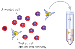
In a previous post, I covered the current options for isolating pure cell populations. One immediate question you will have to ask yourself is whether you would prefer positive selection or negative selection (depletion) for the isolation of your cell type of interest.

Positive selection involves the isolation of a target cell population by using an antibody that specifically binds that population. As an example, a positive selection kit for T cells would use an antibody specific for the CD3 molecule on T cells. Negative selection, however, involves the depletion of all cell types except your cell type of interest. With our T cell isolation example, our negative selection kit would likely involve antibodies specific for B cells (CD19), monocytes (CD14), NK cells (CD56), and so on. With the depletion of these cell types we would only be left with our cells of interest, in this case T cells (CD3).

The Advantages of Positive Selection

Positive selection and negative selection each havepositiveSelection their advantages. Positive selection offers greater purity due to the specificity of the reaction. You know in our example that positive selection of T cells will only yield a high purity of T cells due to the binding of selection antibodies to CD3 molecules. Negative selection, however, is inherently leakier since it is impossible to design a perfect depletion cocktail to target all cells that do not carry CD3 molecules. It is important to point out though that all of the popular cell isolation companies have made quite excellent kits that yield good purity levels when done properly. The difference in purity between positive selection and negative selection is roughly 99% to 95% pure, both of which are more than serviceable.

Another advantage of positive selection is that it offers the ability for a follow-up selection, or sequential isolations. Since negative selection works by binding all cells except the target cells with bead-bound antibodies, there is no way to do further isolations with the negative population. However, the negative flow through population from positive isolation will not have bead-bound antibodies and therefore is available for either another positive selection or a negative selection of your choice.

The Advantages of Negative Selection

The disadvantage of positive selection of course isnegativeSelection that your isolated cells will carry bead-bound antibodies. Not surprisingly, the kit manufacturers will tell you that this is not a concern, but it is something you need to keep and mind and use at your discretion. While neither the antibodies nor the beads should activate your isolated cells, it may in some way affect your downstream experiments. If you feel this could be an issue and you would prefer ‘untouched’ cells, then negative selection may be the right choice for you. First, however, be sure the negative selection kit actually depletes all necessary cells in order to achieve a pure target population. Often these kits are designed for common target tissues, such as peripheral bloods, lymph nodes, and spleens. Unfortunately negative selection kits may not work well for other target tissues. For example, my own work involves isolation of T cells from tumor samples. Since stock negative selection kits do not contain depletion antibodies for tumor cells, negative selection is not an option for our assays, and as a result we are forced to use positive selection.

It is important to choose an optimal cell isolation strategy specific to your assay, your target cells, and your tissue source. In my next post I will offer some tips for sorting through the various kits and technologies many companies offer for cell isolation.

Computers meet T cells: in silico identification of mutated tumor antigens targeted by T cells

It is well accepted that T cells can recognize and kill tumors that arise in individuals but that tumor cells escape immune surveillance due to the immunosuppressive tumor microenvironment that renders these T cells dysfunctional is less understood.  Only a relatively small number of antigens that T cells recognize for tumor-killing have been identified, and the methods used to identify these antigens are quite cumbersome.  In a recent article in Nature Medicine, Robbins et al. utilize informatics methods to identify mutated tumor antigens in melanoma patients that allowed effective targeting by anti-tumor T cells.

Genome sequencing T cells

In an effort to identify clinically relevant mutated tumor cell epitopes recognized by T cells, Robbins et al. first performed whole-exome sequencing of tumor cells and matched normal cells from melanoma patients who demonstrated tumor regression following adoptive transfer of autologous tumor infiltrating lymphocytes (TILs).  Mutations in tumor cells that resulted in amino acid changes were identified and then screened using an MHC binding algorithm that predicts high affinity binding of peptide sequences to specific HLA alleles.  Candidate peptides of 9-10 amino acids in length were synthesized and pulsed with specific HLA-expressing target cell lines to load the peptides into the MHC complex.  Peptide-pulsed target cells or autologous tumor cell lines were then cultured with autologous TILs from the same donor and IFN-gamma production was assessed as a read out of T cell activation.

Three metastatic melanoma patients were assessed using this methodology.  The first patient was homozygous for HLA-A*0201, and thus mutated melanoma cell line peptides predicted to bind to the HLA-A*0201 allele were identified by the MHC-binding algorithm.  From this donor, 4 out of 55 candidate peptides elicited IFN-gamma responses from autologous T cells cultured with peptide-pulsed target cells.  Two of these mutated peptides were found to correspond to the casein kinase1α1 protein (CSNK1A1), one peptide was mapped to the growth arrest specific 7 gene (GAS7) gene, and the fourth was a fragment of the HAUS augmin-like complex, subunit 3 (HAUS3) protein.  The wild-type versions of each of these peptides bound very poorly (100-10,000 fold less) or not at all to the HLA and were not recognized by T cells.  Two other donors were assessed for predicted binding of mutated peptides to HLA-A*0101 and HLA-A*1101.  Autologous T cell responses were found to be activated in response to mutated peptides from pleckstrin homology domain containing, family M member 2 (PLEKHM2), protein phosphatase 1 regulatory subunit 3B (PPP1R3B), matrilin 2 (MATN2), and cyclin-dependent kinase 12 (CDK12) genes, but not their wild-type counterparts.  Furthermore, tumor lines were validated to express these mutated proteins.

Finally, the authors compared the reactivity of peripheral blood mononuclear cells (PBMCs) drawn before and after adoptive TIL transfer into two of these patients to determine if anti-tumor reactive T cell clones persisted in vivo.  T cells that recognized the same tumor antigens as the TILs were identified post-adoptive transfer at greater levels than prior to adoptive transfer.  Thus, T cells that recognize mutated tumor epitopes may play a clinically relevant role in mediating tumor regression.  Many questions remain, including a direct demonstration that such tumor-reactive TILs are responsible for mediating the observed tumor regression in these patients, and whether further mutation of these residues might facilitate immune escape later it the course of disease. 

Mining exomic sequencing data to identify mutated antigens recognized by adoptively transferred tumor-reactive T cells.  Robbins PF, Lu YC, El-Gamil M, Li YF, Gross C, Gartner J, Lin JC, Teer JK, Cliften P, Tycksen E, Samuels Y, Rosenberg SA. Nat Med. 2013 May 5. doi: 10.1038/nm.3161.

NetMHCpan, a method for quantitative predictions of peptide binding to any HLA-A and -B locus protein of known sequence.  Nielsen M, Lundegaard C, Blicher T, Lamberth K, Harndahl M, Justesen S, Røder G, Peters B, Sette A, Lund O, Buus S. PLoS One. 2007 Aug 29;2(8):e796.

A bifunctional FoxP3+ regulatory T cell subset converts to pro-inflammatory helper T cells

Recently a number of studies have arisen characterizing Tregulatory cellvarious functional subsets of CD4+ FoxP3+ regulatory T cells (TREGS), as well as their plasticity and ability to differentiate into other TH subtypes.  For instance, TREGS that express RORγt were found to be the specific TREG subset that promotes pro-tumor immune functions in colorectal cancer patients.  In a recent article in Immunity, Sharma et al. identify another TREG subset: FoxP3+ TREGS that loose expression of Eos convert to a pro-inflammatory helper subtype that promotes naïve CD8+ T cells differentiation into potent effectors.

Eos is a transcription factor in the Ikaros family, and acts as an obligate co-repressor in complex with FoxP3 to inhibit expression of FoxP3-repressed genes.  In a quest to understand why TREGS in inflammatory environments were observed to become pro-inflammatory without losing FoxP3 expression, Sharma et al. examined the expression of Eos in FoxP3+ TREGS under inflammatory conditions.

Conversion of FoxP3+ TREGS into an inflammatory phenotype was demonstrated by acquired expression of IL-2, IL-17, and CD40L in the draining lymph nodes of a vaccination site compared with FoxP3+ TREGS at distant lymph nodes that did not gain this function.  In these converted inflammatory FoxP3+ TREGS, expression of Eos was rapidly lost.  IL-6 was required for downregulation of Eos, as TREGS in mice lacking IL-6 did not lose Eos expression under the same conditions.  However, IL-6 alone was insufficient for Eos downregulation, which also required interactions with MHC class II on activated dendritic cells.  Loss of Eos expression was furthermore shown to be required for acquisition of the pro-inflammatory phenotype, as TREGS with forced overexpression of Eos did not undergo this conversion.

Interestingly, not all FoxP3+ TREGS were equivalent in their propensity to lose Eos expression and become pro-inflammatory.  Thymic FoxP3+ TREGS were assessed for stability of Eos under treatment with cyclohexamide. CD38+CD69+CD103 TREGS were “Eos-labile” and specifically lost Eos expression within one hour of cyclohexamide treatment, while CD38CD69CD103+ TREGS maintained Eos expression.  Expression of other markers associated with FoxP3+ TREGS including CD25 and CTLA-4 were equivalent between these two phenotypes highlighting the inability of using these TREG markers to discriminate between these populations.  When these FoxP3+ TREGS were sorted into CD38+CD103and CD38CD103+ subsets and transferred into mice, followed by the vaccination schema, only CD38+CD103 TREGS lost Eos expression and gained CD40L and IL-2 expression. The Eos-labile TREGS do however have characteristic suppressive functions when examined in several models including protection from colitis in a Rag-deficient CD45RBHI effector cell-driven autoimmune colitis model and in vitro suppression of T cell proliferation driven by anti-CD3.

Because the Eos-labile subset was observed in the thymus as part of the natural TREG repertoire, the authors examined the signals required for development of this subset.  Again, IL-6 was required as this subset did not arise in IL-6-/- mice.  Epigenetic analysis of DNA methylation patterns comparing these FoxP3+ TREGS subsets revealed distinctive patterns of methylation yet these subsets were still much more closely related to each other as compared with FoxP3 CD4+ T cells.  Future studies will be needed to determine the nature of these epigenetic differences and which signals are controlled by IL-6.

Interestingly, the authors explored the functional contribution of the Eos-labile pro-inflammatory TREGS subset on CD8+ priming in the vaccination model.  Depletion of TREGS resulted in loss of CD8+ T cell proliferation and granzyme B expression as well as loss of CD86 upregulation on DCs, while adding back just the Eos-labile subset or IL-2 plus CD40-agonist antibodies rescued these defects.  The Eos-labile subset did not however, contribute to reactivation of memory CD4+ T cells, and thus these cells appear to play a specific role in the initial priming stages of naïve T cell activation.  Thus, despite having regulatory activity, these cells are critical in priming CD8+ T cell responses by supplying IL-2 and CD40L signals.

However, indoleamine 2,3-dioxygenase (IDO) was able to block Eos downregulation and acquisition of IL-2, IL-17, and CD40L expression.  Importantly, in a murine tumor vaccination model, blocking IDO was important for FoxP3+ inflammatory TREG induction and acquisition of anti-tumor effector CD8+ T cell responses.  The mechanism of IDO inhibition of Eos downregulation was found to be at least in part, dependent on the antagonization of the IL-6-STAT3 pathway by IDO-mediated production of kynurenine-pathway metabolites which activate the aryl hydrocarbon receptor (AhR).  Interestingly, different AhR ligands have been previously shown to differentially regulate induction of TH17 cells vs. TREGS (Quintana et al.), and kyenurine was a TREG inducing AhR ligand (Mezrich et al.).  Additionally, the contrasting effects of IL-6 and IDO will be an important factor in priming immune cell responses.

Overall, this thorough investigation identified the mechanisms that induce and inhibit this newly defined Eos-labile TREG subset that maintains FoxP3 expression and has typical suppressive TREG activity, yet is critically important in priming effector T cell immune responses.  Future studies will be needed to address how these cells balance regulatory and priming activities as well as the relationships between this subset and the many other TREG subsets described.


An inherently bifunctional subset of foxp3(+) T helper cells is controlled by the transcription factor eos.   Sharma MD, Huang L, Choi JH, Lee EJ, Wilson JM, Lemos H, Pan F, Blazar BR, Pardoll DM, Mellor AL, Shi H, Munn DH. Immunity. 2013 May 23;38(5):998-1012. doi: 10.1016/j.immuni.2013.01.013. Epub 2013 May 16.

Eos, goddess of treg cell reprogramming.  Rieder SA, Shevach EM. Immunity. 2013 May 23;38(5):849-50. doi: 10.1016/j.immuni.2013.05.001.

Control of T(reg) and T(H)17 cell differentiation by the aryl hydrocarbon receptor.  Quintana FJ, Basso AS, Iglesias AH, Korn T, Farez MF, Bettelli E, Caccamo M, Oukka M, Weiner HL. Nature. 2008 May 1;453(7191):65-71. doi: 10.1038/nature06880. Epub 2008 Mar 23.

An interaction between kynurenine and the aryl hydrocarbon receptor can generate regulatory T cells.  Mezrich JD, Fechner JH, Zhang X, Johnson BP, Burlingham WJ, Bradfield CA. J Immunol. 2010 Sep 15;185(6):3190-8. doi: 10.4049/jimmunol.0903670. Epub 2010 Aug 18.