Highlight: Is too much salt bad for your guts?

salt

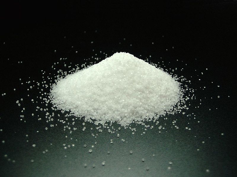
Whether eating too much table salt in our diet is bad for our health has long been debated. Links have been proposed to several cardiovascular diseases. But, a recent expert committee for the Institute of Medicine concluded that the data do not support such a link [1], keeping the discussion going. Two recent publications in Nature, however, suggest that too much dietary salt might impact our immune system instead and potentially increase the likelihood of autoimmune diseases.

CD4 T lymphocytes can differentiate in specialized subsets that promote or help diverse immune responses. Called T helper (Th) cells, particular subsets are named after prominent cytokines they produce, e.g. IL-17 in the case of Th17 T cells. Th17 cells are important for protection of the body against many bacterial and fungal infections and they are prevalent in the intestinal tissue were they are believed to aid the barrier function of the gut to keep the intestinal bacteria were they belong [2]. However, the “too much of a good thing” proverb applies to lymphocytes too and in the case of Th17 T cells this is exemplified by their pathogenic involvement in several autoimmune diseases. Therefore, the control of cell number and function of Th17 cells requires a delicate balance.

It was known that there is a cross talk between the gut lumen and the Th17 cell response. For example, a few years back it was shown that the frequency of a common bacterium within the gut microbiota could influence the prevalence of Th17 cells in the intestinal tissue [3]. The two new studies demonstrate that table salt (sodium chloride, NaCl) is a surprising new factor on the list to influence the frequency and function of Th17 cells [4-6].Lymphocyte activation

Adding 40 mM NaCl – a level found in the intestinal tissue of animals after feeding of a high salt diet – to in vitro cultures augmented the differentiation of Th17 cells [4, 5]. Similar to in vitro, feeding mice with a high salt diet increased the frequency of Th17 cell in the intestinal tissue, but not in the lymph nodes or the spleen. In both settings (in vitro and in vivo) the resulting Th17 cells were capable of producing large amounts of pro-inflammatory cytokines. By analysis of the mRNA expression, both reports characterized the MAP-kinase p38, NFAT5 (nuclear factor of activated T cells 5) and SGK1 (serum glucocorticoid-regulated kinase-1) as critical molecular players in sensing NaCl and mediating its effect. The elimination of any of these factors from the T cells, either by genetic ablation or by impeding the expression by means of RNA-interference (shRNA), blocked the increased Th17 cell differentiation in the presence of NaCl. Although all three proteins are part of the same pathway, SGK1 appeared to be central in the regulation of the NaCl induced effect. Although this finding is surprising, the results are in line with the known function of SGK1 in sodium transport and homeostasis [7]. SGK1 expression was not only induced by increased NaCl concentrations, but also by the cytokine IL-23, which has a critical role in stabilizing and reinforcing the TH17 phenotype [2]. As NaCl also increased the expression of the IL-23 receptor this established a positive feedback loop that strengthened the Th17 cell differentiation. Importantly, both groups also showed that raising the levels of dietary salt could augment the severity of EAE (experimental autoimmune encephalomyelitis), a mouse model for the autoimmune disease multiple sclerosis [4-6].

In summary, these reports demonstrate that high levels of salt in the diet could make mice susceptible to a form of autoimmune disease that involves pathogenic Th17 T cells. The data suggest that high concentration of NaCl might be an environmental risk factor for autoimmune diseases. However, it should be pointed out that high concentration of NaCl did not induce autoimmune responses by itself, as the EAE animal model requires the immunization with a know self-antigen. Autoimmunity is a complex interplay of numerous genetic pre-disposing and environmental factors. In this regard these new reports [4, 5] suggest that high dietary salt concentrations might tilt the balance a bit towards autoimmunity in genetically predisposed individuals.

However, the reality will likely be more complicated – as it usually is. For example, it will be critical to show that the correlation between dietary NaCl and Th17 cells is valid also in humans. Furthermore, with this knew knowledge other factors might come to light soon. For example, SGK1 expression is also stimulated by several hormones including endogenous steroids like stress hormones [7], suggesting that the induction of Th17 cell might be augmented by stress as well. Therefore, these intriguing new reports [4, 5] will surely spur now the required research to clarify these points. Till then, going slow on sodium-laden junk food might be generally a justified suggestion.                                 

References:

[1] Strom, Brian (2013). Sodium Intake in Populations: Assessment of Evidence. Washington, DC: The National Academies Press: The Institute of Medicine.

[2] Weaver, C. T., Elson, C. O., Fouser, L. A. & Kolls, J. K. The Th17 pathway and inflammatory diseases of the intestines, lungs, and skin. Annu Rev Pathol 8, 477–512 (2013).

[3] Ivanov, I. I. et al. Induction of Intestinal Th17 Cells by Segmented Filamentous Bacteria. Cell 139, 485–498 (2009).

[4] Kleinewietfeld, M. et al. Sodium chloride drives autoimmune disease by the induction of pathogenic TH17 cells. Nature (2013). doi:10.1038/nature11868.

[5] Wu, C. et al. Induction of pathogenic TH17 cells by inducible salt-sensing kinase SGK1. Nature (2013). doi:10.1038/nature11984.

[6] O’Shea, J. J. & Jones, R. G. Autoimmunity: Rubbing salt in the wound. Nature 496, 437–439 (2013).

[7] Lang, F. & Shumilina, E. Regulation of ion channels by the serum- and glucocorticoid-inducible kinase SGK1. The FASEB Journal 27, 3–12 (2013).

A unique set of cell surface markers for induced T regulatory cells

Transplantation of antigen-specific T cells during cancer immunotherapy has generated many notable results in the fight against diverse cancers. In these scenarios, the transplanted T cell populations act as a sort of highly mobile army that can track and kill tumor cells wherever they hide. In contrast, the goal of therapy during autoimmunity is to suppress immune activity, not increase its potency. Would transplantation of a “peacekeeper” immune cell-type be able to specifically quell autoimmune reactions? T regulatory cells (Tregs) are attractive candidates for the peacekeeper role based on their ability to dominantly suppress auto-reactive cell populations. As opposed to studies of cancer immunotherapy, clinical trials for Treg adoptive transplant are hampered by the lack of specific cell surface markers for these populations that enable their purification1. The defining characteristics of Treg populations (eg. FoxP3, IL-10, TGF-beta) are intracellular proteins whose analysis and quantification requires permeabilization and, hence, destruction of the cells. Positive selection for markers such as CD4 and CD25 which are expressed on the surface of certain types of Tregs also enriches for effector T cell populations whose functions upon transplantation may serve to further stimulate immune activity in an autoreactive host.Adoptive t cell transfer

In this month’s issue of Nature Medicine, Gagliani et al. sought to address the need for Treg-specific cell surface markers2. The authors focused on a particular type of inducible Treg called Type 1 regulatory T cells (Tr1 cells). These are a highly suppressive population of CD4+ T cells that are thought to control immune reactions both through IL-10 secretion and direct, Granzyme B-mediated destruction of myeloid antigen presenting cells. Galiani et al. were able to isolate Tr1 clones from the peripheral blood of healthy donors using a limiting-dilution assay: CD4+ T cells were plated in wells at a density of 1 cell/well, grown in conditions known to be suitable for Tr1 development, and then assessed for high levels of IL-10 secretion. The isolated Tr1 clones were stimulated in vitro and their gene expression profiles were measured at different time points and compared to that of naïve CD4+ T cells (Th0 cells). Under these conditions, the authors found that Tr1 cells uniquely expressed genes for two cell surface markers, CD49b and LAG-3. Used independently, these markers would enrich for multiple T cell types. But when used in combination, CD49b and LAG-3 allowed the investigators to isolate Tr1 cells from human peripheral blood which expressed high-levels of IL-10 and were able to suppress T cell proliferation in vitro. The authors went on to show that this cell-surface marker combination could also be used to isolate Tr1 cells from well-defined mouse models of Treg function. Finally, authors showed that CD49b and LAG-3 could effectively enrich for Tr1 cells from a highly-expanded, in vitro-polarized bulk population. This raises the possibility of generating large numbers of highly pure IL-10 secreting Tr1 cells for adoptive transplantation during autoimmunity.

Gagliani et al. have effectively used gene profiling of a target cell type to identify cell-surface markers for a previously difficult-to-analyze population. These new markers should facilitate further clinical study of adoptive transplant of Treg populations for autoimmunity. Now that it is possible to identify Tr1 cells from blood, it will be interesting to see how numbers of these cells correlate to different disease states and how they change in response to immune modulatory treatments. Furthermore, coupling the polarization and enrichment of Tr1 cells to tetramer-based identification of antigen-specific T cells may allow for highly-selective targeting of autoimmune reactions.

References:

1. Human T regulatory cell therapy: take a billion or so and call me in the morning. Riley JL, June CH, Blazar BR. Immunity. 2009 May;30(5):656-65. doi: 10.1016/j.immuni.2009.04.006.

2. Coexpression of CD49b and LAG-3 identifies human and mouse T regulatory type 1 cells. Gagliani N, Magnani CF, Huber S, Gianolini ME, Pala M, Licona-Limon P, Guo B, Herbert DR, Bulfone A, Trentini F, Di Serio C, Bacchetta R, Andreani M, Brockmann L, Gregori S, Flavell RA, Roncarolo MG. Nat Med. 2013 Jun;19(6):739-46. doi: 10.1038/nm.3179. Epub 2013 Apr 28.

Computers meet T cells: in silico identification of mutated tumor antigens targeted by T cells

It is well accepted that T cells can recognize and kill tumors that arise in individuals but that tumor cells escape immune surveillance due to the immunosuppressive tumor microenvironment that renders these T cells dysfunctional is less understood.  Only a relatively small number of antigens that T cells recognize for tumor-killing have been identified, and the methods used to identify these antigens are quite cumbersome.  In a recent article in Nature Medicine, Robbins et al. utilize informatics methods to identify mutated tumor antigens in melanoma patients that allowed effective targeting by anti-tumor T cells.

Genome sequencing T cells

In an effort to identify clinically relevant mutated tumor cell epitopes recognized by T cells, Robbins et al. first performed whole-exome sequencing of tumor cells and matched normal cells from melanoma patients who demonstrated tumor regression following adoptive transfer of autologous tumor infiltrating lymphocytes (TILs).  Mutations in tumor cells that resulted in amino acid changes were identified and then screened using an MHC binding algorithm that predicts high affinity binding of peptide sequences to specific HLA alleles.  Candidate peptides of 9-10 amino acids in length were synthesized and pulsed with specific HLA-expressing target cell lines to load the peptides into the MHC complex.  Peptide-pulsed target cells or autologous tumor cell lines were then cultured with autologous TILs from the same donor and IFN-gamma production was assessed as a read out of T cell activation.

Three metastatic melanoma patients were assessed using this methodology.  The first patient was homozygous for HLA-A*0201, and thus mutated melanoma cell line peptides predicted to bind to the HLA-A*0201 allele were identified by the MHC-binding algorithm.  From this donor, 4 out of 55 candidate peptides elicited IFN-gamma responses from autologous T cells cultured with peptide-pulsed target cells.  Two of these mutated peptides were found to correspond to the casein kinase1α1 protein (CSNK1A1), one peptide was mapped to the growth arrest specific 7 gene (GAS7) gene, and the fourth was a fragment of the HAUS augmin-like complex, subunit 3 (HAUS3) protein.  The wild-type versions of each of these peptides bound very poorly (100-10,000 fold less) or not at all to the HLA and were not recognized by T cells.  Two other donors were assessed for predicted binding of mutated peptides to HLA-A*0101 and HLA-A*1101.  Autologous T cell responses were found to be activated in response to mutated peptides from pleckstrin homology domain containing, family M member 2 (PLEKHM2), protein phosphatase 1 regulatory subunit 3B (PPP1R3B), matrilin 2 (MATN2), and cyclin-dependent kinase 12 (CDK12) genes, but not their wild-type counterparts.  Furthermore, tumor lines were validated to express these mutated proteins.

Finally, the authors compared the reactivity of peripheral blood mononuclear cells (PBMCs) drawn before and after adoptive TIL transfer into two of these patients to determine if anti-tumor reactive T cell clones persisted in vivo.  T cells that recognized the same tumor antigens as the TILs were identified post-adoptive transfer at greater levels than prior to adoptive transfer.  Thus, T cells that recognize mutated tumor epitopes may play a clinically relevant role in mediating tumor regression.  Many questions remain, including a direct demonstration that such tumor-reactive TILs are responsible for mediating the observed tumor regression in these patients, and whether further mutation of these residues might facilitate immune escape later it the course of disease. 

Mining exomic sequencing data to identify mutated antigens recognized by adoptively transferred tumor-reactive T cells.  Robbins PF, Lu YC, El-Gamil M, Li YF, Gross C, Gartner J, Lin JC, Teer JK, Cliften P, Tycksen E, Samuels Y, Rosenberg SA. Nat Med. 2013 May 5. doi: 10.1038/nm.3161.

NetMHCpan, a method for quantitative predictions of peptide binding to any HLA-A and -B locus protein of known sequence.  Nielsen M, Lundegaard C, Blicher T, Lamberth K, Harndahl M, Justesen S, Røder G, Peters B, Sette A, Lund O, Buus S. PLoS One. 2007 Aug 29;2(8):e796.

A bifunctional FoxP3+ regulatory T cell subset converts to pro-inflammatory helper T cells

Recently a number of studies have arisen characterizing Tregulatory cellvarious functional subsets of CD4+ FoxP3+ regulatory T cells (TREGS), as well as their plasticity and ability to differentiate into other TH subtypes.  For instance, TREGS that express RORγt were found to be the specific TREG subset that promotes pro-tumor immune functions in colorectal cancer patients.  In a recent article in Immunity, Sharma et al. identify another TREG subset: FoxP3+ TREGS that loose expression of Eos convert to a pro-inflammatory helper subtype that promotes naïve CD8+ T cells differentiation into potent effectors.

Eos is a transcription factor in the Ikaros family, and acts as an obligate co-repressor in complex with FoxP3 to inhibit expression of FoxP3-repressed genes.  In a quest to understand why TREGS in inflammatory environments were observed to become pro-inflammatory without losing FoxP3 expression, Sharma et al. examined the expression of Eos in FoxP3+ TREGS under inflammatory conditions.

Conversion of FoxP3+ TREGS into an inflammatory phenotype was demonstrated by acquired expression of IL-2, IL-17, and CD40L in the draining lymph nodes of a vaccination site compared with FoxP3+ TREGS at distant lymph nodes that did not gain this function.  In these converted inflammatory FoxP3+ TREGS, expression of Eos was rapidly lost.  IL-6 was required for downregulation of Eos, as TREGS in mice lacking IL-6 did not lose Eos expression under the same conditions.  However, IL-6 alone was insufficient for Eos downregulation, which also required interactions with MHC class II on activated dendritic cells.  Loss of Eos expression was furthermore shown to be required for acquisition of the pro-inflammatory phenotype, as TREGS with forced overexpression of Eos did not undergo this conversion.

Interestingly, not all FoxP3+ TREGS were equivalent in their propensity to lose Eos expression and become pro-inflammatory.  Thymic FoxP3+ TREGS were assessed for stability of Eos under treatment with cyclohexamide. CD38+CD69+CD103 TREGS were “Eos-labile” and specifically lost Eos expression within one hour of cyclohexamide treatment, while CD38CD69CD103+ TREGS maintained Eos expression.  Expression of other markers associated with FoxP3+ TREGS including CD25 and CTLA-4 were equivalent between these two phenotypes highlighting the inability of using these TREG markers to discriminate between these populations.  When these FoxP3+ TREGS were sorted into CD38+CD103and CD38CD103+ subsets and transferred into mice, followed by the vaccination schema, only CD38+CD103 TREGS lost Eos expression and gained CD40L and IL-2 expression. The Eos-labile TREGS do however have characteristic suppressive functions when examined in several models including protection from colitis in a Rag-deficient CD45RBHI effector cell-driven autoimmune colitis model and in vitro suppression of T cell proliferation driven by anti-CD3.

Because the Eos-labile subset was observed in the thymus as part of the natural TREG repertoire, the authors examined the signals required for development of this subset.  Again, IL-6 was required as this subset did not arise in IL-6-/- mice.  Epigenetic analysis of DNA methylation patterns comparing these FoxP3+ TREGS subsets revealed distinctive patterns of methylation yet these subsets were still much more closely related to each other as compared with FoxP3 CD4+ T cells.  Future studies will be needed to determine the nature of these epigenetic differences and which signals are controlled by IL-6.

Interestingly, the authors explored the functional contribution of the Eos-labile pro-inflammatory TREGS subset on CD8+ priming in the vaccination model.  Depletion of TREGS resulted in loss of CD8+ T cell proliferation and granzyme B expression as well as loss of CD86 upregulation on DCs, while adding back just the Eos-labile subset or IL-2 plus CD40-agonist antibodies rescued these defects.  The Eos-labile subset did not however, contribute to reactivation of memory CD4+ T cells, and thus these cells appear to play a specific role in the initial priming stages of naïve T cell activation.  Thus, despite having regulatory activity, these cells are critical in priming CD8+ T cell responses by supplying IL-2 and CD40L signals.

However, indoleamine 2,3-dioxygenase (IDO) was able to block Eos downregulation and acquisition of IL-2, IL-17, and CD40L expression.  Importantly, in a murine tumor vaccination model, blocking IDO was important for FoxP3+ inflammatory TREG induction and acquisition of anti-tumor effector CD8+ T cell responses.  The mechanism of IDO inhibition of Eos downregulation was found to be at least in part, dependent on the antagonization of the IL-6-STAT3 pathway by IDO-mediated production of kynurenine-pathway metabolites which activate the aryl hydrocarbon receptor (AhR).  Interestingly, different AhR ligands have been previously shown to differentially regulate induction of TH17 cells vs. TREGS (Quintana et al.), and kyenurine was a TREG inducing AhR ligand (Mezrich et al.).  Additionally, the contrasting effects of IL-6 and IDO will be an important factor in priming immune cell responses.

Overall, this thorough investigation identified the mechanisms that induce and inhibit this newly defined Eos-labile TREG subset that maintains FoxP3 expression and has typical suppressive TREG activity, yet is critically important in priming effector T cell immune responses.  Future studies will be needed to address how these cells balance regulatory and priming activities as well as the relationships between this subset and the many other TREG subsets described.


An inherently bifunctional subset of foxp3(+) T helper cells is controlled by the transcription factor eos.   Sharma MD, Huang L, Choi JH, Lee EJ, Wilson JM, Lemos H, Pan F, Blazar BR, Pardoll DM, Mellor AL, Shi H, Munn DH. Immunity. 2013 May 23;38(5):998-1012. doi: 10.1016/j.immuni.2013.01.013. Epub 2013 May 16.

Eos, goddess of treg cell reprogramming.  Rieder SA, Shevach EM. Immunity. 2013 May 23;38(5):849-50. doi: 10.1016/j.immuni.2013.05.001.

Control of T(reg) and T(H)17 cell differentiation by the aryl hydrocarbon receptor.  Quintana FJ, Basso AS, Iglesias AH, Korn T, Farez MF, Bettelli E, Caccamo M, Oukka M, Weiner HL. Nature. 2008 May 1;453(7191):65-71. doi: 10.1038/nature06880. Epub 2008 Mar 23.

An interaction between kynurenine and the aryl hydrocarbon receptor can generate regulatory T cells.  Mezrich JD, Fechner JH, Zhang X, Johnson BP, Burlingham WJ, Bradfield CA. J Immunol. 2010 Sep 15;185(6):3190-8. doi: 10.4049/jimmunol.0903670. Epub 2010 Aug 18.

Highlight: How TNF knocks out Tregs!

A healthy and functional immune system requires a delicate balance of pro- and contra-inflammatory signals. Whereas, it is important to induce a strong and efficient immune response against pathogens, it is similarly important to dampen these responses after the pathogen is fought off to revert the immune system to a calm steady state. If the balance is disturbed, diseases can on the one hand, become chronic/overwhelming or, on the other hand, inflammatory responses that cannot terminate can result in autoimmune responses.

Crucial elements in the regulation of excessive immune responses are regulatory T (Treg) cells. Tregs are known to inhibit the response of other immune cells. Their essential role in limiting overwhelming immune responses is demonstrated by the detrimental consequences of their loss. Mice or humans lacking Tregs develop widespread and lethal autoimmune diseases. Besides several surface markers, Tregs are best characterized by the expression of the transcription factor FoxP3. This factor is essential for Treg function and its artificial expression in other T cells can induce a regulatory potential. Therefore, the expression of FoxP3 is required for a T cell to have regulatory potential (Buckner; Josefowicz et al.). However, it was known for many years that in cases of numerous autoimmune diseases FoxP3+ Tregs could be found in high numbers at the sides of inflammation, but that they did not demonstrate any or not sufficient regulatory activity. This enigmatic observation was so far poorly understood (Buckner; Josefowicz et al.).Treg balance

In the March 2013 issue of Nature Medicine Nie and colleagues shed new light on the underlying mechanism that impairs Treg function at the sites of inflammation. Studying Treg cells from rheumatoid arthritis (RA) patients the authors demonstrated that phosphorylation of FoxP3 of the serine at position 418 (S418) is required for its regulatory action. If FoxP3 lacks this particular phosphorylation the Treg cell is not suppressive! FoxP3 S418 in Tregs is usually phosphorylated and hence Tregs are regulatory by default. However, the authors show that due to the action of the enzyme ‘protein phosphatase 1’ (PP1) FoxP3 can lose its S418 phosphorylation. Intriguingly, the presence of the cytokine TNF lead to an up-regulation of PP1 expression in the Tregs in a dose-dependent manner, and this lead to de-phosphorylation of FoxP3 S418. Treg cells expressing a mutant FoxP3 that replaced the serine at position 418 with an alanine retained their suppressive potential even in the presence of TNF, demonstrating the importance of the phosphorylation of S418. With this finding, the authors were able to link the pro-inflammatory milieu (TNF) to a specific effect inside of the Tregs (de-phosphorylation of S418) that lead to the observed loss of the regulatory function of Treg cells. Importantly, the authors were also able to demonstrate the therapeutic potential of this knowledge. They monitored RA patients that underwent treatment with blocking anti-TNF antibodies (infliximab) and found that Tregs from patient PBMCs restored S418 phosphorylation and regained regulatory potential!

This is the second case for a post-transcriptional regulation of FoxP3 that can influence Treg function. Deacetylation of FoxP3 has been linked to impaired Treg function previously (Tao et al.). Additionally, the work of Nie et al. now adds mechanistic information to previous reports on the negative effect of TNF on Tregs (Valencia et al.; Zanin-Zhorov et al.).

Given the ubiquitous role of TNF during inflammation, it is very likely that the mechanism described by Nie et al. applies to many if not all cases of ongoing inflammation where Treg function is impaired. Furthermore, their data on the effects of anti-TNF antibody treatment in RA suggest a similar therapeutic potential in other autoimmune diseases. Surely, this report will ignite further investigation in this direction and will aid the development of better treatments for patients suffering from autoimmune diseases.

References:

Bromberg, J., 2013. TNF-α trips up Treg cells in rheumatoid arthritis. Nat Med, 19(3), pp.269–270.

Buckner, J.H., 2010. Mechanisms of impaired regulation by CD4(+)CD25(+)FOXP3(+) regulatory T cells in human autoimmune diseases. Nat Rev Immunol, 10(12), pp.849–859.

Josefowicz, S.Z., Lu, L.-F. & Rudensky, A.Y., 2012. Regulatory T cells: mechanisms of differentiation and function. Annual Review of Immunology, 30, pp.531–564.

Nie, H. et al., 2013. Phosphorylation of FOXP3 controls regulatory T cell function and is inhibited by TNF-α in rheumatoid arthritis. Nat Med, 19(3), pp.322–328.

Tao, R. et al., 2007. Deacetylase inhibition promotes the generation and function of regulatory T cells. Nature Medicine, 13(11), pp.1299–1307.

Valencia, X. et al., 2006. TNF downmodulates the function of human CD4+CD25hi T-regulatory cells. Blood, 108(1), pp.253–261.

Zanin-Zhorov, A. et al., 2010. Protein kinase C-theta mediates negative feedback on regulatory T cell function. Science, 328(5976), pp.372–376.