Cell-based therapy for Parkinson’s disease: past, present and future.

Parkinson’s disease (PD) is a chronic neurodegenerative condition effecting dopaminergic neurons of the midbrain. PD manifests itself around age 50 with mainly motor symptoms, such as tremor (shaking), slowness of movement, rigidity and postural instability. Number of pharmaceutical agents (e.g., L-Dopa and MAO-B inhibitors) has been used for symptomatic relief in PD patients, but the ultimate therapy target is the replacement of degenerating dopaminergic neurons with new, healthy neurons.

describe the imageCell replacement therapy for PD dates back to mid 80s with the transplantation of adrenal medullary tissue into patients’ striatum [1-3], which resulted only moderate improvements. At the same time, researchers in Sweden performed transplantation of fetal ventral mesencephalic tissue from aborted fetuses [4, 5]. These early studies observed important and persistent improvement based on numerous clinical outcomes. Moreover, postmortem examination of the brains of PD patients, who received ventral mesencephalic tissue transplantation, showed sustained survival of the graft and re-innervation of the striatum [6]. With the lift of federal funding ban on using fetal tissue for research and therapy by President Clinton in 1993, United States also began clinical trials utilizing fetal ventral mesencephalic tissue [7, 8]. Unfortunately, not only the patients didn’t display any significant improvements following transplantation in these trials, they developed additional abnormal, involuntary movements (i.e., graft-induced dyskinesia), due to surgery, which was also observed in other trials.

Close examination of the transplantation studies using fetal ventral mesencephalic tissue revealed few noteworthy outcomes:

1. Younger patients with newly developed pathology showed significant improvements over older patients with severe PD pathology.

2. Some patients showed continued improvements 3-4 years after surgery, while they did not display any benefits during the first year, indicating that the improvement in clinical parameters may take a while to appear over time. Regardless, it is clear that patients respond differently to the transplants of dopaminergic neurons, making the clinical outcomes fluctuate considerably.

3. Preparation of the fetal tissues, as well as selection of patients for transplantation, varied significantly from center to center carrying out the clinical trials, further indicating the need for standardizing tissue preparation, patient selection and implantation site.

Compared to the aforementioned points, the use of fetal ventral mesencephalic tissue for grafting constitutes one of the biggest problems in cell based therapy for PD. It has been challenging to standardize the number and the quality of the fetal dopaminergic cells in graft preparations. Furthermore, the purity of the preparations also varies from batch to batch. Lastly, many ethical -and sometimes legal- issues surround fetal tissues/cells significantly limiting their clinical applicability. Do we have an alternative source that is free of these concerns/problems? The answer is yes, but not at the moment. With the isolation of human embryonic stem cells (hESCs) in 1998 and the introduction of human induced pluripotent stem cells (iPSCs) in 2007, stem cell derived dopaminergic neurons are at the top of everyone’s list when it comes to replacing degenerating neurons in PD. hESCs have been the primary source to produce dopaminergic neurons so far [9-11], but with the popularity and the advantages of iPSCs, the focus is more likely to shift to iPSC-derived dopaminergic neurons in future transplantation efforts.

Number of studies utilizing stem cell derived dopaminergic neurons in animal models of PD reported promising results over the years. However, we are far from using these cells in clinical trials. Many issues, such as long-term stability of the transplanted cells, sustained functional recovery, ability to re-innervate the host striatum, generation of GMP grade cells and long-terms safety especially with regards to tumor formation, remain to be determined. To be able to answer these concerns are critical for successful clinical translation of stem cell derived dopaminergic neurons. Nevertheless, the target is in front of everyone, and the field of regenerative medicine is moving at an incredible speed to reach it.  It should also be noted that an increasing number of novel therapeutic approaches (e.g., gene therapy and growth factor infusions) have been under development -in addition to cell transplantations- with the aim of restoring dopaminergic function in PD patients.

While we are looking ahead with the promise of stem cell derived dopaminergic neurons for future of cell-based therapy in PD, there are many lessons to be learnt from the early clinical trials using fetal ventral mesencephalic tissue. There is no question that fetal dopamine neurons will serve as a reference and a standard against stem cell derived neurons for future clinical trials, since we know that the transplants survived, re-innervated the striatum, and generated adequate symptomatic relief in some patients for more than a decade following surgery. For PD patients, who are interested in cell-based therapy now, the decision of whether to wait for clinical trials utilizing stem cell derived neurons or to proceed with currently available fetal tissue grafts remains a somewhat difficult question and should take into consideration the aforementioned strengths and weaknesses of each approach.


References:

[1] Backlund EO, Granberg PO, Hamberger B, et al. Transplantation of adrenal medullary tissue to striatum in parkinsonism. First clini- cal trials. J Neurosurg 1985;62:169–173.

[2] Herrera-Marschitz M, Stromberg I, Olsson D, Ungerstedt U, Olson L. Adrenal medullary implants in the dopamine-denervated rat striatum. II. Acute behavior as a function of graft amount and location and its modulation by neuroleptics. Brain Res 1984;297:53–61.

[3] Madrazo I, Drucker-Colin R, Diaz V, Martinez-Mata J, Torres C, Becerril JJ. Open microsurgical autograft of adrenal medulla to the right caudate nucleus in two patients with intractable Parkinson’s disease. N Engl J Med 1987;316:831–834.

[4] Lindvall O, Brundin P, Widner H, et al. Grafts of fetal dopamine neurons survive and improve motor function in Parkinson’s dis- ease. Science 1990;247:574–577.

[5] Widner H, Tetrud J, Rehncrona S, et al. Bilateral fetal mesence- phalic grafting in two patients with parkinsonism induced by 1- methyl-4-phenyl-1,2,3,6-tetrahydropyridine (MPTP). N Engl J Med 1992;327:1556–1563.

[6] Kordower JH, Rosenstein JM, Collier TJ, et al. Functional fetal nigral grafts in a patient with Parkinson’s disease: chemoanatomic, ultrastructural, and metabolic studies. J Comp Neurol 1996;370:203–230.

[7] Freed CR, Greene PE, Breeze RE, et al. Transplantation of embry- onic dopamine neurons for severe Parkinson’s disease. N Engl J Med 2001;344:710–719.

[8] Olanow CW, Goetz CG, Kordower JH, et al. A double-blind con- trolled trial of bilateral fetal nigral transplantation in Parkinson’s disease. Ann Neurol 2003;54:403–414.

[9] Lee SH, Lumelsky N, Studer L, Auerbach JM, McKay RD. Effi- cient generation of midbrain and hindbrain neurons from mouse embryonic stem cells. Nat Biotechnol 2000;18:675–679.

[10] Cho MS, Lee YE, Kim JY, et al. Highly efficient and large-scale generation of functional dopamine neurons from human embryonic stem cells. Proc Natl Acad Sci U S A 2008;105:3392–3397.

[11] Kawasaki H, Suemori H, Mizuseki K, et al. Generation of dopami- nergic neurons and pigmented epithelia from primate ES cells by stromal cell-derived inducing activity. Proc Natl Acad Sci U S A 2002;99:1580–1585.

Identification of Type I Innate Lymphoid Cells that functionally resemble TH1 and NK cells

Innate lymphoid cells (ILCs) are subsets of lymphoid cells that do not rearrange their antigen receptors like T cells and B cells but have other features of lymphocytes.  ILCs include the Natural Killer (NK) cell subset, as well as cells that behave similarly to T helper cell subsets by producing similar characteristic cytokines.  Type 2 ILCs (ILC2) resemble TH2 describe the image
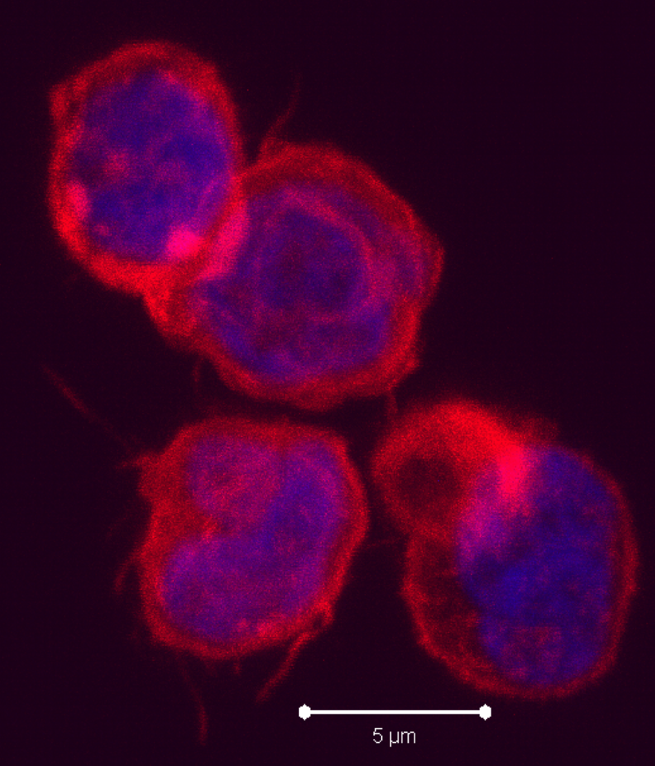
cells in that they produce IL-5 and IL-13.  RORγt+ ILCs aka ILC3s, include subsets that resemble TH17 and TH22 cells by producing IL-17 and IL-22, respectively, as well as a subset that produces both cytokines.   Several recent articles have identified another class of ILCs in both humans and mice.  These cells resemble TH1 cells in that they express T-bet/TBX21 and produce IFN-gamma, and are distinct from conventional NK cells found among peripheral blood mononuclear cells (PBMC).  These newly characterized cellular subsets have been denoted as Type 1 ILCs (ILC1).

In the April 2013 issue of Immunity, Fuchs et. al sought to more fully characterize the ILC subsets present in human mucosal lymphoid tissues.  In human tonsils, a CD3CD56+ NKp44+CD103+ subset was identified that expressed T-bet and produced IFN-gamma when stimulated with either PMA/ionomycin, IL-12, or IL-15.  These cells also expressed perforin and granzyme and had cytolytic activity.  Although these cells express CD56+and NKp44+, which are markers characteristic of NK cells, they appear to be related to but distinct from prototypical CD56hi NK cells found in PBMC.  For instance, unlike CD56hi PBMC NK cells, these ILC1 did not exhibit a response to IL-18, as measured by a synergistic production of IFN-gamma when stimulated with IL-12+ IL-18 vs. IL-12 alone.

In a separate study, published in the March 2013 issue of Nature Immunology, Bernink et. al identified a mucosal human ILC1 subset in tonsils that differs from that found by Fuchs et. al, being CD56NKp44as well asCD127+ and c-Kit.  Similarly to the cells described by Fuchs et. al, these cells expressed T-bet and produced  IFN-gamma when stimulated with PMA/ionomycin or IL-12.  However, they did not express perforin and granzyme.  Additional characterizations differentiated these cells from NK cells including the lack of the KIR3DL1 and IL-15Rα markers expressed by NK cells.

ILCs have been found to reside in mucosal associated lymphoid tissues include the oral, lung, and gastrointestinal mucosa, and are thought to function in immune responses to pathogens as well as in tissue repair.  ILCs including ILC3s have also been found to participate in inflammatory disease pathogenesis.  Both types of ILC1 cells were shown to be increased in the intestinal mucosa of Crohn’s disease patients, although their exact locations differed.  CD56+ NKp44+CD103+ cells were found to accumulate in the intraepithelial layer while CD127+CD56c-KitNKp44cells were found in the lamina propria.  Thus, these two subsets of ILC1 cells differ in multiple aspects including tissue localization.

In conclusion, both types of ILC1 cells identified in these studies are distinct from conventional PBMC CD56hi NK cells, express T-bet, and produce IFN-gamma in response to IL-12 and IL-15 stimulation.  Notably, ILC3 cells also heterogeneously express CD56, IFN-gamma, granzymes and perforin.  Thus, many questions remain as to the functional and developmental differences between different ILC subsets and between CD56+ ILC1 cells and PBMC NK cells that reside in various tissues.

Reading:

Intraepithelial Type 1 Innate Lymphoid Cells Are a Unique Subset of IL-12- and IL-15-Responsive IFN-γ-Producing Cells.  Fuchs A, Vermi W, Lee JS, Lonardi S, Gilfillan S, Newberry RD, Cella M, Colonna M. Immunity. 2013 Apr 18;38(4):769-81.

Human type 1 innate lymphoid cells accumulate in inflamed mucosal tissues.  Bernink JH, Peters CP, Munneke M, te Velde AA, Meijer SL, Weijer K, Hreggvidsdottir HS, Heinsbroek SE, Legrand N, Buskens CJ, Bemelman WA, Mjösberg JM, Spits H.  Nat Immunol. 2013 Mar;14(3):221-9.

ILC1 Populations Join the Border Patrol.  Maloy KJ, Uhlig HH. Immunity. 2013 Apr 18;38(4):630-2. doi: 10.1016/j.immuni.2013.03.005.

Innate lymphoid cells: emerging insights in development, lineage relationships, and function.  Spits H, Cupedo T. Annu Rev Immunol. 2012;30:647-75.

A T-bet gradient controls the fate and function of CCR6-RORγt+ innate lymphoid cells.  Klose CS, Kiss EA, Schwierzeck V, Ebert K, Hoyler T, d’Hargues Y, Göppert N, Croxford AL, Waisman A, Tanriver Y, Diefenbach A.  Nature. 2013 Feb 14;494(7436):261-5.

Reprogramming of old HSCs reverses functional defects associated with aging

HSCs must continuously self-renew to replenish the pool of mature blood cells throughout the life an adult.  One requirement for extensive self-renewal is high telomerase activity to prevent telomere shortening.  HSCs isolated from adult bone marrow have shorter telomeres than cells from fetal liver or umbilical cord blood 1, suggesting that proliferative potential may decrease with age.  Also, HSC aging is associated with decreased lymphoid potential, as well as an up-regulation of genes involved in leukemic transformation 2.  Consequently, “aging” HSCs may have functional defects that might be detrimental for therapeutic strategies involving genetic manipulation and transplantation of HSCs for the treatment of various blood disorders.

describe the image

Previous studies have demonstrated that during reprogramming, developing iPSCs express epigenetic features of ESCs, and lose those of the starting somatic cell 3.  In addition, a previous study reported a significant elongation of telomeres in derived iPSCs compared to the starting somatic cell 4.  Thus, it may be possible that reprogramming reverses functional defects associated with HSC aging.

Recently, in Blood, Wahlestedt et al examined whether characteristics of aging HSCs are reversible 5.  First, they derived iPSCs from young and aged murine HSCs.  To examine their differentiation potential, they injected the derived iPSCs into murine blastocysts and analyzed the engraftment of the donor cells in the developing chimeric embryos.  Overall, iPSCs derived from aged HSCs demonstrated similar differentiation potential compared to that of younger HSCs.  The engraftment of bone marrow mononuclear cells from primary chimeric mice in a competitive transfer experiment was comparable to that of young HSCs.  Aged HSCs, on the other hand, demonstrated a significant reduction in repopulation capacity.  Interestingly, aged iPSC-derived HSCs also generated naïve T cells at similar levels as young HSCs.

Next, the authors examined telomere length following re-differentiation of the young and aged iPSCs.  Telomeres in aged HSCs were ~11% shorter compared to young HSCs.  However, telomeres of the HSCs derived from the aged iPSCs demonstrated a 2-fold elongation compared to blastocyst control HSCs.  This 2-fold elongation was maintained even after transplantation.  Overall, these results indicate that iPSC induction from HSCs results in elongation of telomeres.

In short, Wahlestedt et al demonstrated that reprogramming does indeed reverse some of the functional defects associated with chronologically aged HSCs, including decreased differentiation potential and shortened telomeres.  However, the study did not address whether the iPSCs derived from aged HSCs had an increased DNA mutation frequency, since HSC aging is also associated with a higher mutation rate.  It would also be interesting to determine whether the above phenomena are also observed in reprogramming of aged human HSCs.  If iPSC induction does indeed result in an “epigenetic reset,” then HSCs derived from iPSCs may have unique characteristics favorable for use in clinical settings.


References

1          Vaziri, H. et al. Evidence for a mitotic clock in human hematopoietic stem cells: loss of telomeric DNA with age. Proc Natl Acad Sci U S A 91, 9857-9860 (1994).

2          Rossi, D. J. et al. Cell intrinsic alterations underlie hematopoietic stem cell aging. Proc Natl Acad Sci U S A 102, 9194-9199, doi:10.1073/pnas.0503280102 (2005).

3          Maherali, N. et al. Directly reprogrammed fibroblasts show global epigenetic remodeling and widespread tissue contribution. Cell Stem Cell 1, 55-70, doi:10.1016/j.stem.2007.05.014 (2007).

4          Marion, R. M. et al. Telomeres acquire embryonic stem cell characteristics in induced pluripotent stem cells. Cell Stem Cell 4, 141-154, doi:10.1016/j.stem.2008.12.010 (2009).

5          Wahlestedt, M. et al. An epigenetic component of hematopoietic stem cell aging amenable to reprogramming into a young state. Blood, doi:10.1182/blood-2012-11-469080 (2013).

ACTIVATED MAPK PATHWAY LIMITS EFFICACY OF ROMIDEPSIN

Histone deacetylase (HDAC) inhibitors (HDIs), a new class of epigenetic anti-tumor agents, have shown promise so far in the treatment of hematologic malignancies. Many, if not most, cell lines tested in vitro show sensitivity to HDIs, and synergy in combinations is also often noted. Yet, Phase I and II trials of HDIs have uniformly been disappointing in solid tumors. Even preclinical models in which HDIs exhibited potent anti-tumor effects in vivo have not succeeded in the clinic. Therefore, a detailed understanding of the mechanisms of resistance to HDIs may lead to strategies designed to increase clinical efficacy. Studies have proposed several mechanisms of resistance which include increased expression of the P-glycoprotein (Pgp) encoding multidrug-resistance gene ABCB1, increased expressresistance to HDIS resized 600
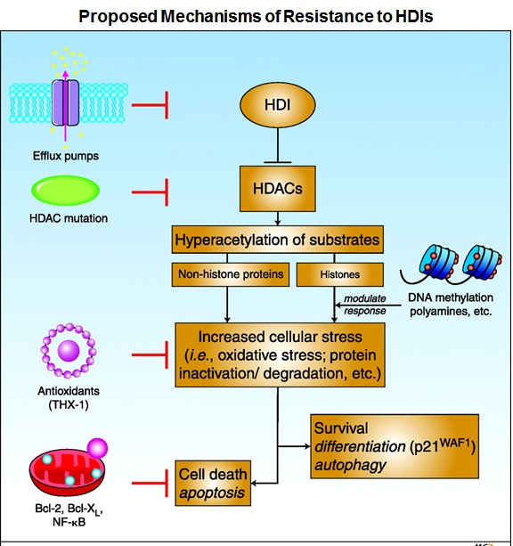
ion of reactive oxygen species (ROS) scavenger protein thioredoxin, elevated expression of anti-apoptotic proteins Bcl-2 and Bcl-xL, increased expression of histone HDAC enzymes, and activation of several signaling pathways including MAPK, phosphoinositide 3-kinase and signal transducer and activator of transcription.

The FDA approved histone deacetylase inhibitor romidepsin (istodax®) for the treatment of cutaneous T-cell lymphoma (CTCL) in 2009 and for the treatment of peripheral T-cell lymphoma (PTCL) in 2011. Disease progression was noted in some patients who initially responded to therapy, while disease in other patients did not respond to therapy suggesting that both de novo and acquired resistance to romidepsin were observed during the trial. Thus, studies are needed to determine the mechanisms of resistance to the HDI romidepsin particularly in T-cell lymphoma.

Several studies have noted selection of Pgp overexpression in vitro as a mechanism of drug resistance to romidepsin. The ease of selection of Pgp is in part because romidepsin is a substrate for Pgp, and in part based on ABCB1 gene induction as a consistent cellular response to HDIs. While induction of ABCB1 has been noted in normal and malignant peripheral blood mononuclear cells of patients treated with romidepsin, no evidence for Pgp-mediated resistance emerged in clinical samples obtained at the time of disease progression from patients with CTCL or PTCL. Therefore, identification of non-Pgp resistance mechanisms for romidepsin is warranted. A recent study published in the peer reviewed journal Blood by Chakraborty et al. (Blood. 2013 Mar 26) reported that activation of the mitogen activated protein kinase (MAPK) pathway conferred resistance to romidepsin through degradation of the pro-apoptotic BH-3 only protein Bim. To explore Pgp-independent mechanisms of resistance to romidpesin, a CTCL model consisting of the HuT78 cell line and its romidepsin-selected sublines were used. These sublines were separately selected in romidepsin in the presence of the Pgp-inhibitors verapamil or valspodar (PSC833) to avoid overexpression of Pgp. The resulting cell lines were resistant to romidepsin and inhibition of Pgp could reverse the resistance of only the cells that had not been selected in the presence of the Pgp inhibitors. The failure to reverse the resistance of the romidepsin-selected cells with a Pgp inhibitor suggested a different mechanism of action, which had not been identified in the earlier studies. A gene microarray study detected increased expression of the insulin receptor in the romidepsin-selected cells compared to the parental HuT78 cells. In addition, romidepsin-resistant cells also exhibited increased activation of MEK protein, a downstream component of the MAPK signaling pathway. Treating these resistant cells with the allosteric MEK inhibitors resulted in exquisite sensitivity while the parental HuT78 cells did not respond to the MEK inhibition. Restoration of the pro-apoptotic protein Bim was noted in romidepsin-resistant cells following MEK inhibition which was otherwise found to be degraded in the resistant cells. Combined treatment of MEK inhibitor with romidepsin also caused increased death of resistant cells. In their study, Chakraborty and colleagues also noted loss of Bim in the skin biopsy samples obtained from CTCL patients who experienced disease progression after romidepsin treatment. In addition, this study also reported perturbation of the MAPK regulated genes in the CTCL patients treated with romidepsin. Collectively these observations suggested that activation of the MAPK pathway may limit efficacy of the HDI romidepsin through degradation of the pro-apoptotic protein Bim, and in future clinical trials combination of romidepsin with MEK inhibitor may exhibit promising results.


References:

1.Fantin VR, Richon VM. Mechanisms of resistance to histone deacetylase inhibitors and their therapeutic implications. Clin Cancer Res. 2007;13(24):7237-7242.

2.Chakraborty AR, Robey RW, Luchenko VL, et al. MAPK pathway activation leads to Bim loss and histone deacetylase inhibitor resistance: rationale to combine romidepsin with a MEK inhibitor. Blood. 2013.

The Hippo-YAP Pathway: New Connection between Cancer and Stem Cells.

First discovered  by laboratories studying  Drosophila development 18 years ago1-3, the Hippo-YAP signaling pathway (also known as the Salvador-Warts-Hippo Pathway) is a novel pathway implicated in organism development, stem cell biology, and cancer biology4. While not much is known about the Hippo-YAP pathway, this signaling mechanism could lead to a promising paradigm in regenerative medicine and treating cancer.

While we are far from fully discovering every aspect Hippo-YAP signaling pathway, some components of this novel pathway have been uncovered. In mammalian cells, the first signal modulator to be stimulated is mammalian STE-20 protein kinase 1 & 2(Mst1/2). This stimulation causes autophosphorylation, which in turn starts a kinase cascade; phosphorylating the proteins Salvador homolog 1 (Sav1), MOB kinase activator 1 (Mob1), and large tumor suppressor 1 & 2 (Lats1/2). Once Lats1/2 is activated, it phosphorylates YAP (Yes-associated protein)4. This phosphorylation of YAP sequesters it outside of the cell and leads to its proteosomal degradation and thus blocking its ability to complex with the protumor TEAD transcription factors, which in turn inhibits proliferation and blocks inhibition of apoptosis4. Interestingly, other alternative mechanisms, such as directly targeting YAP via the WNT pathway, or activation of YAP/TAZ via the SMAD signaling pathway by TGFβ and BMP, have been demonstrated5. While the stimulation of the Hippo pathway is still being revealed, researchers have discovered two mechanisms hippo stimulation: cell-cell contact and activation of G-protein coupled receptors4,5.  Stimulation of G-protein Coupled Receptors (GPCRs), Go with the ligands LPA or S1P and Gs with glucagon and epinephrine, have been shown to activate the Hippo-YAP signaling pathway, causing phosphorylation of Mst1/26. On the other hand, the cell-cell contact method of hippo activation most likely phosphorylates Mst1/2 through the upstream component: Merlin6.  However, while there is phosphorylation of Mst1/2 in both stimulation methods, neither stimulation pathway is known, save one or two components, upstream of Mst1/2. Furthermore, the complexity of this signaling pathway is certain and upstream signals may be redundant6.

The Hippo-YAP signaling pathway plays a crucial role in embryological development. At the middle of this is the Hippo signaling component is transcriptional co-activator with PDZ-binding motif (TAZ, also known as WWTR1). TAZ is able to regulate the signaling mechanisms of the SMAD2/3-4 signaling pathway4; a pathway that regulates the TGF-beta signaling cascade that is important in early embryogenesis7. Furthermore, it has been demonstrated that functional loss of the TAZ protein, and not YAP, will lead to uncontrolled differentiation of human embryonic stem cells (hESCs) as well as loss of self-renewal of hESCs4.  Surprisingly, although YAP is not as important as TAZ to block differentiation, YAP is inactivated during normal hESC differentiation4. In addition to stem cell differentiation, the Hippo-YAP signaling pathway has been shown to be important for polarization of tissues8 in both planar and apicobasal cell polarity5, tissue shape and patterning9, and overall tissue homeostasis9.

While the activation of the Hippo-YAP pathway seems to be important for embryogenesis, the dysregulation of the Hippo-YAP pathway seems to play a striking role in tumorigenesis9. Deletion of the upstream Mst1/2 component of the Hippo-YAP pathway has been shown to cause uncontrolled liver growth. Microscopic analysis of liver biopsies revealed that these tissues were full of hepatocellular carcinoma and cholangiocarcinoma4. Likewise, overactivation of the YAP protein caused uncontrolled, extreme thickening of epidermal layer4. However, the pathway that is thought to be involved in this process of YAP activation is not the canonical Hippo-YAP pathway, but a signaling through alpha catenin. The catenin family and the Hippo-YAP signaling pathway were further shown to interact when overexpression of YAP facilitated the expression of Notch/Wnt signaling pathway indirectly by YAP-driven overexpression of beta catenin4. Because the Notch/Wnt pathways are important for cancer stem cell phenotype10 and cancer metastasis11, further investigation into the roles of the Hippo-YAP signaling pathway could bring a lot of clinical significance.

describe the imageAt the writing of this blog, no proposed drug has been proposed that directly targets the Hippo signaling pathway.  However, many possible targets for therapy are being investigated that would also affect the Hippo signaling pathway5. One of these is targeting the homeodomain-interacting protein kinase 2 (HIPK2) which has been demonstrated to activate YAP 5.In addition, using GPCR antagonists, such as Dobutamine, have been shown to decrease activation levels of YAP5. Also promising, researchers have solved many domains of the YAP structure, which may lead to specific inhibition of this oncogene by potential inhibitors12. Because of the role that the Hippo signaling pathway may play on tumor growth inhibition, it may not be long before candidate drugs targeting this pathway will start to enter the FDA drug pipeline.


Further Reading:

1              Justice, R. W., Zilian, O., Woods, D. F., Noll, M. & Bryant, P. J. The Drosophila tumor suppressor gene warts encodes a homolog of human myotonic dystrophy kinase and is required for the control of cell shape and proliferation. Genes & development 9, 534-546 (1995).

2              Xu, T., Wang, W., Zhang, S., Stewart, R. A. & Yu, W. Identifying tumor suppressors in genetic mosaics: the Drosophila lats gene encodes a putative protein kinase. Development 121, 1053-1063 (1995).

3              Wu, S., Huang, J., Dong, J. & Pan, D. hippo encodes a Ste-20 family protein kinase that restricts cell proliferation and promotes apoptosis in conjunction with salvador and warts. Cell 114, 445-456 (2003).

4              Ramos, A. & Camargo, F. D. The Hippo signaling pathway and stem cell biology. Trends in cell biology 22, 339-346, doi:10.1016/j.tcb.2012.04.006 (2012).

5              Harvey, K. F., Zhang, X. & Thomas, D. M. The Hippo pathway and human cancer. Nature reviews. Cancer 13, 246-257, doi:10.1038/nrc3458 (2013).

6              Yu, F. X. et al. Regulation of the Hippo-YAP pathway by G-protein-coupled receptor signaling. Cell 150, 780-791, doi:10.1016/j.cell.2012.06.037 (2012).

7              Massague, J. TGFbeta signalling in context. Nature reviews. Molecular cell biology 13, 616-630, doi:10.1038/nrm3434 (2012).

8              Yu, F. X. & Guan, K. L. The Hippo pathway: regulators and regulations. Genes & development 27, 355-371, doi:10.1101/gad.210773.112 (2013).

9              Pan, D. The hippo signaling pathway in development and cancer. Developmental cell 19, 491-505, doi:10.1016/j.devcel.2010.09.011 (2010).

10           Takebe, N., Harris, P. J., Warren, R. Q. & Ivy, S. P. Targeting cancer stem cells by inhibiting Wnt, Notch, and Hedgehog pathways. Nature reviews. Clinical oncology 8, 97-106, doi:10.1038/nrclinonc.2010.196 (2011).

11           Fodde, R. & Brabletz, T. Wnt/beta-catenin signaling in cancer stemness and malignant behavior. Current opinion in cell biology 19, 150-158, doi:10.1016/j.ceb.2007.02.007 (2007).

12           Sudol, M., Shields, D. C. & Farooq, A. Structures of YAP protein domains reveal promising targets for development of new cancer drugs. Seminars in cell & developmental biology 23, 827-833, doi:10.1016/j.semcdb.2012.05.002 (2012).

Going Serum-Free in Cryopreserving PBMCs: Better Immunoassay Performance?

Probably the most common way to cryopreserve cells, including human peripheral blood mononuclear cells (PBMC) is using a mixture of 90% serum with 10% DMSO.  However, serum is very expensive, and every new lot must first be tested for its effects on the background and performance of the various cellular assays performed.  A recent article in Cancer, Immunology, Immunotherapy, by Filbert et. al, reports on the results of an effort led by the Cancer Immunotherapy Immunoguiding Program to compare the viability, recovery, and performance in IFN-gamma ELISPOT assays of PBMCs cryopreserved in serum-containing versus various serum-free mediums.

This was a large-scale study which engaged 31 labs across ten countries.  This study is part of a larger concerted effort by the Immunoguiding Program of the Cancer Immunotherapy Association and the Cancer Research Institute’s Cancer Immunotherapy Consortium to assess the importance of harmonizing the most commonly utilized immunological assays across institutions, such that standardized results can be obtained.  The major inertia driving this effort is to establish a platform for standardized evaluation of patient immune responses to support the growing field of clinical immunotherapeutics.

In this study, three different freezing media were compared in 31 labs and seven freezing media were compared in a single center.  Human PBMCs from HLA-A*0201 donors were cryopreserved in these various freezing mediums and sent to the different labs for evaluation of viability, recovery, and performance in IFN-gamma ELISPOT protocols against several HLA-A*0201-restricted epitopes from HCMV, Influenza, and EBV viruses.  Each lab used its own established ELISPOT protocol.

All 31 labs compared PBMCs cryopreserved in (1) 90 % heat-inactivated human AB serum + 10 % DMSO, (2) CryoMaxx II, and (3) 10 % human serum albumin (HSA) + 10 % DMSO + 80 % RPMI.  Interestingly, the viability of cells after thawing as well as the number of cells recovered after thawing and after a 1-24 hour rest, were found to be significantly higher in both serum-free mediums compared to the human AB serum-containing media.  The overall cell loss from the number of cells initially cryopreserved ended up being an average of 35.2 % for PBMCs cryopreserved in the human AB serum-containing media, and roughly 22% for both of the serum-free mediums.  Thus, these assays suggest that these serum-free mediums provide more optimal freezing conditions compared with the human AB serum-containing media.  The performance in ELISPOT assays however, was not found to be significantly different for cells frozen in these different mediums.

In addition to those three mediums, a single laboratory made the same assessments for PBMCs cryopreserved in an additional four mediums: (4) CryoKit ABC, (5) 90 % heat-inactivated FCS + 10 % DMSO, (6) 12.5 % BSA + 77.5 % RPMI + 10 % DMSO, and (7) 12.5 % BSA + 77.5 % RPMI + 5 % DMSO + 5 % hydroxyethyl starch. In this comparison however, serum-free and serum-containing mediums had similar effects on viability, cell recovery, and in the ELISPOT assay, although the BSA-containing mediums had the worst performance overall.

In conclusion, although commonly used FBS and FCS-containing mediums were not compared in the multi-lab test, the strong performance of cells cryopreserved in serum-free media regarding subsequent viability, recovery, and in ELISPOT assays recommends that further consideration be given to cryopreservation in such serum-free media. Long term storage quality of cells frozen in various serum-free media is still an issue to be addressed as well as the comparative performance of PBMCs in the many other immunological assays.  Using defined serum-free media as opposed to lot-variant serum-containing media may allow for more robust standardization of immunological assays.

Serum-free freezing media support high cell quality and excellent ELISPOT assay performance across a wide variety of different assay protocols.  Filbert H, Attig S, Bidmon N, Renard BY, Janetzki S, Sahin U, Welters MJ, Ottensmeier C, van der Burg SH, Gouttefangeas C, Britten CM. Cancer Immunol Immunother. 2013 Apr;62(4):615-27. doi: 10.1007/s00262-012-1359-5. Epub 2012 Nov 9.

The impact of harmonization on ELISPOT assay performance.  Janetzki S, Britten CM. Methods Mol Biol. 2012;792:25-36. doi: 10.1007/978-1-61779-325-7_2.

Harmonization of immune biomarker assays for clinical studies.  van der Burg SH, Kalos M, Gouttefangeas C, Janetzki S, Ottensmeier C, Welters MJ, Romero P, Britten CM, Hoos A. Sci Transl Med. 2011 Nov 9;3(108):108ps44. doi: 10.1126/scitranslmed.3002785.

Standardized Serum-Free Cryomedia Maintain Peripheral Blood Mononuclear Cell Viability, Recovery, and Antigen-Specific T-Cell Response Compared to Fetal Calf Serum-Based Medium.  Germann A, Schulz JC, Kemp-Kamke B, Zimmermann H, von Briesen H. Biopreserv Biobank. 2011 Sep;9(3):229-236.

Receptor Tyrosine Kinase “Hijacking” in Glioblastoma

Francis Collins, the director of the National Institutes of Health’s Human Genome Research Institute, commented in a “Brave New Pharmacy” (Time Magazine, June 2001) that a new era of drug discovery was upon us where “if you understand the genetic basis of a disease, then you can predict what protein it produces and set about developing a drug to block it.”  One such success is the development of Trastuzumab, an antibody against the extracellular domain of HER-2 (Human Epidermal Growth Factor Receptor 2 also known as ErbB-2) which was found to be over-expressed in 15-30% of breast cancers.  However, targeting other ErbBs that are found in cancer has not been successful.

ErbB1 (also know as Epidermal Growth Factor Receptor or EGFR) has also been found to be over-expressed in a variety of tumors.  EGFR is a 170,000 dalton transmembrane glycoprotein with intrinsic tyrosine kinase activity and family members include EGFR, ErbB2 (HER-2), ErbB3 and ErbB4.  The predominant ligand for EGFR is epidermal growth factor (EGF), a 53-amino acid polypeptide, as well as the EGF family members transforming growth factor a (TGF-a), amphiregulin, heparin-binding EGF, β-cellulin, neuregulin and epiregulin.  These proteins share a high binding affinity for EGFR and, upon binding to the receptor, induce EGFR dimerization, internalization and auto-phosphorylation which triggers signaling events involved in proliferation, migration, survival, and angiogenesis.  Since EGFR signaling induces numerous mitogenic effects, EGFR over-expression and/ or gain-of-function mutations (EGFRvIII) can promote oncogenic transformation.

EGFR inhibitors have been developed to treat cancers that are caused by EGFR up-regulation such as breast, colorectal, head and neck, non-small cell lung carcinoma, pancreatic renal cell, squamous cell and thyroid cancer.  EGFR inhibitors are either protein-tyrosine-kinase (PTK) inhibitors that bind to the tyrosine kinase domain or monoclonal antibodies that bind to the extracellular component of EGFR, preventing actual substrates from binding to the receptors and therefore preventing activation of EGFR.  These drugs include Iressa (Gefitinib), Tarceva (Erlotinib), Erbitux (Cetuximab), Tykerb (Lapatinib), Vectibix (Panitumumab), and Caprelsa (Vandetanib).

However, there are numerous genetic mechanisms of resistance to anti-EGFR therapy including acquisition and/ or selection for secondary EGFR mutations, additional mutations in effectors resulting in constitutive activation of signaling pathways downstream of EGFR and co-occurrence of other amplified or mutated RTKs that bypass the EGFR pathway.  EGFR mutations, which have been found in gliomas, non-small cell lung cancer, breast and ovarian cancer, have diminished response to EGFR therapy most likely due to conformational changes that affect intracellular domains involved in ATP binding sites.  These mutations may also overwhelm the contribution of other signaling pathways for cell survival, thus allowing the cancer cells to increase their dependence on the EGFR signaling pathway for survival.

In the March issue of Cancer Discovery, a team of researchers identified a unique mechanism by which glioblastomEGFR switch pic
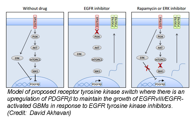
a (GBM) cells develop resistance to anti-EGFR therapy.  They demonstrate for the first time that an EGFR-dependent cancer can escape targeted therapy by developing dependence on another non-amplified, non-mutated RTK.  Specifically, they show that GBMs with EGFR mutations evade EGFR tyrosine kinase inhibitors (TKI) by transcriptionally de-repressing platelet-derived growth factor receptor β (PDGFRβ).  Cell lines, patient-derived tumor cultures, and xenotransplants showed that the persistently active EGFR mutation (EGFRvIII) suppressed PDGFRβ expression via mTORC1 and ERK-dependent mechanisms but that EGFR TKI treatment de-repressed PDGFRβ allowing the tumors to become “addicted” to a non-amplified, non-mutated RTK for continued growth and resistance to targeted treatment.

Tumor tissue from GBM patients in a phase II clinical trial for an EGFR TKI (Lapatinib) revealed a reciprocal relationship between the activation of PDGFRβ and EGFRvIII.  Tissue analysis from one patient before and after therapy revealed that Lapatinib treatment significantly reduced EGFR activation, but with a concomitant increase in PDGFRβ expression, supporting their in vitro and in vivo data that pharmacologic inhibition of EGFR results in RTK switching to PDGFRβ signaling.

We have targets and we have drugs, but RTK inhibitors have resulted in unfulfilled promises.  Acquired drug resistance has presented a significant challenge for personalized cancer therapy.  Despite being able to identify druggable RTK mutations in patients as well as second site mutations, non-genetic adaptive resistance mechanisms are able to “rewire” their circuitry through pathway crosstalk and release of inhibitory feedback loops.  To further develop kinase cancer drugs, scientists need to combine RTK inhibitors with other agents (chemotherapy, radiation, other small molecules etc.) as well as target multiple tumor-promoting signaling pathways, either with drug combinations or with a single multi-targeted compound.


Further reading:

Akhavan D, Pourzia AL, Nourian AA, Williams KJ, Nathanson D, Babic I, Villa GR, Tanaka K, Nael A, Yang H, Dang J, Vinters HV, Yong WH, Flagg M, Tamanoi F, Sasayama T, James CD, Kornblum HI, Cloughesy TF, Cavenee WK, Bensinger SJ, Mischel PS.  De-repression of PDGFRβ transcription promotes acquired resistance to EGFR tyrosine kinase inhibitors in glioblastoma patients.  Cancer Discovery. 2013 Mar 27. [Epub ahead of print]

Deric L. Wheeler, Emily F. Dunn, and Paul M. Harari.  Understanding resistance to EGFR inhibitors—impact on future treatment strategies.  Nature Reviews Clinical Oncology. 2010 September; 7(9): 493–507.

James Perry, Masahiko Okamoto, Michael Guiou, Katsuyuki Shirai, Allison Errett, and Arnab Chakravarti.  Novel Therapies in Glioblastoma.  Neurology Research International.  Volume 2012 (2012), Article ID 428565, 14 pages

Mechanical Properties of 3D Cell Culture Affect Stem Cell Phenotype

The field of regenerative medicine holds great promise as we gain greater understanding of how stem cells differentiate into the many cell types found in our bodies.  However, the clinical applications of these stem cells have been hampered by the challenges in replicating the in vitro capabilities of these cells once transplanted into the body.  This is partially due to the fact that most stem cell work is based on culturing cells in a flat two-dimensional (2D) format.

Cell culture in 2D has been routinely used in laboratories for over 40 years.  Cells are grown on flat dishes made of coated or uncoated polystyrene plastic or glass that are very stiff and arguably primitive.  These cells attach and spread on the surface to form unnatural cell attachments to other cells and to deposited proteins that are denatured on this synthetic surface.  Thus, the culturing of cells in 2D does not accurately reproduce the extracellular matrix (ECM) found in native tissue resulting in an alteration of many complex biological responses.  The native microenvironment provides mechanical signals, soluble factors, communication between neighboring cells and communication between the cell and its matrix.  This spatial and temporal organization affects normal cell fate including division, proliferation, migration, differentiation and apoptosis.

To overcome these challenges scientists have developed several three-dimensional (3D) culture methods such as cell spheroids, micro-carrier cultures, scaffolds, or tissue-engineered models.  Cell spheroids, self-assembled spherical clusters of cell colonies, are simple, reproducible and similar to physiological tissues compared to other methods involving ECM scaffolds and hydrogel systems (water-swollen polymer networks).  They are created from single culture or co-culture techniques such as hanging drop, rotating culture, non-adhesive surfaces or concave plate methods.  A similar method is the development of epithelial tissues to form polarized sheets.  However, as the size and complexity of the 3D model increases, so does the requirement for a scaffold which will ideally produce features naturally found within the ECM required for native cell function.

In this 3D cell culture environment, cells synthesize and secrete a flexible and pliable extracellular matrix in their native configuration.  Gap junctions are increased in 3D culture allowing cells to communicate with each other via exchange of ions, small molecules and electrical currents.  Surface adhesion molecules and receptors critical for cell function are also maximized.  The various effects of 3D culture versus 2D culture on differentiation, drug metabolism, expression, cell function, morphology, proliferation, viability, response to stimuli and in vivo relevance are vast and ever expanding (JUST THE FACTS: Specific effects of 3D vs. 2D cell culture).

In a recent study published April 11, 2013 online in Advanced Functional Materials, researchers at Case Western University describe how micropatterning technology influences stem cell fate decisions.  Micropatterning technology is the use of a technique to influence the network pattern of microgels aka intelligent hydrogel systems.  While this technology has recently become an important tool for spatially controlling stem cell microenvironment, very little is known about the effect of the size of the micropatterned regions, which influences hydrogel stiffness and transport properties, on stem cell behavior.  The researchers developed a 3D micropatterned hydrogel system that was either single-crosslinked or dual-crosslinked and evaluated human adipose-derived stem cell (hASC) behavior.  The cells grew into clusters in the singly-crosslinked regions where the size of the hASC clusters depended on the micropattern size (increased cell-cell interactions may have promoted cell proliferation), while hASCs encapsulated in the dual-crosslinked regions remained mostly isolated and had lower proliferation rates.  Interestingly, osteogenic (bone) and chondrogenic (cartilage) differentiation of the hASCs increased as the micropattern size increased but there was no effect on adipogenic (fat cells) differentiation.  The researchers believe that controlling local biomaterial properties may allow them to guide the formation of complex tissues.

Another study published March 24, 2013 in Nature Materials describes how mechanotransduction (how cells take information about its physical environment and t3D Traction image
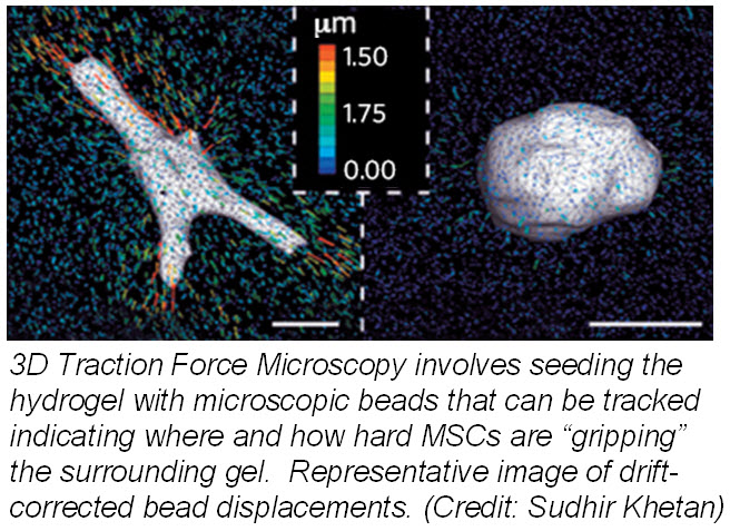
ranslate that into chemical signals) can influence stem cell fate.  Researchers from University of Pennsylvania showed that cell fate is regulated by cell-generated tension that is enabled through cell-mediated degradation of the covalently crosslinked matrix.  When cultured on “softer” 2D covalently crosslinked gels (RGD-modified methacrylated hyaluronic acid hydrogels), mesenchymal stem cells (MSCs) differentiated into adipocytes when cultured in bipotential adipogenic/ osteogenic media.  In contrast, MSCs cultured on “harder” 2D alginate gels differentiated into chondrocytes.  This phenomenon was not present in 3D hydrogels and was attributed to the inability of cells to degrade the covalent cross-linked bonds resulting in MSCs differentiating into adipocytes.  Introduction of proteolytically cleavable crosslinks and utilizing 3D traction force microscopy, revealed that MSC differentiation into bone cells was dependent on the cells to better anchor themselves into the environment and degradation signals.

These two studies provide insight into how the microenvironment can affect the fate of stem cells.  Understanding how the microenvironment influences stem cell behavior is important for tissue engineering approaches.  Cell-based assays have the potential to provide reliable data for regenerative medicine but scientists need to bridge the in vitro and in vivo gap by growing cells within a microenvironment that establishes physiological cell-cell and cell-substrate interactions that regulate proliferation and differentiation.  Hence, 3D models will provide more reliable and meaningful therapeutic results compared to 2D tests.


Further reading:

Haycock JW.  3D cell culture: a review of current approaches and techniques.  Methods Mol Biol. 2011; 695:1-15

Oju Jeon, Eben Alsberg.  Regulation of Stem Cell Fate in a Three-Dimensional Micropatterned Dual-Crosslinked Hydrogel System.  Advanced Functional Materials.  Article first published online: 11 April 2013

Wei Song, Naoki Kawazoe, and Guoping Chen.  Dependence of Spreading and Differentiation of Mesenchymal Stem Cells on Micropatterned Surface Area.  Journal of Nanomaterials.  Volume 2011 (2011), 9 pages

Sudhir Khetan, Murat Guvendiren, Wesley R. Legant, Daniel M. Cohen, Christopher S. Chen and Jason A. Burdick.  Degradation-mediated cellular traction directs stem cell fate in covalently crosslinked three-dimensional hydrogels. Nature Materials. Published online 24 March 2013


DELAYING DRUG RESISTANCE AND PROLONGING SURVIVAL IN MELANOMA

With the increasing knowledge about the role of V600E B-RAF mutation in melanoma progression, efforts have been made to target and inhibit this kinase and its downstream signaling. The ATP-competitive type I B-RAF inhibitors vemurafenib and dabrafenib (GSK2118436) exhibit remarkable anti-cancer activity in patients with V600E B-RAF mutant melanomas. Targeted inhibition of BRAF with vemurafenib causes tumor regression and extends survival in many patients with BRAF-mutant metastatic melanoma. In 2011, the Food and Drug Administration (FDA) approved vemurafenib tablets (ZELBORAF) for the treatment of patients with unresectable or metastatic melanoma with the V600EBRAF mutation. Vemurafenib is not recommended for use in patients with wild-type BRAF melanoma. Even though a very high percentage of patients respond to vemurafenib, resistance to this drug develops relatively quickly. With continued treatment, the emergence of resistance can be seen as soon as 6-8 weeks following initial documentation of response. However, a subset of patients maintains drug responsiveness beyond 18 months. Overall, the median duration of responsiveness to vemurafenib is 8 months. Some studies described involvement of certain molecular mechanisms associated with vemurafenib resistance in melanoma. Reactivation of the MAPK pathway, NRAS mutation, overexpression of platelet derived growth factor beta receptor, activation of PI3K/AKT signaling, genomic amplification of V600EBRAF are some of the mMech. of action of vemurafenib resized 600
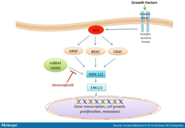
echanisms of acquired resistance to BRAF inhibitors (for detail please refer to my blog post titled ” RESISTANCE TO B-RAF INHIBITORS IN MELANOMA’’). Due to heterogeneous nature of cancer cells, it is crucial to gain a thorough understanding of the underlying drug-resistance mechanisms so that we can develop novel strategies to circumvent resistance and achieve more-prolonged responses. A recent study published in the journal Nature by Das Thakur and colleagues reported that intermittent treatment of vemurafenib prevented resistance in primary human melanoma xenografts. To study mechanisms of resistance to vemurafenib, Das Thakur et al. developed an animal model by continuously treating mice bearing a vemurafenib-naive, patient-derived BRAF-mutant melanoma with vemurafenib until drug resistance developed. Exome sequence analysis did not detect any secondary mutations in the coding sequences of BRAF, NRAS, KRAS, HRAS, and MEK1 in the resistant tumors. No alternatively spliced isoform of V600EBRAF, another known mechanism of vemurafenib resistance in melanoma was also detected in the resistant tumors. However, increased expression of V600EBRAF protein was noted in the resistant tumors and inhibition of V600EBRAF gene by RNA interference resulted in suppression of proliferation. These data suggested that the tumor cells were BRAF oncogene dependent and the observed drug resistance was due to the increased expression of V600EBRAF protein. In addition to these, another interesting observation was noted in this study when Das Thakur et al. tried to establish cell lines derived from the drug-resistant tumors. Cell lines derived from the drug-resistant tumors could not be developed without vemurafenib, where withdrawal of vemurafenib from the newly established cell lines changed cell morphology and decreased proliferation. This suggested that vemurafenib-resistant tumor cells in melanoma suffer a fitness deficit in the absence of vemurafenib. A similar type of vemurafenib dependency was also observed in SK-MEL239-C3 melanoma cells in which resistance is due to expression of a splice variant of V600EBRAF, and also in tumor cells isolated from a BRAF-mutated vemurafenib-resistant melanoma patient. Consistent with these findings, Das Thakur et al. observed tumor regression within 10 days in mice bearing vemurafenib resistant melanoma following cessation of vemurafenib treatment, although tumors eventually started re-growing. Collectively these results suggested that withdrawal of vemurafenib might create a hostile environment for drug-resistant cells and detain the onset of drug resistance. A comparison study made between continuous and intermittent vemurafenib treatment in human melanoma xenografts bearing mice further validated these observations. Drug resistance was developed in mice with 100 days receiving continuous treatment, whereas none of the mice on the intermittent treatment schedule exhibited drug resistance after 200 days of treatment. Therefore, these findings recommend that discontinuous treatment of vemurafenib may select against drug-resistant cells and prolong the responses to vemurafenib in melanoma. Future studies are needed especially in clinical trials to validate this proposal.


References:

1.         Das Thakur M, Salangsang F, Landman AS, et al. Modelling vemurafenib resistance in melanoma reveals a strategy to forestall drug resistance. Nature. 2013;494(7436):251-255.

2.         Sullivan RJ, Flaherty KT. Resistance to BRAF-targeted therapy in melanoma. Eur J Cancer. 2013;49(6):1297-1304.

Identification of a Novel Eomesodermin Expressing T cell Subset

41BB (CD137) is a costimulatory receptor transiently upregulated on T cells following activation.  41BB is activated by its ligand 41BBL (TNFSF9), a TNF receptor superfamily member expressed by activated antigen presenting cells and anti-41BB agonistic antibodies are in clinical trials for cancer immunotherapy.  In a recent article in The Journal of Experimental Medicine, Curran et. al demonstrate that 41BB activation of T cells leads to the generation of a novel subset of CD4+ and CD8+ T cells dependant on the master transcription factor Eomesodermin (Eomes).

describe the imageActivation of 41BB on T cells leads to enhanced T cell survival.  Anti-41BB-agonistic antibodies have demonstrated significant anti-tumor activity in mice by enhancing anti-tumor cytotoxic T cell responses.  Thus, there are currently several clinical trials underway exploring the efficacy of anti-41BB-agonist antibodies in several types of cancers, including melanoma, renal carcinoma, ovarian cancer, and lymphoma.  In a previous study by the same group (Curran et. al, PLoS One, 2011), an observation was made that a unique subset of T cells infiltrated B16 melanoma tumors in mice after anti-41BB-agonistic antibody treatment.  These T cells expressed the inhibitory receptor KLRG1, and elicited strong anti-tumor activity.  Thus, in the current study, the authors sought to further characterize this T cell subset in mice.

To define the phenotype and functions of tumor-associated KLRG1+ versus KLRG1T cells types, T cells were isolated from B16 tumors established in mice, following treatment with anti-41BB antibodies plus irradiated Flt3-ligand–expressing B16 cells (FVAX) or FVAX alone. The addition of FVAX further enhanced the tumor-infiltrating frequency of KLRG1+ T cells elicited by anti-41BB antibodies.  Gene expression analysis revealed that KLRG1+ CD4+ and CD8+ T cells expressed significantly higher levels of cytoxicity genes: multiple granzymes, perforin, and FasL, than KLRG1T cells.  In vitro cytotoxicity assays with B16 melanoma cell targets demonstrated enhanced killing capacity of KLRG1+ compared with KLRG1 CD4+ and CD8+ T cells.

Superior cytotoxic functions are generally associated with CD4+ TH1 and CD8+ TC1 T cell subsets, dependant on the transcription factor T-bet (TBX21).  However, analysis of expression of the known master transcription factors governing different T cell subsets, found that expression of Eomes but not T-bet was elevated in KLRG1+ T cells.  Runx3 expression was also slightly elevated in KLRG1+ versus KLRG1T cells.  Furthermore, transgenic mice lacking Eomes expression in CD4+ cells (CD4-CRE/Eomesflox/flox) did not develop tumor-infiltrating KLRG1+ T cells after anti-41BB antibody treatment, demonstrating the necessity of Eomes for development of these cells, even when Eomes expression is only absent in the CD4+ T cell compartment.  Thus, these novel subsets of KLRG1+ T cells were termed CD4+ THEO and CD8+ TCEO T cells.

Interestingly, KLRG1+ T cells play a role not only in anti-tumor immunity, but were induced and found at significant levels in spleens and livers from mice infected with Listeria Monocytogenes or LCMV.

As this is a newly described T cell subset, many questions remain.  However, most relevant is whether equivalents of these cells exist in humans, and the roles they play in human diseases.

Further Reading:

Systemic 4-1BB activation induces a novel T cell phenotype driven by high expression of Eomesodermin.  Curran MA, Geiger TL, Montalvo W, Kim M, Reiner SL, Al-Shamkhani A, Sun JC, Allison JP. J Exp Med. 2013 Apr 8;210(4):743-55.

Combination CTLA-4 blockade and 4-1BB activation enhances tumor rejection by increasing T-cell infiltration, proliferation, and cytokine production.  Curran MA, Kim M, Montalvo W, Al-Shamkhani A, Allison JP. PLoS One. 2011 Apr 29;6(4):e19499.

Immunotherapy of cancer with 4-1BB.  Vinay DS, Kwon BS. Mol Cancer Ther. 2012 May;11(5):1062-70. doi: 10.1158/1535-7163.MCT-11-0677. Epub 2012 Apr 24.

Immune regulation by 4-1BB and 4-1BBL: complexities and challenges.  Wang C, Lin GH, McPherson AJ, Watts TH. Immunol Rev. 2009 May;229(1):192-215.