Progranulin Antibodies a Common Link in Vasculitis, Lupus, and RA

Patients with autoimmune rheumatic diseases (ARD) such as rheumatoid arthritis (RA) and systemic lupus erythematosus (SLE) have a significantly increased risk of developing cardiovascular disease (CVD) and often develop CVD earlier than those without underlying autoimmunity, although it is not clear whether CVD is a general consequence of RA and SLE or only affects a subgroup of patients.  Control of autoimmune inflammation by disease-modifying anti-rheumatic drugs (DMARD), especially those that target immune factors also involved in vasculitis (e.g., T and B cells), is believed to have a protective effect.  One area of current research is focused on identifying commonalities across multiple ARD that suggest specific mechanisms of ARD-related CVD in order to develop diagnostics, preventatives, and treatments for those at greatest risk.

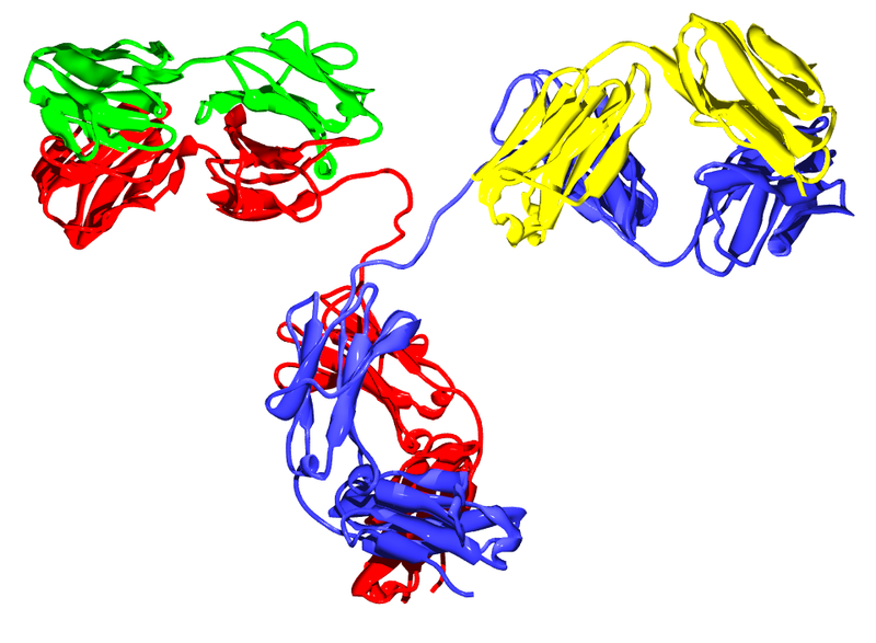
IgG2 antibody
IgG2 antibody

A recent article in the Journal of Autoimmunity suggests anti-progranulin antibodies as one potential mechanism.  Thurner and colleagues used a protein macro-array to screen serum from patients with anti-neutrophil cytoplasmic antibody (ANCA)-associated systemic vasculitides for novel autoantibodies specific to these diseases.  Of the six candidate autoantigens reactive with pooled vasculitis patient serum, progranulin was the only autoantigen appearing in every one of the vasculitides studied.  However, extended screenings showed that a positive progranulin antibody titer was not specific for vasculitides; although the prevalence was low in healthy controls (1/97 or 1%) and patients with melanoma (0/98) or sepsis (0/22), progranulin antibodies were also detectedin serum from patients with RA (16/44 or 36%) and SLE (39/91 or 43%).

Progranulin, also called proepithelin, granulin-epithelin precursor, or acrogranin, is a glycoprotein secreted by epithelial cells, neurons, and certain leukocytes.  In addition to growth factor-like activity, progranulin has immunomodulatory effects in vitro and in vivo.  Full-length progranulin decreases oxidant production by activated neutrophils, blocks TNFα-induced immune responses via binding to TNFR-1 and -2, and promotes up-regulation of IL-4, IL-5, and IL-10.  Progranulin deficiency in mice results in greater inflammation in collagen-induced arthritis (CIA) and collagen antibody-induced arthritis models of human RA; treatment of either progranulin-deficient or wild-type mice with recombinant human progranulin ameliorates CIA inflammation.

Progranulin is cleaved by several proteases into mature granulins.  Neither recombinant nor proteolytically released granulins antagonize TNFα.  Rather, granulins increase expression of pro-inflammatory cytokines IL-1β, IL-8, and TNFα.  SLPI and apolipoprotein A-I binding to progranulin protects it from cleavage by matrix metalloproteinases and other proteases.  However, during inflammation, neutrophils and macrophages release serine proteases that increase progranulin digestion.  In the context of ongoing inflammation in ARD, this may result in increased cleavage of anti-inflammatory progranulin to pro-inflammatory granulin.

Thurner et al. are the first to report the presence of neutralizing anti-progranulin antibodies in RA, SLE, and small- and medium-vessel vasculitides, which may represent a pro-inflammatory mechanism common to several autoimmune diseases.  Their findings provide additional support for exploration of the progranulin/granulin pathway as a therapeutic target and suggest the potential use of anti-progranulin antibodies as a diagnostic and/or prognostic tool in ARD.  Further studies using sera of patients with known autoimmune disease states are needed to confirm these findings and address the additional questions raised, such as –  What causes the failure of self-tolerance to progranulin and the generation of anti-progranulin antibodies, as seen in ~20-40% of the patients in this study?  Are these anti-progranulin antibodies common to all autoimmune diseases?  Could the development of progranulin-neutralizing antibodies even become a biomarker in ARD, for example as a predictor of responsiveness to DMARD therapy, or an indicator of future progression to ARD-related CVD?  We await the results of these and other studies in this area with great interest.

Further Reading:

Progranulin antibodies in autoimmune diseases.  Thurner L, Preuss KD, Fadle N, Regitz E, Klemm P, Zaks M, Kemele M, Hasenfus A, Csernok E, Gross WL, Pasquali JL, Martin T, Bohle RM, Pfreundschuh M.  J Autoimmun. 2013 May; 42:29-38.

Insights into the role of progranulin in immunity, infection, and inflammation.  Jian J, Konopka J, Liu C.  J Leukoc Biol. 2013 Feb; 93(2):199-208.

Cardiovascular disease in autoimmune rheumatic diseases.  Hollan I, Meroni PL, Ahearn JM, Cohen Tervaert JW, Curran S, Goodyear CS, Hestad KA, Kahaleh B, Riggio M, Shields K, Wasko MC.  Autoimmun Rev. 2013 Aug; 12(10):1004–1015.

CD20-Negative Circulating Plasmablasts Are Target for New B Cell Therapies in Anti-CCP Positive RA

Diagnosis of rheumatoid arthritis (RA) is based on meeting several of the criteria established by the American College of Rheumatology and the European League Against Rheumatism.  One of these criteria is the presence of anti-citrullinated protein antibodies (ACPA).  ACPA seropositivity is currently tested using a filaggrin-derived peptide (anti-cyclic citrullinated peptide [anti-CCP]) ELISA, although other ELISAs in development such as mutated citrullinated vimentin (MCV) show promising results.

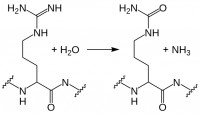
citrullination
Citrullination

While myelin basic protein, filaggrin, and several histone proteins are naturally citrullinated, other proteins such as fibrin and vimentin can become citrullinated during an inflammatory response.  Citrullination, enzymatic conversion of arginine residues into citrulline, increases protein hydrophobicity, which can change its structure.  In RA, these citrullinated proteins are recognized as “non-self” by immune cells, leading to production of ACPA.  Recent studies suggest these autoantibodies are not merely convenient diagnostic markers resulting from the autoimmune response, but instead may play a role in RA pathogenesis. Current research in this area includes identifying subtypes of RA based on ACPA positivity and specificity, and determining the roles and mechanisms of action for ACPA in RA autoimmunity.

Little is known about the B cells which produce these ACPA.  However, in a recent report in Annals of Rheumatic Disease, Kerkman and her colleagues used B cells isolated from the peripheral blood of ACPA-positive and -negative RA patients, as well as healthy individuals, to examine ACPA production in vitro and identify the ACPA-producing cell populations.

Initially, the authors stimulated peripheral B cells with B cell activating factor (BAFF) and anti-IgM F(ab′)2-fragments to induce ACPA production.  Although total IgG production was equivalent across the cultures, only B cells from ACPA-positive RA patients produced ACPA.  There was good correlation of ACPA titers obtained from in vitro culture with in vivo patient ACPA titers, underscoring the utility of this model system.  Next, the authors examined spontaneous ACPA production in unstimulated peripheral blood mononuclear cells (PBMC) from ACPA-positive RA patients.  In this case, total IgG was up to 100x lower than that seen in their studies with stimulated B cells; however, the amount of ACPA produced was equivalent.

Were ACPA in PBMC cultures generated solely by circulating plasmablasts, or were antigen presenting cells (APC) present in the PBMC population also stimulating production of ACPA by memory or even naïve B cells?  Kerkman et al. used FACS to selectively deplete ACPA-positive RA patient PBMC of plasmablast/plasma cell or naïve/memory populations, as well as to sort the CD19+ B cell subpopulations.  Naïve B cells (CD20+CD27-) did not produce any ACPA, even when stimulated with BAFF and IgM F(ab′)2.  Memory B cells (CD20+CD27+) produced ACPA upon stimulation, indicating CCP-specific memory cells are present in the circulation of ACPA-positive RA patients; however, ACPA production in CD20-depleted PBMC remained essentially unchanged, while unstimulated PBMCs depleted of plasmablasts/plasma cells produced significantly less ACPA.

acpa b cells

This study demonstrates the presence of circulating ACPA-producing plasmablasts/plasma cells in the peripheral blood of patients with ACPA-positive RA.  This is a novel and unexpected finding, since the plasmablast population is typically a transient population within PBMCs following antigen exposure, with antibody production continuing from mature plasma cells in the spleen and lymph nodes.
Circulating ACPA-producing B cells may persist in RA due to plasmablast replication and/or to memory B cell activation in response to persistent systemic citrullinated antigens.  Currently approved RA therapies which target the CD20+ B cell population, such as rituximab, would affect the memory B cell population, but not CD20- plasmablasts.  New therapies targeting circulating plasmablasts/plasma cells in addition to memory B cells could significantly limit ACPA production and subsequent immunological damage in RA, including that due to ACPA-induced TNFα production and complement activation.  Delineating circulating plasmablasts as a major source of ACPA is therefore a step forward in the quest to determine the roles and mechanisms of action for ACPA in RA pathogenesis, and underscores the possibility of developing effective new therapies by targeting specific B cell populations in RA.

Further Reading:

Circulating plasmablasts/plasma cells as a source of anti-citrullinated protein antibodies in patients with rheumatoid arthritis.  Kerkman PF, Rombouts Y, van der Voort EIH, Trouw LA, Huizinga TWJ, Toes REM, Scherer HU.  Ann Rheum Dis 2013 Jul; 72:1259–1263.

The effect of targeted rheumatoid arthritis therapies on anti-citrullinated protein autoantibody levels and B cell responses.  Modi S, Soejima M, Levesque MC.  Clin Exp Immunol 2013 Jul; 173(1):8-17.

B effector cells in rheumatoid arthritis and experimental arthritis.  Finnegan A, Ashaye S, Hamel KM.  Autoimmunity 2012 Aug; 45(5):353-63.

From bedside to bench: linking autoimmunity-associated gene variants to immune function

Autoimmunity results when the immune system, normally tasked to defend against infections and cancer, attacks the body’s own tissues. There are over 80 clinically-distinct autoimmune diseases that differ in terms of which tissues are targeted and which therapies are most effective. Rheumatoid arthritis (RA) and inflammatory bowel disease (IBD) result in the destruction of joints and the intestinal tract, respectively. These diseases respond well to agents such as adalimumab (Humira) and etanercept (Enbrel) that block the action of TNF-alpha, a cytokine that can promote inflammation. During multiple sclerosis (MS) the immune system attacks the central nervous system resulting in progressive neurologic deficits. Despite being an inflammatory disease, multiple sclerosis is actually worsened through the use of anti-TNF-alpha therapies.

Identifying the fundamental dysfunction at the root of an autoimmune disease would aid in choosing the best of available therapies or devising new ones. Advances in genome sequencing technology have allowed researchers to generate an expanding list of genetic differences present in individuals with various autoimmune diseases compared to healthy people. Although several of these disease-associated gene variants have known roles in the immune system, how they contribute to specific autoimmune processes is largely unknown. There is a need for functional characterization of these gene variants in order to determine how they alter immunity and to stratify them as therapeutic targets.

Dr. David Rawlings’ group at Seattle Children’s Hospital sought to address this challenge in a recent paper published in the May 2013 issue of the Journal of Clinical Investigation. The group, with lead author Dr. Xuezhi Dai, investigated a genetic variant of protein tyrosine phosphatase non-receptor 22 (PTPN22) which had previously been linked to several autoimmune diseases, including type 1 diabetes (T1D), RA, Graves’ Disease, and systemic lupus erythematosus (SLE). PTPN22 encodes an enzyme called LYP, a protein tyrosine phosphatase whose general function is to modulate the intensity of certain signals within cellular signaling networks. The disease-linked variant results in an amino acid switch from arginine to tryptophan at position 620 (LYP-R620W). How LYP or LYP-R620W work to modulate immune activity is incompletely understood.

transgenic mouse

To gain insight into the role of LYP-R620W in autoimmune patients, Dai et al. created a genetically engineered mouse with an analogous arginine to tryptophan switch in the mouse version of LYP (called PEP-R619W). The “knock-in” mice expressing PEP-R619W were viable but had slightly shorter life spans compared to their counterparts with normal PEP. As the engineered mice aged they manifested signs of autoimmunity, such as inflamed lung tissue and blood vessels, as well as signs of chronic kidney damage. In addition, PEP-R619W rendered the mice more susceptible to an experimental form of type 1 diabetes.  These mice also produced numerous auto-antibodies, a hallmark of certain autoimmune diseases.

The PEP-R619W knock-in mouse allowed the authors to look in close detail at the effect of this gene variant on specific immune cell populations. Dai et al. found that the knock-in mice had larger numbers of activated/memory T cells than their normal counterparts, indicating a chronically active immune system. T cells from the knock-in mice were shown to be hyper-responsive to stimulation of their antigen receptors indicating augmentation of the intracellular signals that dictate T cell activation. Similarly, the authors found that knock-in mice had larger numbers of specific B cell populations that occur in active immune states. B cells from the knock-in mice proliferated more than those from normal animals in response to stimulation and were more easily induced to secrete antibody. These findings led the authors to conclude that expression of PEP-R619W results in a lower threshold for activation in both T and B cells which contributed to the autoimmune phenotype. Interestingly, the authors discovered that expression of the disease-linked variant exclusively in B cells was sufficient to generate mice with signs of autoimmunity.

Dai et al. provide a great example of how the tools of bench science can be used to deepen the knowledge gained from analysis of patient specimens. Further determination of PEP/LYP substrate specificity and the dynamics of its phosphatase activity during lymphocyte activation could generate targets for the development of highly selective immune suppressants. In addition, the autoimmune phenotype generated with this knock-in mouse is relatively mild. It would be interesting to see how other disease-linked gene variants would cooperate with PEP-R619W to generate either a more aggressive disease or one that resembles a particular autoimmune syndrome. Finally, the ability of B-cell-specific PEP-R619W expression to stimulate autoimmunity suggests that B cells are a critical component of the autoimmune process in patients with this genetic variant. This model provides the opportunity to compare different therapeutic modalities in the PEP-R619W background (for example, B cell depletion versus anti-TNF agents). Such studies could provide the basis for predicting clinical responses to autoimmune therapies based on genotype.

References:

A disease-associated PTPN22 variant promotes systemic autoimmunity in murine models. Dai X, James RG, Habib T, Singh S, Jackson S, Khim S, Moon RT, Liggitt D, Wolf-Yadlin A, Buckner JH, Rawlings DJ. J Clin Invest. 2013 May 1;123(5):2024-36. doi: 10.1172/JCI66963. Epub 2013 Apr 24.

Highlight: How TNF knocks out Tregs!

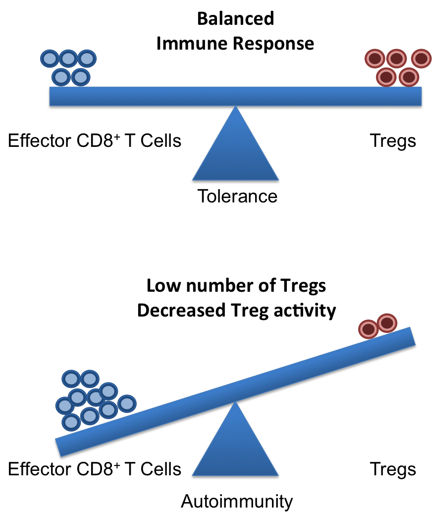
A healthy and functional immune system requires a delicate balance of pro- and contra-inflammatory signals. Whereas, it is important to induce a strong and efficient immune response against pathogens, it is similarly important to dampen these responses after the pathogen is fought off to revert the immune system to a calm steady state. If the balance is disturbed, diseases can on the one hand, become chronic/overwhelming or, on the other hand, inflammatory responses that cannot terminate can result in autoimmune responses.

Crucial elements in the regulation of excessive immune responses are regulatory T (Treg) cells. Tregs are known to inhibit the response of other immune cells. Their essential role in limiting overwhelming immune responses is demonstrated by the detrimental consequences of their loss. Mice or humans lacking Tregs develop widespread and lethal autoimmune diseases. Besides several surface markers, Tregs are best characterized by the expression of the transcription factor FoxP3. This factor is essential for Treg function and its artificial expression in other T cells can induce a regulatory potential. Therefore, the expression of FoxP3 is required for a T cell to have regulatory potential (Buckner; Josefowicz et al.). However, it was known for many years that in cases of numerous autoimmune diseases FoxP3+ Tregs could be found in high numbers at the sides of inflammation, but that they did not demonstrate any or not sufficient regulatory activity. This enigmatic observation was so far poorly understood (Buckner; Josefowicz et al.).Treg balance

In the March 2013 issue of Nature Medicine Nie and colleagues shed new light on the underlying mechanism that impairs Treg function at the sites of inflammation. Studying Treg cells from rheumatoid arthritis (RA) patients the authors demonstrated that phosphorylation of FoxP3 of the serine at position 418 (S418) is required for its regulatory action. If FoxP3 lacks this particular phosphorylation the Treg cell is not suppressive! FoxP3 S418 in Tregs is usually phosphorylated and hence Tregs are regulatory by default. However, the authors show that due to the action of the enzyme ‘protein phosphatase 1’ (PP1) FoxP3 can lose its S418 phosphorylation. Intriguingly, the presence of the cytokine TNF lead to an up-regulation of PP1 expression in the Tregs in a dose-dependent manner, and this lead to de-phosphorylation of FoxP3 S418. Treg cells expressing a mutant FoxP3 that replaced the serine at position 418 with an alanine retained their suppressive potential even in the presence of TNF, demonstrating the importance of the phosphorylation of S418. With this finding, the authors were able to link the pro-inflammatory milieu (TNF) to a specific effect inside of the Tregs (de-phosphorylation of S418) that lead to the observed loss of the regulatory function of Treg cells. Importantly, the authors were also able to demonstrate the therapeutic potential of this knowledge. They monitored RA patients that underwent treatment with blocking anti-TNF antibodies (infliximab) and found that Tregs from patient PBMCs restored S418 phosphorylation and regained regulatory potential!

This is the second case for a post-transcriptional regulation of FoxP3 that can influence Treg function. Deacetylation of FoxP3 has been linked to impaired Treg function previously (Tao et al.). Additionally, the work of Nie et al. now adds mechanistic information to previous reports on the negative effect of TNF on Tregs (Valencia et al.; Zanin-Zhorov et al.).

Given the ubiquitous role of TNF during inflammation, it is very likely that the mechanism described by Nie et al. applies to many if not all cases of ongoing inflammation where Treg function is impaired. Furthermore, their data on the effects of anti-TNF antibody treatment in RA suggest a similar therapeutic potential in other autoimmune diseases. Surely, this report will ignite further investigation in this direction and will aid the development of better treatments for patients suffering from autoimmune diseases.

References:

Bromberg, J., 2013. TNF-α trips up Treg cells in rheumatoid arthritis. Nat Med, 19(3), pp.269–270.

Buckner, J.H., 2010. Mechanisms of impaired regulation by CD4(+)CD25(+)FOXP3(+) regulatory T cells in human autoimmune diseases. Nat Rev Immunol, 10(12), pp.849–859.

Josefowicz, S.Z., Lu, L.-F. & Rudensky, A.Y., 2012. Regulatory T cells: mechanisms of differentiation and function. Annual Review of Immunology, 30, pp.531–564.

Nie, H. et al., 2013. Phosphorylation of FOXP3 controls regulatory T cell function and is inhibited by TNF-α in rheumatoid arthritis. Nat Med, 19(3), pp.322–328.

Tao, R. et al., 2007. Deacetylase inhibition promotes the generation and function of regulatory T cells. Nature Medicine, 13(11), pp.1299–1307.

Valencia, X. et al., 2006. TNF downmodulates the function of human CD4+CD25hi T-regulatory cells. Blood, 108(1), pp.253–261.

Zanin-Zhorov, A. et al., 2010. Protein kinase C-theta mediates negative feedback on regulatory T cell function. Science, 328(5976), pp.372–376.