Progranulin Antibodies a Common Link in Vasculitis, Lupus, and RA

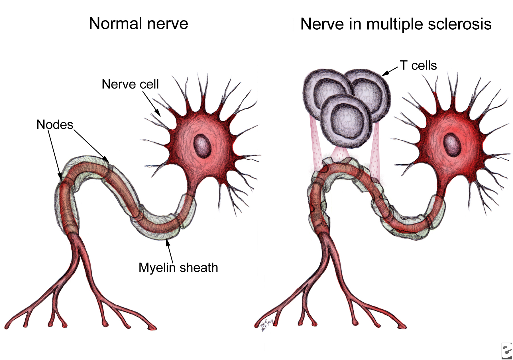
Patients with autoimmune rheumatic diseases (ARD) such as rheumatoid arthritis (RA) and systemic lupus erythematosus (SLE) have a significantly increased risk of developing cardiovascular disease (CVD) and often develop CVD earlier than those without underlying autoimmunity, although it is not clear whether CVD is a general consequence of RA and SLE or only affects a subgroup of patients.  Control of autoimmune inflammation by disease-modifying anti-rheumatic drugs (DMARD), especially those that target immune factors also involved in vasculitis (e.g., T and B cells), is believed to have a protective effect.  One area of current research is focused on identifying commonalities across multiple ARD that suggest specific mechanisms of ARD-related CVD in order to develop diagnostics, preventatives, and treatments for those at greatest risk.

IgG2 antibody
IgG2 antibody

A recent article in the Journal of Autoimmunity suggests anti-progranulin antibodies as one potential mechanism.  Thurner and colleagues used a protein macro-array to screen serum from patients with anti-neutrophil cytoplasmic antibody (ANCA)-associated systemic vasculitides for novel autoantibodies specific to these diseases.  Of the six candidate autoantigens reactive with pooled vasculitis patient serum, progranulin was the only autoantigen appearing in every one of the vasculitides studied.  However, extended screenings showed that a positive progranulin antibody titer was not specific for vasculitides; although the prevalence was low in healthy controls (1/97 or 1%) and patients with melanoma (0/98) or sepsis (0/22), progranulin antibodies were also detectedin serum from patients with RA (16/44 or 36%) and SLE (39/91 or 43%).

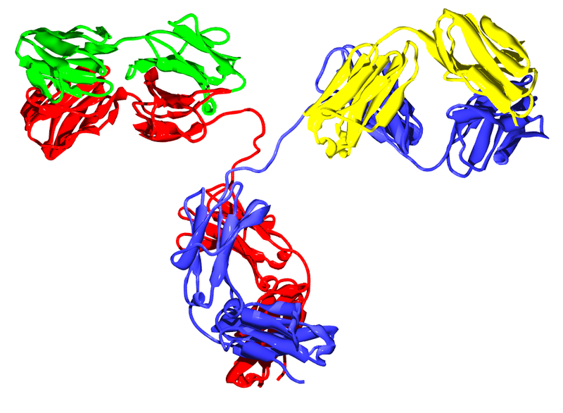
Progranulin, also called proepithelin, granulin-epithelin precursor, or acrogranin, is a glycoprotein secreted by epithelial cells, neurons, and certain leukocytes.  In addition to growth factor-like activity, progranulin has immunomodulatory effects in vitro and in vivo.  Full-length progranulin decreases oxidant production by activated neutrophils, blocks TNFα-induced immune responses via binding to TNFR-1 and -2, and promotes up-regulation of IL-4, IL-5, and IL-10.  Progranulin deficiency in mice results in greater inflammation in collagen-induced arthritis (CIA) and collagen antibody-induced arthritis models of human RA; treatment of either progranulin-deficient or wild-type mice with recombinant human progranulin ameliorates CIA inflammation.

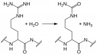
Progranulin is cleaved by several proteases into mature granulins.  Neither recombinant nor proteolytically released granulins antagonize TNFα.  Rather, granulins increase expression of pro-inflammatory cytokines IL-1β, IL-8, and TNFα.  SLPI and apolipoprotein A-I binding to progranulin protects it from cleavage by matrix metalloproteinases and other proteases.  However, during inflammation, neutrophils and macrophages release serine proteases that increase progranulin digestion.  In the context of ongoing inflammation in ARD, this may result in increased cleavage of anti-inflammatory progranulin to pro-inflammatory granulin.

Thurner et al. are the first to report the presence of neutralizing anti-progranulin antibodies in RA, SLE, and small- and medium-vessel vasculitides, which may represent a pro-inflammatory mechanism common to several autoimmune diseases.  Their findings provide additional support for exploration of the progranulin/granulin pathway as a therapeutic target and suggest the potential use of anti-progranulin antibodies as a diagnostic and/or prognostic tool in ARD.  Further studies using sera of patients with known autoimmune disease states are needed to confirm these findings and address the additional questions raised, such as –  What causes the failure of self-tolerance to progranulin and the generation of anti-progranulin antibodies, as seen in ~20-40% of the patients in this study?  Are these anti-progranulin antibodies common to all autoimmune diseases?  Could the development of progranulin-neutralizing antibodies even become a biomarker in ARD, for example as a predictor of responsiveness to DMARD therapy, or an indicator of future progression to ARD-related CVD?  We await the results of these and other studies in this area with great interest.

Further Reading:

Progranulin antibodies in autoimmune diseases.  Thurner L, Preuss KD, Fadle N, Regitz E, Klemm P, Zaks M, Kemele M, Hasenfus A, Csernok E, Gross WL, Pasquali JL, Martin T, Bohle RM, Pfreundschuh M.  J Autoimmun. 2013 May; 42:29-38.

Insights into the role of progranulin in immunity, infection, and inflammation.  Jian J, Konopka J, Liu C.  J Leukoc Biol. 2013 Feb; 93(2):199-208.

Cardiovascular disease in autoimmune rheumatic diseases.  Hollan I, Meroni PL, Ahearn JM, Cohen Tervaert JW, Curran S, Goodyear CS, Hestad KA, Kahaleh B, Riggio M, Shields K, Wasko MC.  Autoimmun Rev. 2013 Aug; 12(10):1004–1015.

CD20-Negative Circulating Plasmablasts Are Target for New B Cell Therapies in Anti-CCP Positive RA

Diagnosis of rheumatoid arthritis (RA) is based on meeting several of the criteria established by the American College of Rheumatology and the European League Against Rheumatism.  One of these criteria is the presence of anti-citrullinated protein antibodies (ACPA).  ACPA seropositivity is currently tested using a filaggrin-derived peptide (anti-cyclic citrullinated peptide [anti-CCP]) ELISA, although other ELISAs in development such as mutated citrullinated vimentin (MCV) show promising results.

citrullination
Citrullination

While myelin basic protein, filaggrin, and several histone proteins are naturally citrullinated, other proteins such as fibrin and vimentin can become citrullinated during an inflammatory response.  Citrullination, enzymatic conversion of arginine residues into citrulline, increases protein hydrophobicity, which can change its structure.  In RA, these citrullinated proteins are recognized as “non-self” by immune cells, leading to production of ACPA.  Recent studies suggest these autoantibodies are not merely convenient diagnostic markers resulting from the autoimmune response, but instead may play a role in RA pathogenesis. Current research in this area includes identifying subtypes of RA based on ACPA positivity and specificity, and determining the roles and mechanisms of action for ACPA in RA autoimmunity.

Little is known about the B cells which produce these ACPA.  However, in a recent report in Annals of Rheumatic Disease, Kerkman and her colleagues used B cells isolated from the peripheral blood of ACPA-positive and -negative RA patients, as well as healthy individuals, to examine ACPA production in vitro and identify the ACPA-producing cell populations.

Initially, the authors stimulated peripheral B cells with B cell activating factor (BAFF) and anti-IgM F(ab′)2-fragments to induce ACPA production.  Although total IgG production was equivalent across the cultures, only B cells from ACPA-positive RA patients produced ACPA.  There was good correlation of ACPA titers obtained from in vitro culture with in vivo patient ACPA titers, underscoring the utility of this model system.  Next, the authors examined spontaneous ACPA production in unstimulated peripheral blood mononuclear cells (PBMC) from ACPA-positive RA patients.  In this case, total IgG was up to 100x lower than that seen in their studies with stimulated B cells; however, the amount of ACPA produced was equivalent.

Were ACPA in PBMC cultures generated solely by circulating plasmablasts, or were antigen presenting cells (APC) present in the PBMC population also stimulating production of ACPA by memory or even naïve B cells?  Kerkman et al. used FACS to selectively deplete ACPA-positive RA patient PBMC of plasmablast/plasma cell or naïve/memory populations, as well as to sort the CD19+ B cell subpopulations.  Naïve B cells (CD20+CD27-) did not produce any ACPA, even when stimulated with BAFF and IgM F(ab′)2.  Memory B cells (CD20+CD27+) produced ACPA upon stimulation, indicating CCP-specific memory cells are present in the circulation of ACPA-positive RA patients; however, ACPA production in CD20-depleted PBMC remained essentially unchanged, while unstimulated PBMCs depleted of plasmablasts/plasma cells produced significantly less ACPA.

acpa b cells

This study demonstrates the presence of circulating ACPA-producing plasmablasts/plasma cells in the peripheral blood of patients with ACPA-positive RA.  This is a novel and unexpected finding, since the plasmablast population is typically a transient population within PBMCs following antigen exposure, with antibody production continuing from mature plasma cells in the spleen and lymph nodes.
Circulating ACPA-producing B cells may persist in RA due to plasmablast replication and/or to memory B cell activation in response to persistent systemic citrullinated antigens.  Currently approved RA therapies which target the CD20+ B cell population, such as rituximab, would affect the memory B cell population, but not CD20- plasmablasts.  New therapies targeting circulating plasmablasts/plasma cells in addition to memory B cells could significantly limit ACPA production and subsequent immunological damage in RA, including that due to ACPA-induced TNFα production and complement activation.  Delineating circulating plasmablasts as a major source of ACPA is therefore a step forward in the quest to determine the roles and mechanisms of action for ACPA in RA pathogenesis, and underscores the possibility of developing effective new therapies by targeting specific B cell populations in RA.

Further Reading:

Circulating plasmablasts/plasma cells as a source of anti-citrullinated protein antibodies in patients with rheumatoid arthritis.  Kerkman PF, Rombouts Y, van der Voort EIH, Trouw LA, Huizinga TWJ, Toes REM, Scherer HU.  Ann Rheum Dis 2013 Jul; 72:1259–1263.

The effect of targeted rheumatoid arthritis therapies on anti-citrullinated protein autoantibody levels and B cell responses.  Modi S, Soejima M, Levesque MC.  Clin Exp Immunol 2013 Jul; 173(1):8-17.

B effector cells in rheumatoid arthritis and experimental arthritis.  Finnegan A, Ashaye S, Hamel KM.  Autoimmunity 2012 Aug; 45(5):353-63.

New Research Points the Way Towards Mechanism of Action, Receptor for MS Copolymer Drugs

Multiple_sclerosis_T_cellsNeurological damage in multiple sclerosis (MS) is caused by autoreactive immune cells, which attack myelin sheathing on axons of the brain and spinal cord, leading to inflammation and myelin loss.  The MS drug Copaxone is a copolymer of glutamic acid, lysine, alanine, and tyrosine (YEAK) that is thought to impair myelin attack by inhibiting MBP self antigen presentation to autoreactive T cells; a related copolymer in which phenylalanine replaces glutamic acid (YFAK) has been developed based on MBP binding to class II MHC.  However, little is known about these drugs’ molecular targets or mechanisms of actionIn vitro studies suggest IL-10 secretion by B cells or regulatory and Th2-CD4+ T cells is involved.  Recent data demonstrate YEAK and YFAK also have MHC-independent effects on macrophages and dendritic cells, although the receptor which mediates these effects is unknown.  In a recent article in The Journal of Immunology, Koenig and colleagues isolated YEAK- and YFAK-interacting proteins from macrophage lysates and identified structures required for copolymer interaction with cells.

Following incubation with RAW264.7 macrophage lysate, biotinylated copolymers were recovered using avidin-coated beads and the associated cellular proteins were identified using mass spectroscopy.  One high-frequency hit with known surface expression and involvement in immune signaling was gp96.  Cell surface gp96 directly activates innate immune cell cytokine production, acts as a class I MHC antigen chaperone, and has been proposed as a Th2-specific co-stimulatory molecule.  CD91 has been implicated in gp96 stimulation of antigen-presenting cells (APC) and is involved in signaling and endocytosis of several ligands.  App was also identified as a surface protein that interacts with YEAK and YFAK.  A β-amyloid species precursor in Alzheimer’s disease, its function on myeloid cells is not well understood.

Macrophages secrete CCL22, a chemoattractant for regulatory and Th2 T cells, in response to YEAK or YFAK.  Koenig et al. studied this response in wild-type versus gp96-, CD91-, and App-deficient cells and found no impairment in any of the knock-out cell lines, indicating that despite their interactions with YEAK and YFAK, neither gp96, CD91, nor App are involved in cell signaling by these copolymers.

Lysine confers a positive charge on these copolymers, leading Koenig et al. to propose that cellular binding may be mediated by electrostatic interaction rather than conformation.  Indeed, increasing salt concentration reduced protein interactions with biotinylated copolymer.  Importantly, using cell lines lacking specific sulfation enzymes, the authors demonstrated that YEAK and YFAK bind to negatively charged heparan sulfate proteoglycans (HSPG).  This interaction is functional: RAW264.7 cells stimulated with YFAK in the presence of heparin sulfate, a structurally similar competitor of HSPG, did not produce CCL22.

HSPG are glycoproteins which contain one or more covalently attached heparin sulfate (HS) chains.  Membrane HSPG are known to act as co-receptors for many growth factors and could therefore play a role in the cellular effects of YEAK by activating cell signaling through an associated receptor or preventing signaling by that receptor’s natural ligand.  For example, YEAK binding to HS, a co-receptor for gp96 binding to CD91, may alter cellular uptake of gp96-peptide complexes via CD91, affecting self antigen cross-presentation and T cell activation by APC.

Alternatively, HSPG also function as receptors for constitutive as well as ligand-induced endocytosis.  YEAK interaction with HSPG may promote its fluid-phase uptake and delivery to intracellular target(s) in a manner similar to that of cationic cell-penetrating peptides.  In fact, gene ontology term enrichment analysis highlights “RNA binding” as a molecular function of YEAK- and YFAK-interacting proteins identified in this study, supporting a potential cytosolic or nuclear site of action.

Significant work remains to define the receptors and molecular mechanisms of action for these copolymers and aid rational design of future immune-modulating drugs.  The authors’ list of 222 copolymer-interacting proteins and characterization of sulfated glycosaminoglycans as the moieties responsible for functional interaction of these copolymers with innate immune cells serve as a solid foundation for further research in this area.

Further Reading:

Amino acid copolymers that alleviate experimental autoimmune encephalomyelitis in vivo interact with heparan sulfates and glycoprotein 96 in APCs.  Koenig PA, Spooner E, Kawamoto N, Strominger JL, Ploegh HL.  J Immunol.  2013 Jul 1; 191(1):XXX.  Epub ahead of print 2013 June 5.

Heparan sulphate proteoglycans fine-tune mammalian physiology.  Bishop JR, Schuksz M, Esko JD.  Nature. 2007 Apr 26; 446(7139):1030-7.

Interactions between heparan sulfate and proteins – design and functional implications.  Lindahl U, Li JP.  Int Rev Cell Mol Biol. 2009; 276:105-59.

Cell surface heparan sulfate proteoglycans influence MHC class II-restricted antigen presentation.  Léonetti M, Gadzinski A, Moine G.  J Immunol. 2010 Oct 1; 185(7):3847-56.


A theory of everything: commensal gut bacteria link environmental exposures to sex hormones in modulating autoimmunity

Whether an individual develops autoimmunity depends on how environmental and genetic factors interact to influence immune function. Genome wide association studies have revealed numerous risk alleles associated with diverse autoimmune diseases and small animal models have helped parse the effects of some of these gene variants on specific components of the immune system (see previous post). These approaches, however, largely ignore two of the more perplexing aspects of autoimmunity: (1) the strong female predominance of many autoimmune diseases and (2) the high incidence of autoimmunity in industrialized areas of the world compared to poorer, rural areas. The issue of sexual dimorphism in autoimmunity has led investigators to surmise that sex hormones are potent modulators of immune function. Indeed, altering estrogen, progesterone, or testosterone levels can influence the progression of autoimmune reactions in animals and people, although the specific mechanisms underpinning these phenomena remain controversial1. The increasing incidence of autoimmunity in the industrialized world has given rise to the “hygiene hypothesis”: exposure to specific microbes early in life is necessary for a fully-functioning immune system. These beneficial exposures are lacking in modernized, clean cities which contributes to immunological derangement and self-reactivity. Evidence supporting the hygiene hypothesis has accumulated in recent years to the point that clinical trials are in progress testing whether purposeful infestation with parasitic worms (thought to be the major microbial exposure missing from a modern upbringing) can control inflammatory bowel disease and multiple sclerosis2.

Microbiota

In a recent paper in Science Magazine, Dr. Jayne Danska’s group at the University of Toronto present an intriguing hypothesis that unifies the roles of early-life microbial exposure and sex hormone levels in driving an autoimmune response3. The group, with lead author Dr. Janet Markle, used a well-established mouse model of type-1 diabetes (T1D)—the non-obese diabetic (NOD) mouse—that is genetically predisposed to develop spontaneous, immune-mediated destruction of beta-islets at around 15 weeks of age, causing diabetes. Similar to several human autoimmune diseases, NOD mice have a 2:1 female-to-male sex bias in progressing to diabetes. In agreement with this, Markle et al. found that male mice housed under standard laboratory conditions were protected from developing diabetes compared to female mice. However, if NOD mice were born and raised in germ-free conditions (i.e. their intestines were never colonized by commensal bacteria) males developed the disease at the same rate as females. In addition, the environment in which the mice were raised impacted their levels of sex hormones: male mice raised in germ-free conditions had lower serum testosterone levels than those raised in standard conditions while germ free female mice had higher testosterone levels than those kept in standard cages. These data supported the conclusion that colonization with commensal bacteria was responsible for the protection of male NOD mice against T1D and that bacterial colonization somehow regulated the production and/or use of testosterone.

To get a sense of the extent to which gut microbiota influenced general physiology of adult male and female NOD mice, the authors used mass spectrometry to profile almost 200 unique small-molecule metabolites in serum. They found that male and female NOD mice raised in standard conditions had distinct profiles of serum metabolites. In contrast, there were few detectable differences between the metabolite profile of males and females raised in germ free conditions. This data suggested two hypotheses: (1) male and female mice had different physiologic responses to the same commensal bacteria, or (2) male and female mice had different commensal communities influencing their hormone and metabolite levels. The authors then sequenced bacterial 16S ribosomal RNA from the intestines of NOD mice at various stages of maturation (just after weaning, at puberty, and as adults). While male and female NOD mice had indistinguishable gut microbiota after weaning, sex-based differences in commensal bacteria were apparent at puberty and became even more pronounced in adulthood.

Having established that adult male and female NOD mice have distinct bacterial populations in their intestines, Markle et al. showed that transplanting the “male” gut microbiota into pre-pubescent female NOD mice altered the composition of the recipient’s commensal populations for several weeks and resulted in increased serum testosterone levels. Importantly, transplantation of male commensal bacteria protected the female recipients from T1D. Markers of T1D disease activity, such as inflammation of beta-islets and production of auto-antibodies, were reduced in recipients of male gut bacteria compared to unmanipulated females. This effect was abrogated upon treatment with the anti-androgen Flutamide, indicating that testosterone levels were a critical regulator of autoimmune pathogenesis.

Markle et al. have put forth an interesting model in which sexual maturation results in sex-specific programming of intestinal commensal bacteria. These distinct populations have differential effects on host physiology and hormonal balance which, in turn, modulate immune function. The centrality of gut microbiota to immunity raises the intriguing possibility of treating a dysfunctional immune system by altering the make-up of the intestine’s commensal communities. Such studies are already underway for intestinal disorders using “fecal transplants”, but the concept may extend to more systemic autoimmune disorders4. This approach would benefit from knowing the specific effects of different bacterial species on host physiology in order to identify the critical components of effective therapeutic microbial regimens.  Similarly, it will be interesting to see which of the changes in metabolite and hormone levels that accompany shifts in commensal populations are most impactful on immune function. The metabolite profiling approach shown in this article could be useful for identifying endogenous compounds that serve as immune modulators.

Reference:

1. Sex differences in spontaneous versus induced animal models of autoimmunity. Lee TP, Chiang BL. Autoimmun Rev. 2012 May;11(6-7):A422-9. doi: 10.1016/j.autrev.2011.11.020. Epub 2011 Dec 4.

2. Vaccine against autoimmune disease: can helminths or their products provide a therapy? Zaccone P, Cooke A. Curr Opin Immunol. 2013 Mar 2. pii: S0952-7915(13)00027-7. doi: 10.1016/j.coi.2013.02.006.

3. Sex differences in the gut microbiome drive hormone-dependent regulation of autoimmunity. Markle JG, Frank DN, Mortin-Toth S, Robertson CE, Feazel LM, Rolle-Kampczyk U, von Bergen M, McCoy KD, Macpherson AJ, Danska JS. Science. 2013 Mar 1;339(6123):1084-8. doi: 10.1126/science.1233521. Epub 2013 Jan 17.

4. Stool transplants: ready for prime time? Weissman JS, Coyle W. Curr Gastroenterol Rep. 2012 Aug;14(4):313-6. doi: 10.1007/s11894-012-0263-7.


From bedside to bench: linking autoimmunity-associated gene variants to immune function

Autoimmunity results when the immune system, normally tasked to defend against infections and cancer, attacks the body’s own tissues. There are over 80 clinically-distinct autoimmune diseases that differ in terms of which tissues are targeted and which therapies are most effective. Rheumatoid arthritis (RA) and inflammatory bowel disease (IBD) result in the destruction of joints and the intestinal tract, respectively. These diseases respond well to agents such as adalimumab (Humira) and etanercept (Enbrel) that block the action of TNF-alpha, a cytokine that can promote inflammation. During multiple sclerosis (MS) the immune system attacks the central nervous system resulting in progressive neurologic deficits. Despite being an inflammatory disease, multiple sclerosis is actually worsened through the use of anti-TNF-alpha therapies.

Identifying the fundamental dysfunction at the root of an autoimmune disease would aid in choosing the best of available therapies or devising new ones. Advances in genome sequencing technology have allowed researchers to generate an expanding list of genetic differences present in individuals with various autoimmune diseases compared to healthy people. Although several of these disease-associated gene variants have known roles in the immune system, how they contribute to specific autoimmune processes is largely unknown. There is a need for functional characterization of these gene variants in order to determine how they alter immunity and to stratify them as therapeutic targets.

Dr. David Rawlings’ group at Seattle Children’s Hospital sought to address this challenge in a recent paper published in the May 2013 issue of the Journal of Clinical Investigation. The group, with lead author Dr. Xuezhi Dai, investigated a genetic variant of protein tyrosine phosphatase non-receptor 22 (PTPN22) which had previously been linked to several autoimmune diseases, including type 1 diabetes (T1D), RA, Graves’ Disease, and systemic lupus erythematosus (SLE). PTPN22 encodes an enzyme called LYP, a protein tyrosine phosphatase whose general function is to modulate the intensity of certain signals within cellular signaling networks. The disease-linked variant results in an amino acid switch from arginine to tryptophan at position 620 (LYP-R620W). How LYP or LYP-R620W work to modulate immune activity is incompletely understood.

transgenic mouse

To gain insight into the role of LYP-R620W in autoimmune patients, Dai et al. created a genetically engineered mouse with an analogous arginine to tryptophan switch in the mouse version of LYP (called PEP-R619W). The “knock-in” mice expressing PEP-R619W were viable but had slightly shorter life spans compared to their counterparts with normal PEP. As the engineered mice aged they manifested signs of autoimmunity, such as inflamed lung tissue and blood vessels, as well as signs of chronic kidney damage. In addition, PEP-R619W rendered the mice more susceptible to an experimental form of type 1 diabetes.  These mice also produced numerous auto-antibodies, a hallmark of certain autoimmune diseases.

The PEP-R619W knock-in mouse allowed the authors to look in close detail at the effect of this gene variant on specific immune cell populations. Dai et al. found that the knock-in mice had larger numbers of activated/memory T cells than their normal counterparts, indicating a chronically active immune system. T cells from the knock-in mice were shown to be hyper-responsive to stimulation of their antigen receptors indicating augmentation of the intracellular signals that dictate T cell activation. Similarly, the authors found that knock-in mice had larger numbers of specific B cell populations that occur in active immune states. B cells from the knock-in mice proliferated more than those from normal animals in response to stimulation and were more easily induced to secrete antibody. These findings led the authors to conclude that expression of PEP-R619W results in a lower threshold for activation in both T and B cells which contributed to the autoimmune phenotype. Interestingly, the authors discovered that expression of the disease-linked variant exclusively in B cells was sufficient to generate mice with signs of autoimmunity.

Dai et al. provide a great example of how the tools of bench science can be used to deepen the knowledge gained from analysis of patient specimens. Further determination of PEP/LYP substrate specificity and the dynamics of its phosphatase activity during lymphocyte activation could generate targets for the development of highly selective immune suppressants. In addition, the autoimmune phenotype generated with this knock-in mouse is relatively mild. It would be interesting to see how other disease-linked gene variants would cooperate with PEP-R619W to generate either a more aggressive disease or one that resembles a particular autoimmune syndrome. Finally, the ability of B-cell-specific PEP-R619W expression to stimulate autoimmunity suggests that B cells are a critical component of the autoimmune process in patients with this genetic variant. This model provides the opportunity to compare different therapeutic modalities in the PEP-R619W background (for example, B cell depletion versus anti-TNF agents). Such studies could provide the basis for predicting clinical responses to autoimmune therapies based on genotype.

References:

A disease-associated PTPN22 variant promotes systemic autoimmunity in murine models. Dai X, James RG, Habib T, Singh S, Jackson S, Khim S, Moon RT, Liggitt D, Wolf-Yadlin A, Buckner JH, Rawlings DJ. J Clin Invest. 2013 May 1;123(5):2024-36. doi: 10.1172/JCI66963. Epub 2013 Apr 24.