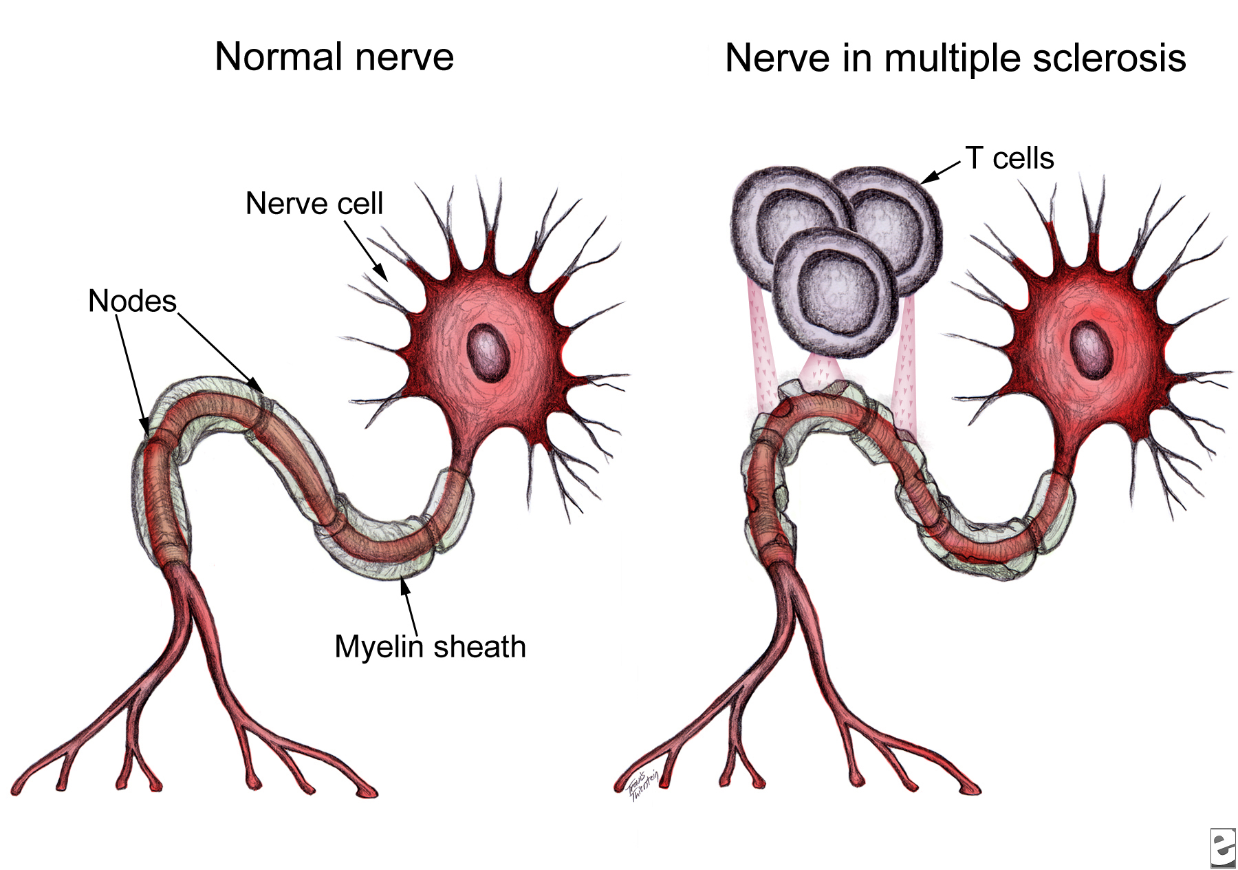
 Neurological damage in multiple sclerosis (MS) is caused by autoreactive immune cells, which attack myelin sheathing on axons of the brain and spinal cord, leading to inflammation and myelin loss. The MS drug Copaxone is a copolymer of glutamic acid, lysine, alanine, and tyrosine (YEAK) that is thought to impair myelin attack by inhibiting MBP self antigen presentation to autoreactive T cells; a related copolymer in which phenylalanine replaces glutamic acid (YFAK) has been developed based on MBP binding to class II MHC. However, little is known about these drugs’ molecular targets or mechanisms of action. In vitro studies suggest IL-10 secretion by B cells or regulatory and Th2-CD4+ T cells is involved. Recent data demonstrate YEAK and YFAK also have MHC-independent effects on macrophages and dendritic cells, although the receptor which mediates these effects is unknown. In a recent article in The Journal of Immunology, Koenig and colleagues isolated YEAK- and YFAK-interacting proteins from macrophage lysates and identified structures required for copolymer interaction with cells.
Following incubation with RAW264.7 macrophage lysate, biotinylated copolymers were recovered using avidin-coated beads and the associated cellular proteins were identified using mass spectroscopy. One high-frequency hit with known surface expression and involvement in immune signaling was gp96. Cell surface gp96 directly activates innate immune cell cytokine production, acts as a class I MHC antigen chaperone, and has been proposed as a Th2-specific co-stimulatory molecule. CD91 has been implicated in gp96 stimulation of antigen-presenting cells (APC) and is involved in signaling and endocytosis of several ligands. App was also identified as a surface protein that interacts with YEAK and YFAK. A β-amyloid species precursor in Alzheimer’s disease, its function on myeloid cells is not well understood.
Macrophages secrete CCL22, a chemoattractant for regulatory and Th2 T cells, in response to YEAK or YFAK. Koenig et al. studied this response in wild-type versus gp96-, CD91-, and App-deficient cells and found no impairment in any of the knock-out cell lines, indicating that despite their interactions with YEAK and YFAK, neither gp96, CD91, nor App are involved in cell signaling by these copolymers.
Lysine confers a positive charge on these copolymers, leading Koenig et al. to propose that cellular binding may be mediated by electrostatic interaction rather than conformation. Indeed, increasing salt concentration reduced protein interactions with biotinylated copolymer. Importantly, using cell lines lacking specific sulfation enzymes, the authors demonstrated that YEAK and YFAK bind to negatively charged heparan sulfate proteoglycans (HSPG). This interaction is functional: RAW264.7 cells stimulated with YFAK in the presence of heparin sulfate, a structurally similar competitor of HSPG, did not produce CCL22.
HSPG are glycoproteins which contain one or more covalently attached heparin sulfate (HS) chains. Membrane HSPG are known to act as co-receptors for many growth factors and could therefore play a role in the cellular effects of YEAK by activating cell signaling through an associated receptor or preventing signaling by that receptor’s natural ligand. For example, YEAK binding to HS, a co-receptor for gp96 binding to CD91, may alter cellular uptake of gp96-peptide complexes via CD91, affecting self antigen cross-presentation and T cell activation by APC.
Alternatively, HSPG also function as receptors for constitutive as well as ligand-induced endocytosis. YEAK interaction with HSPG may promote its fluid-phase uptake and delivery to intracellular target(s) in a manner similar to that of cationic cell-penetrating peptides. In fact, gene ontology term enrichment analysis highlights “RNA binding” as a molecular function of YEAK- and YFAK-interacting proteins identified in this study, supporting a potential cytosolic or nuclear site of action.
Significant work remains to define the receptors and molecular mechanisms of action for these copolymers and aid rational design of future immune-modulating drugs. The authors’ list of 222 copolymer-interacting proteins and characterization of sulfated glycosaminoglycans as the moieties responsible for functional interaction of these copolymers with innate immune cells serve as a solid foundation for further research in this area.
Further Reading:
Amino acid copolymers that alleviate experimental autoimmune encephalomyelitis in vivo interact with heparan sulfates and glycoprotein 96 in APCs. Koenig PA, Spooner E, Kawamoto N, Strominger JL, Ploegh HL. J Immunol. 2013 Jul 1; 191(1):XXX. Epub ahead of print 2013 June 5.
Heparan sulphate proteoglycans fine-tune mammalian physiology. Bishop JR, Schuksz M, Esko JD. Nature. 2007 Apr 26; 446(7139):1030-7.
Interactions between heparan sulfate and proteins – design and functional implications. Lindahl U, Li JP. Int Rev Cell Mol Biol. 2009; 276:105-59.
Cell surface heparan sulfate proteoglycans influence MHC class II-restricted antigen presentation. Léonetti M, Gadzinski A, Moine G. J Immunol. 2010 Oct 1; 185(7):3847-56.


