Tumor Immunotherapies Combine Big for Synergy in Phase I Trials

There are currently two immune cell targeting cancer_immunotherapyimmunotherapeutic agents that have received FDA approval for treatment of various malignancies.  The first approved in 2010 was the autologous dendritic cell vaccine Provenge (Sipuleucel-T) by Dendreon Corporation, for hormone refractory metastatic prostate cancer.  A second immunotherapeutic gaining FDA approval in 2011 for late stage melanoma, was an antibody called Ipilimumab, which inhibits CTLA-4, a major negative regulator of T cell activation.  Antagonists to PD-1 such as Nivolumab and/or PD-L1, another receptor/ligand pair of T cell negative regulators, are expected to join this crowd by 2015.  However, early results from combinatorial immunotherapeutics trials have demonstrated that significant synergy may be achieved by combining several immunotherapeutic modalities. A report in the June edition of The New England Journal of Medicine by Wolchok et al., demonstrates impressive synergistic results with combination therapy of Ipilimumab and Nivolumab in achieving deep and durable tumor regression in patients with advanced metastatic melanoma.

CTLA-4 and PD-1 are negative regulatory immune checkpoint inhibitors expressed on activated T cells and share homology with the TCR co-stimulatory receptor CD28.  CTLA-4 strongly competes with CD28 for binding to CD80 (B7-1) and CD86 (B7-2) on antigen presenting cells thereby limiting CD28-activation signals, and furthermore recruits inhibitory molecules into the TCR signaling synapse.  PD-1 interacts with ligands homologous with the B7 family, PD-L1 (B7H1) and PD-L2 (B7-DC), which are upregulated on tumor and stromal cells and activated antigen presenting cells.  PD-1 interaction with its ligands leads to recruitment of SHP1 and SHP2 phosphatases to the immune synapse, resulting in inhibition of TCR-mediated signaling.  PD-1 and CTLA-4 are considered non-redundant in their functions, and thus blocking both of these has been proposed to have a synergistic effect on T cell function in cancer, which was previously demonstrated in murine tumor models.

In previous clinical trials, Bristol-Myers Squibb’s Ipilimumab (MDX-010, Yervoy, IgG1) with or without a gp100 peptide vaccine extended overall survival of previously treated metastatic melanoma patients by almost 4 months versus gp100 peptide alone.  In a second study, Ipilimumab plus darcarbazine versus darcarbazine alone was tested in previously untreated metastatic melanoma patients.  The addition of Ipilimumab to the darcarbazine regimen extended overall survival by approximately two months, and lent to significantly higher survival rates at 1, 2, and 3 years later.  Bristol-Myers Squibb’s PD-1 blocking antibody (BMS-936558, IgG4) showed significant clinical efficiency in metastatic or advanced non–small-cell lung cancer, melanoma, and renal-cell cancer.

In this dose-escalation phase I trial reported on by Wolchok et al., 53 patients with advanced melanoma were concurrently treated with Ipilimumab and Nivolumab, and 33 patients received sequenced treatment.  Although the primary goal of a phase I trial is to evaluate safety, the clinical responses of the concurrently treated patients were exciting enough to garner much attention.  In the concurrent regimen, 40% of patients had an objective response (modified WHO criteria), and 16 patients had a reduction in their tumor burden by 80% or more at 12 weeks, 5 being complete responses. Not only were responses faster and more pronounced than the previous clinical trials evaluating these inhibitors alone, but in the responding patients, the reduction in tumor burden was quite durable over the course of the study.  Thus, results from phase III clinical trials comparing the combination to the inhibitors alone will be eagerly awaited.  As an interesting note, tumor expression of PD-L1 has been proposed to be an indication of efficacy for Nivolumab.  However, even in patients with PD-L1-negative tumors, responses to this combination regimen were observed.

Despite the strong promise of these inhibitors in combination, adverse events were notably higher than the inhibitors had exhibited alone in previous trials.  Although no treatment-related deaths were reported, 72% of patients exhibited grade 3 or 4 adverse events, 53% of patients exhibited treatment-related grade 3 or 4 adverse events, and 21% of patients discontinued therapy due to treatment-related adverse events.  Adverse events were however manageable with either immunosuppressant or hormone-replacement therapies.

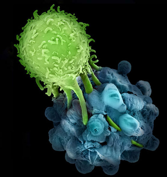
Many different cancer immunotherapeutics are now being tested in clinical trials, including a number of therapies combining immunotherapeutic modalities in the hopes to achieve synergy.  The results from the current trial indicate that anti-tumor T cells are not only present in tumor-bearing patients, but when uninhibited, can lend significantly to tumor-killing.  This is truly an exciting time for cancer immunology.

Further Reading:

Nivolumab plus Ipilimumab in Advanced Melanoma.  Wolchok JD, Kluger H, Callahan MK, Postow MA, Rizvi NA, Lesokhin AM, Segal NH, Ariyan CE, Gordon RA, Reed K, Burke MM, Caldwell A, Kronenberg SA, Agunwamba BU, Zhang X, Lowy I, Inzunza HD, Feely W, Horak CE, Hong Q, Korman AJ, Wigginton JM, Gupta A, Sznol M. N Engl J Med. 2013 Jun 2.

Safety, Activity, and Immune Correlates of Anti–PD-1 Antibody in Cancer. Topalian, S.L. et al. N. Engl. J. Med. 366, 2443–2454 (2012).

Improved Survival with Ipilimumab in Patients with Metastatic Melanoma.  Hodi, F.S. et al. N. Engl. J. Med. 363, 711–723 (2010).

Ipilimumab plus dacarbazine for previously untreated metastatic melanoma.  Robert C, Thomas L, Bondarenko I, O’Day S, M D JW, Garbe C, Lebbe C, Baurain JF, Testori A, Grob JJ, Davidson N, Richards J, Maio M, Hauschild A, Miller WH Jr, Gascon P, Lotem M, Harmankaya K, Ibrahim R, Francis S, Chen TT, Humphrey R, Hoos A, Wolchok JD. N Engl J Med. 2011 Jun 30;364(26):2517-26.

Ipilimumab: an anti-CTLA-4 antibody for metastatic melanoma.  Lipson EJ, Drake CG. Clin Cancer Res. 2011 Nov 15;17(22):6958-62.

A unique set of cell surface markers for induced T regulatory cells

Transplantation of antigen-specific T cells during cancer immunotherapy has generated many notable results in the fight against diverse cancers. In these scenarios, the transplanted T cell populations act as a sort of highly mobile army that can track and kill tumor cells wherever they hide. In contrast, the goal of therapy during autoimmunity is to suppress immune activity, not increase its potency. Would transplantation of a “peacekeeper” immune cell-type be able to specifically quell autoimmune reactions? T regulatory cells (Tregs) are attractive candidates for the peacekeeper role based on their ability to dominantly suppress auto-reactive cell populations. As opposed to studies of cancer immunotherapy, clinical trials for Treg adoptive transplant are hampered by the lack of specific cell surface markers for these populations that enable their purification1. The defining characteristics of Treg populations (eg. FoxP3, IL-10, TGF-beta) are intracellular proteins whose analysis and quantification requires permeabilization and, hence, destruction of the cells. Positive selection for markers such as CD4 and CD25 which are expressed on the surface of certain types of Tregs also enriches for effector T cell populations whose functions upon transplantation may serve to further stimulate immune activity in an autoreactive host.Adoptive t cell transfer

In this month’s issue of Nature Medicine, Gagliani et al. sought to address the need for Treg-specific cell surface markers2. The authors focused on a particular type of inducible Treg called Type 1 regulatory T cells (Tr1 cells). These are a highly suppressive population of CD4+ T cells that are thought to control immune reactions both through IL-10 secretion and direct, Granzyme B-mediated destruction of myeloid antigen presenting cells. Galiani et al. were able to isolate Tr1 clones from the peripheral blood of healthy donors using a limiting-dilution assay: CD4+ T cells were plated in wells at a density of 1 cell/well, grown in conditions known to be suitable for Tr1 development, and then assessed for high levels of IL-10 secretion. The isolated Tr1 clones were stimulated in vitro and their gene expression profiles were measured at different time points and compared to that of naïve CD4+ T cells (Th0 cells). Under these conditions, the authors found that Tr1 cells uniquely expressed genes for two cell surface markers, CD49b and LAG-3. Used independently, these markers would enrich for multiple T cell types. But when used in combination, CD49b and LAG-3 allowed the investigators to isolate Tr1 cells from human peripheral blood which expressed high-levels of IL-10 and were able to suppress T cell proliferation in vitro. The authors went on to show that this cell-surface marker combination could also be used to isolate Tr1 cells from well-defined mouse models of Treg function. Finally, authors showed that CD49b and LAG-3 could effectively enrich for Tr1 cells from a highly-expanded, in vitro-polarized bulk population. This raises the possibility of generating large numbers of highly pure IL-10 secreting Tr1 cells for adoptive transplantation during autoimmunity.

Gagliani et al. have effectively used gene profiling of a target cell type to identify cell-surface markers for a previously difficult-to-analyze population. These new markers should facilitate further clinical study of adoptive transplant of Treg populations for autoimmunity. Now that it is possible to identify Tr1 cells from blood, it will be interesting to see how numbers of these cells correlate to different disease states and how they change in response to immune modulatory treatments. Furthermore, coupling the polarization and enrichment of Tr1 cells to tetramer-based identification of antigen-specific T cells may allow for highly-selective targeting of autoimmune reactions.

References:

1. Human T regulatory cell therapy: take a billion or so and call me in the morning. Riley JL, June CH, Blazar BR. Immunity. 2009 May;30(5):656-65. doi: 10.1016/j.immuni.2009.04.006.

2. Coexpression of CD49b and LAG-3 identifies human and mouse T regulatory type 1 cells. Gagliani N, Magnani CF, Huber S, Gianolini ME, Pala M, Licona-Limon P, Guo B, Herbert DR, Bulfone A, Trentini F, Di Serio C, Bacchetta R, Andreani M, Brockmann L, Gregori S, Flavell RA, Roncarolo MG. Nat Med. 2013 Jun;19(6):739-46. doi: 10.1038/nm.3179. Epub 2013 Apr 28.

A theory of everything: commensal gut bacteria link environmental exposures to sex hormones in modulating autoimmunity

Whether an individual develops autoimmunity depends on how environmental and genetic factors interact to influence immune function. Genome wide association studies have revealed numerous risk alleles associated with diverse autoimmune diseases and small animal models have helped parse the effects of some of these gene variants on specific components of the immune system (see previous post). These approaches, however, largely ignore two of the more perplexing aspects of autoimmunity: (1) the strong female predominance of many autoimmune diseases and (2) the high incidence of autoimmunity in industrialized areas of the world compared to poorer, rural areas. The issue of sexual dimorphism in autoimmunity has led investigators to surmise that sex hormones are potent modulators of immune function. Indeed, altering estrogen, progesterone, or testosterone levels can influence the progression of autoimmune reactions in animals and people, although the specific mechanisms underpinning these phenomena remain controversial1. The increasing incidence of autoimmunity in the industrialized world has given rise to the “hygiene hypothesis”: exposure to specific microbes early in life is necessary for a fully-functioning immune system. These beneficial exposures are lacking in modernized, clean cities which contributes to immunological derangement and self-reactivity. Evidence supporting the hygiene hypothesis has accumulated in recent years to the point that clinical trials are in progress testing whether purposeful infestation with parasitic worms (thought to be the major microbial exposure missing from a modern upbringing) can control inflammatory bowel disease and multiple sclerosis2.

Microbiota

In a recent paper in Science Magazine, Dr. Jayne Danska’s group at the University of Toronto present an intriguing hypothesis that unifies the roles of early-life microbial exposure and sex hormone levels in driving an autoimmune response3. The group, with lead author Dr. Janet Markle, used a well-established mouse model of type-1 diabetes (T1D)—the non-obese diabetic (NOD) mouse—that is genetically predisposed to develop spontaneous, immune-mediated destruction of beta-islets at around 15 weeks of age, causing diabetes. Similar to several human autoimmune diseases, NOD mice have a 2:1 female-to-male sex bias in progressing to diabetes. In agreement with this, Markle et al. found that male mice housed under standard laboratory conditions were protected from developing diabetes compared to female mice. However, if NOD mice were born and raised in germ-free conditions (i.e. their intestines were never colonized by commensal bacteria) males developed the disease at the same rate as females. In addition, the environment in which the mice were raised impacted their levels of sex hormones: male mice raised in germ-free conditions had lower serum testosterone levels than those raised in standard conditions while germ free female mice had higher testosterone levels than those kept in standard cages. These data supported the conclusion that colonization with commensal bacteria was responsible for the protection of male NOD mice against T1D and that bacterial colonization somehow regulated the production and/or use of testosterone.

To get a sense of the extent to which gut microbiota influenced general physiology of adult male and female NOD mice, the authors used mass spectrometry to profile almost 200 unique small-molecule metabolites in serum. They found that male and female NOD mice raised in standard conditions had distinct profiles of serum metabolites. In contrast, there were few detectable differences between the metabolite profile of males and females raised in germ free conditions. This data suggested two hypotheses: (1) male and female mice had different physiologic responses to the same commensal bacteria, or (2) male and female mice had different commensal communities influencing their hormone and metabolite levels. The authors then sequenced bacterial 16S ribosomal RNA from the intestines of NOD mice at various stages of maturation (just after weaning, at puberty, and as adults). While male and female NOD mice had indistinguishable gut microbiota after weaning, sex-based differences in commensal bacteria were apparent at puberty and became even more pronounced in adulthood.

Having established that adult male and female NOD mice have distinct bacterial populations in their intestines, Markle et al. showed that transplanting the “male” gut microbiota into pre-pubescent female NOD mice altered the composition of the recipient’s commensal populations for several weeks and resulted in increased serum testosterone levels. Importantly, transplantation of male commensal bacteria protected the female recipients from T1D. Markers of T1D disease activity, such as inflammation of beta-islets and production of auto-antibodies, were reduced in recipients of male gut bacteria compared to unmanipulated females. This effect was abrogated upon treatment with the anti-androgen Flutamide, indicating that testosterone levels were a critical regulator of autoimmune pathogenesis.

Markle et al. have put forth an interesting model in which sexual maturation results in sex-specific programming of intestinal commensal bacteria. These distinct populations have differential effects on host physiology and hormonal balance which, in turn, modulate immune function. The centrality of gut microbiota to immunity raises the intriguing possibility of treating a dysfunctional immune system by altering the make-up of the intestine’s commensal communities. Such studies are already underway for intestinal disorders using “fecal transplants”, but the concept may extend to more systemic autoimmune disorders4. This approach would benefit from knowing the specific effects of different bacterial species on host physiology in order to identify the critical components of effective therapeutic microbial regimens.  Similarly, it will be interesting to see which of the changes in metabolite and hormone levels that accompany shifts in commensal populations are most impactful on immune function. The metabolite profiling approach shown in this article could be useful for identifying endogenous compounds that serve as immune modulators.

Reference:

1. Sex differences in spontaneous versus induced animal models of autoimmunity. Lee TP, Chiang BL. Autoimmun Rev. 2012 May;11(6-7):A422-9. doi: 10.1016/j.autrev.2011.11.020. Epub 2011 Dec 4.

2. Vaccine against autoimmune disease: can helminths or their products provide a therapy? Zaccone P, Cooke A. Curr Opin Immunol. 2013 Mar 2. pii: S0952-7915(13)00027-7. doi: 10.1016/j.coi.2013.02.006.

3. Sex differences in the gut microbiome drive hormone-dependent regulation of autoimmunity. Markle JG, Frank DN, Mortin-Toth S, Robertson CE, Feazel LM, Rolle-Kampczyk U, von Bergen M, McCoy KD, Macpherson AJ, Danska JS. Science. 2013 Mar 1;339(6123):1084-8. doi: 10.1126/science.1233521. Epub 2013 Jan 17.

4. Stool transplants: ready for prime time? Weissman JS, Coyle W. Curr Gastroenterol Rep. 2012 Aug;14(4):313-6. doi: 10.1007/s11894-012-0263-7.


From bedside to bench: linking autoimmunity-associated gene variants to immune function

Autoimmunity results when the immune system, normally tasked to defend against infections and cancer, attacks the body’s own tissues. There are over 80 clinically-distinct autoimmune diseases that differ in terms of which tissues are targeted and which therapies are most effective. Rheumatoid arthritis (RA) and inflammatory bowel disease (IBD) result in the destruction of joints and the intestinal tract, respectively. These diseases respond well to agents such as adalimumab (Humira) and etanercept (Enbrel) that block the action of TNF-alpha, a cytokine that can promote inflammation. During multiple sclerosis (MS) the immune system attacks the central nervous system resulting in progressive neurologic deficits. Despite being an inflammatory disease, multiple sclerosis is actually worsened through the use of anti-TNF-alpha therapies.

Identifying the fundamental dysfunction at the root of an autoimmune disease would aid in choosing the best of available therapies or devising new ones. Advances in genome sequencing technology have allowed researchers to generate an expanding list of genetic differences present in individuals with various autoimmune diseases compared to healthy people. Although several of these disease-associated gene variants have known roles in the immune system, how they contribute to specific autoimmune processes is largely unknown. There is a need for functional characterization of these gene variants in order to determine how they alter immunity and to stratify them as therapeutic targets.

Dr. David Rawlings’ group at Seattle Children’s Hospital sought to address this challenge in a recent paper published in the May 2013 issue of the Journal of Clinical Investigation. The group, with lead author Dr. Xuezhi Dai, investigated a genetic variant of protein tyrosine phosphatase non-receptor 22 (PTPN22) which had previously been linked to several autoimmune diseases, including type 1 diabetes (T1D), RA, Graves’ Disease, and systemic lupus erythematosus (SLE). PTPN22 encodes an enzyme called LYP, a protein tyrosine phosphatase whose general function is to modulate the intensity of certain signals within cellular signaling networks. The disease-linked variant results in an amino acid switch from arginine to tryptophan at position 620 (LYP-R620W). How LYP or LYP-R620W work to modulate immune activity is incompletely understood.

transgenic mouse

To gain insight into the role of LYP-R620W in autoimmune patients, Dai et al. created a genetically engineered mouse with an analogous arginine to tryptophan switch in the mouse version of LYP (called PEP-R619W). The “knock-in” mice expressing PEP-R619W were viable but had slightly shorter life spans compared to their counterparts with normal PEP. As the engineered mice aged they manifested signs of autoimmunity, such as inflamed lung tissue and blood vessels, as well as signs of chronic kidney damage. In addition, PEP-R619W rendered the mice more susceptible to an experimental form of type 1 diabetes.  These mice also produced numerous auto-antibodies, a hallmark of certain autoimmune diseases.

The PEP-R619W knock-in mouse allowed the authors to look in close detail at the effect of this gene variant on specific immune cell populations. Dai et al. found that the knock-in mice had larger numbers of activated/memory T cells than their normal counterparts, indicating a chronically active immune system. T cells from the knock-in mice were shown to be hyper-responsive to stimulation of their antigen receptors indicating augmentation of the intracellular signals that dictate T cell activation. Similarly, the authors found that knock-in mice had larger numbers of specific B cell populations that occur in active immune states. B cells from the knock-in mice proliferated more than those from normal animals in response to stimulation and were more easily induced to secrete antibody. These findings led the authors to conclude that expression of PEP-R619W results in a lower threshold for activation in both T and B cells which contributed to the autoimmune phenotype. Interestingly, the authors discovered that expression of the disease-linked variant exclusively in B cells was sufficient to generate mice with signs of autoimmunity.

Dai et al. provide a great example of how the tools of bench science can be used to deepen the knowledge gained from analysis of patient specimens. Further determination of PEP/LYP substrate specificity and the dynamics of its phosphatase activity during lymphocyte activation could generate targets for the development of highly selective immune suppressants. In addition, the autoimmune phenotype generated with this knock-in mouse is relatively mild. It would be interesting to see how other disease-linked gene variants would cooperate with PEP-R619W to generate either a more aggressive disease or one that resembles a particular autoimmune syndrome. Finally, the ability of B-cell-specific PEP-R619W expression to stimulate autoimmunity suggests that B cells are a critical component of the autoimmune process in patients with this genetic variant. This model provides the opportunity to compare different therapeutic modalities in the PEP-R619W background (for example, B cell depletion versus anti-TNF agents). Such studies could provide the basis for predicting clinical responses to autoimmune therapies based on genotype.

References:

A disease-associated PTPN22 variant promotes systemic autoimmunity in murine models. Dai X, James RG, Habib T, Singh S, Jackson S, Khim S, Moon RT, Liggitt D, Wolf-Yadlin A, Buckner JH, Rawlings DJ. J Clin Invest. 2013 May 1;123(5):2024-36. doi: 10.1172/JCI66963. Epub 2013 Apr 24.