Highlight: A silver bullet against bacteria?

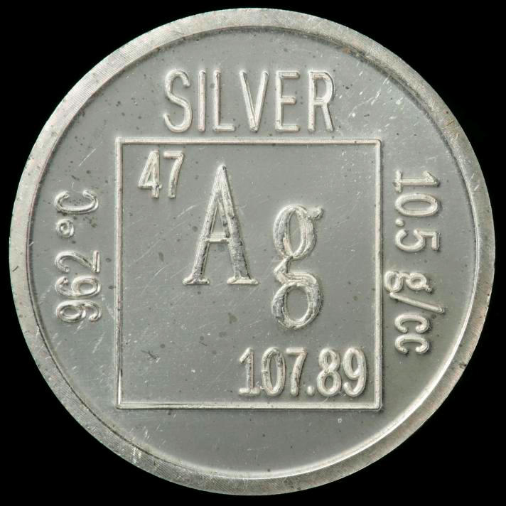
The development of multi-resistant bacterial strains is a major medical problem, especially in hospitals. New antibiotics are constantly under development, but the success rate seems to have slowed down in recent years. However, two recent publications suggest that instead of finding novel antibiotics, an alternative strategy could be to increase the sensitivity of the bacteria to the antibiotics already in use.s12sIn the first report [1], the authors demonstrated that silver ions interfered with several metabolic pathways and increased the permeability of the cell membrane, both made the bacteria much more susceptible to antibiotics. The use of silver to combat bacteria is not unprecedented and actually has been used at least since Grecian times to treat wounds and to preserve water. However, with the advent of potent antibiotics in the 1940s their use fell out of favor.

The authors uncovered two independent mechanisms of how silver is beating up bacteria. They studied E. coli a Gram-negative bacteria that is especially difficult to treat with many drugs due to its thick cell wall.

First, when exposed to silver ions the bacteria produced more reactive oxygen species (ROS). ROS are chemically highly reactive molecules that can bind unspecifically to closeby proteins and DNA and thereby irreversibly alter or damage those structures. Small amounts of ROS are constantly produced during several chemical reactions of the normal metabolism, but cell stress in any of its forms can greatly increase their production. The widespread damage that ROS can inflict weakens the cell and if the damage is too severe it can ultimately lead to cell death.

The second mechanism of silver is its ability to affect two metabolic pathways of the bacterial cells. On the one hand, the ability of the bacteria to maintain their iron level was disturbed. On the other hand, the formation of disulfide bonds, which are crucial for the structural integrity and function of many proteins, was affected in the presence of the silver ions. Both of these actions can be understood as severe forms of cell stress.

As a result of the ROS production and the metabolic impairment, the bacterial cell wall became more permeable. This is important as Gram-negative bacteria have a thick extra cell coating that prevents large molecules to enter. Many antibiotics are too big to enter through this bacterial cell wall, but the silver treatment allowed the antibiotic to enter the cells. By gaining access to the cell, the gram-negative bacteria became sensitive to large molecule antibiotics that usually work only with Gram-positive bacteria that lack such a thick cell coating. This finding greatly expands the arsenal of antibiotics that can be used against Gram-negative bacteria.

Importantly, bacteria that were weakened and made permeable by the silver ions became highly susceptible to even low amounts of antibiotics. The authors tested this in vivo with a mouse model of urinary tract infection. When the antibiotic treatment of the infected mice was supplemented with small amounts of silver ions, the silver greatly augmented the efficiency of the antibiotic: 10 fold to up to 1,000 fold. In one experiment, only 10% of the infected mice that were treated with the antibiotic alone survived, but when treated additional with the silver ions 90% survived!

This silver sensitization was also effective with two types of infections that are particular difficult to treat: dormant bacteria that remain inactive during the antibiotic treatment and rebound afterwards, and bacteria that produce slime layers, called biofilms. Biofilms can be visualized as huge amounts of extra coating produced by the bacteria that make them stick to surfaces (e.g. catheters in the clinic) and provides them with an extra shielding against antibiotics.

However, before somebody now starts grinding his silver spoons into his food, the caveat has to be noted that silver has some side effects: it can accumulate in your body, e.g. in the skin and when it is then exposed to sun can turn you into a smurf, quite literally, as the skin turns irreversible blue-grayish. The medical term is ‘Argyria’ and one stunning example is Paul Karason. Although the concentrations of silver used by Morones-Ramirez et al. were much lower, it still shows that the use of silver will likely be very limited in humans.

In a similar vein to the report by Morones-Ramirez et al., another recent study [2] showed that very high doses of vitamin C also could trigger the production of above-mentioned ROS in the bacterium Mycobacterium tuberculosis, the causative agent of tuberculosis. Thereby, vitamin C was able to kill the bacteria, either directly or in concert with antibiotics. Similar to the case above, the efficiency of the antibiotic was greatly increased when applied together with the vitamin C. However, starting to eat now vitamin C in bucket loads might be a bit premature too.

Vitamin C structure
Vitamin C structure

Nonetheless, both reports can be viewed as proof-of-principle studies. In both studies agents that by themselves are rather harmless to bacteria could massively increase their sensitivity towards antibiotics! Having established such potential it is likely that other substances will be described in the near future that are safer and still have this prominent potential to boost the efficiency of antibiotics.

References:

[1] Morones-Ramirez, J. R., Winkler, J. A., Spina, C. S. & Collins, J. J. Silver Enhances Antibiotic Activity Against Gram-Negative Bacteria. Science Translational Medicine 5, 190ra81–190ra81 (2013).

[2] Vilchèze, C., Hartman, T., Weinrick, B. & Jacobs, W. R. Mycobacterium tuberculosis is extraordinarily sensitive to killing by a vitamin C-induced Fenton reaction. Nat Commun 4, 1881 (2013).

Highlight: Is too much salt bad for your guts?

salt

Whether eating too much table salt in our diet is bad for our health has long been debated. Links have been proposed to several cardiovascular diseases. But, a recent expert committee for the Institute of Medicine concluded that the data do not support such a link [1], keeping the discussion going. Two recent publications in Nature, however, suggest that too much dietary salt might impact our immune system instead and potentially increase the likelihood of autoimmune diseases.

CD4 T lymphocytes can differentiate in specialized subsets that promote or help diverse immune responses. Called T helper (Th) cells, particular subsets are named after prominent cytokines they produce, e.g. IL-17 in the case of Th17 T cells. Th17 cells are important for protection of the body against many bacterial and fungal infections and they are prevalent in the intestinal tissue were they are believed to aid the barrier function of the gut to keep the intestinal bacteria were they belong [2]. However, the “too much of a good thing” proverb applies to lymphocytes too and in the case of Th17 T cells this is exemplified by their pathogenic involvement in several autoimmune diseases. Therefore, the control of cell number and function of Th17 cells requires a delicate balance.

It was known that there is a cross talk between the gut lumen and the Th17 cell response. For example, a few years back it was shown that the frequency of a common bacterium within the gut microbiota could influence the prevalence of Th17 cells in the intestinal tissue [3]. The two new studies demonstrate that table salt (sodium chloride, NaCl) is a surprising new factor on the list to influence the frequency and function of Th17 cells [4-6].Lymphocyte activation

Adding 40 mM NaCl – a level found in the intestinal tissue of animals after feeding of a high salt diet – to in vitro cultures augmented the differentiation of Th17 cells [4, 5]. Similar to in vitro, feeding mice with a high salt diet increased the frequency of Th17 cell in the intestinal tissue, but not in the lymph nodes or the spleen. In both settings (in vitro and in vivo) the resulting Th17 cells were capable of producing large amounts of pro-inflammatory cytokines. By analysis of the mRNA expression, both reports characterized the MAP-kinase p38, NFAT5 (nuclear factor of activated T cells 5) and SGK1 (serum glucocorticoid-regulated kinase-1) as critical molecular players in sensing NaCl and mediating its effect. The elimination of any of these factors from the T cells, either by genetic ablation or by impeding the expression by means of RNA-interference (shRNA), blocked the increased Th17 cell differentiation in the presence of NaCl. Although all three proteins are part of the same pathway, SGK1 appeared to be central in the regulation of the NaCl induced effect. Although this finding is surprising, the results are in line with the known function of SGK1 in sodium transport and homeostasis [7]. SGK1 expression was not only induced by increased NaCl concentrations, but also by the cytokine IL-23, which has a critical role in stabilizing and reinforcing the TH17 phenotype [2]. As NaCl also increased the expression of the IL-23 receptor this established a positive feedback loop that strengthened the Th17 cell differentiation. Importantly, both groups also showed that raising the levels of dietary salt could augment the severity of EAE (experimental autoimmune encephalomyelitis), a mouse model for the autoimmune disease multiple sclerosis [4-6].

In summary, these reports demonstrate that high levels of salt in the diet could make mice susceptible to a form of autoimmune disease that involves pathogenic Th17 T cells. The data suggest that high concentration of NaCl might be an environmental risk factor for autoimmune diseases. However, it should be pointed out that high concentration of NaCl did not induce autoimmune responses by itself, as the EAE animal model requires the immunization with a know self-antigen. Autoimmunity is a complex interplay of numerous genetic pre-disposing and environmental factors. In this regard these new reports [4, 5] suggest that high dietary salt concentrations might tilt the balance a bit towards autoimmunity in genetically predisposed individuals.

However, the reality will likely be more complicated – as it usually is. For example, it will be critical to show that the correlation between dietary NaCl and Th17 cells is valid also in humans. Furthermore, with this knew knowledge other factors might come to light soon. For example, SGK1 expression is also stimulated by several hormones including endogenous steroids like stress hormones [7], suggesting that the induction of Th17 cell might be augmented by stress as well. Therefore, these intriguing new reports [4, 5] will surely spur now the required research to clarify these points. Till then, going slow on sodium-laden junk food might be generally a justified suggestion.                                 

References:

[1] Strom, Brian (2013). Sodium Intake in Populations: Assessment of Evidence. Washington, DC: The National Academies Press: The Institute of Medicine.

[2] Weaver, C. T., Elson, C. O., Fouser, L. A. & Kolls, J. K. The Th17 pathway and inflammatory diseases of the intestines, lungs, and skin. Annu Rev Pathol 8, 477–512 (2013).

[3] Ivanov, I. I. et al. Induction of Intestinal Th17 Cells by Segmented Filamentous Bacteria. Cell 139, 485–498 (2009).

[4] Kleinewietfeld, M. et al. Sodium chloride drives autoimmune disease by the induction of pathogenic TH17 cells. Nature (2013). doi:10.1038/nature11868.

[5] Wu, C. et al. Induction of pathogenic TH17 cells by inducible salt-sensing kinase SGK1. Nature (2013). doi:10.1038/nature11984.

[6] O’Shea, J. J. & Jones, R. G. Autoimmunity: Rubbing salt in the wound. Nature 496, 437–439 (2013).

[7] Lang, F. & Shumilina, E. Regulation of ion channels by the serum- and glucocorticoid-inducible kinase SGK1. The FASEB Journal 27, 3–12 (2013).

Highlight: How TNF knocks out Tregs!

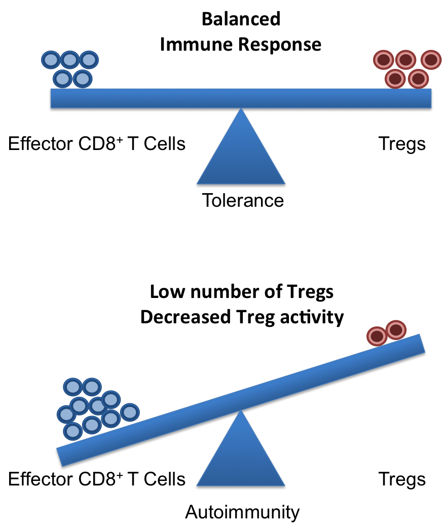
A healthy and functional immune system requires a delicate balance of pro- and contra-inflammatory signals. Whereas, it is important to induce a strong and efficient immune response against pathogens, it is similarly important to dampen these responses after the pathogen is fought off to revert the immune system to a calm steady state. If the balance is disturbed, diseases can on the one hand, become chronic/overwhelming or, on the other hand, inflammatory responses that cannot terminate can result in autoimmune responses.

Crucial elements in the regulation of excessive immune responses are regulatory T (Treg) cells. Tregs are known to inhibit the response of other immune cells. Their essential role in limiting overwhelming immune responses is demonstrated by the detrimental consequences of their loss. Mice or humans lacking Tregs develop widespread and lethal autoimmune diseases. Besides several surface markers, Tregs are best characterized by the expression of the transcription factor FoxP3. This factor is essential for Treg function and its artificial expression in other T cells can induce a regulatory potential. Therefore, the expression of FoxP3 is required for a T cell to have regulatory potential (Buckner; Josefowicz et al.). However, it was known for many years that in cases of numerous autoimmune diseases FoxP3+ Tregs could be found in high numbers at the sides of inflammation, but that they did not demonstrate any or not sufficient regulatory activity. This enigmatic observation was so far poorly understood (Buckner; Josefowicz et al.).Treg balance

In the March 2013 issue of Nature Medicine Nie and colleagues shed new light on the underlying mechanism that impairs Treg function at the sites of inflammation. Studying Treg cells from rheumatoid arthritis (RA) patients the authors demonstrated that phosphorylation of FoxP3 of the serine at position 418 (S418) is required for its regulatory action. If FoxP3 lacks this particular phosphorylation the Treg cell is not suppressive! FoxP3 S418 in Tregs is usually phosphorylated and hence Tregs are regulatory by default. However, the authors show that due to the action of the enzyme ‘protein phosphatase 1’ (PP1) FoxP3 can lose its S418 phosphorylation. Intriguingly, the presence of the cytokine TNF lead to an up-regulation of PP1 expression in the Tregs in a dose-dependent manner, and this lead to de-phosphorylation of FoxP3 S418. Treg cells expressing a mutant FoxP3 that replaced the serine at position 418 with an alanine retained their suppressive potential even in the presence of TNF, demonstrating the importance of the phosphorylation of S418. With this finding, the authors were able to link the pro-inflammatory milieu (TNF) to a specific effect inside of the Tregs (de-phosphorylation of S418) that lead to the observed loss of the regulatory function of Treg cells. Importantly, the authors were also able to demonstrate the therapeutic potential of this knowledge. They monitored RA patients that underwent treatment with blocking anti-TNF antibodies (infliximab) and found that Tregs from patient PBMCs restored S418 phosphorylation and regained regulatory potential!

This is the second case for a post-transcriptional regulation of FoxP3 that can influence Treg function. Deacetylation of FoxP3 has been linked to impaired Treg function previously (Tao et al.). Additionally, the work of Nie et al. now adds mechanistic information to previous reports on the negative effect of TNF on Tregs (Valencia et al.; Zanin-Zhorov et al.).

Given the ubiquitous role of TNF during inflammation, it is very likely that the mechanism described by Nie et al. applies to many if not all cases of ongoing inflammation where Treg function is impaired. Furthermore, their data on the effects of anti-TNF antibody treatment in RA suggest a similar therapeutic potential in other autoimmune diseases. Surely, this report will ignite further investigation in this direction and will aid the development of better treatments for patients suffering from autoimmune diseases.

References:

Bromberg, J., 2013. TNF-α trips up Treg cells in rheumatoid arthritis. Nat Med, 19(3), pp.269–270.

Buckner, J.H., 2010. Mechanisms of impaired regulation by CD4(+)CD25(+)FOXP3(+) regulatory T cells in human autoimmune diseases. Nat Rev Immunol, 10(12), pp.849–859.

Josefowicz, S.Z., Lu, L.-F. & Rudensky, A.Y., 2012. Regulatory T cells: mechanisms of differentiation and function. Annual Review of Immunology, 30, pp.531–564.

Nie, H. et al., 2013. Phosphorylation of FOXP3 controls regulatory T cell function and is inhibited by TNF-α in rheumatoid arthritis. Nat Med, 19(3), pp.322–328.

Tao, R. et al., 2007. Deacetylase inhibition promotes the generation and function of regulatory T cells. Nature Medicine, 13(11), pp.1299–1307.

Valencia, X. et al., 2006. TNF downmodulates the function of human CD4+CD25hi T-regulatory cells. Blood, 108(1), pp.253–261.

Zanin-Zhorov, A. et al., 2010. Protein kinase C-theta mediates negative feedback on regulatory T cell function. Science, 328(5976), pp.372–376.


Artifacts and non-specific staining in flow cytometry, Part II

flow cytometryIn Part I, I talked about un-specific binding and Fc-receptor binding. Besides these cases of non-specific binding, there are also other cases of antibody/fluorochrome binding that appears non-specific but that actually represents a real specific interaction – even though it is usually one that is not welcomed. I call these ‘pseudo-artifacts’ and you will read about some really odd stuff here.


(1) Binding of fluorochromes to Fc-receptors

The fact that Fc-receptors (FcR) bind antibodies is obvious, but lesser known is the fact that some of the fluorochrome linked to your antibody can also bind some FcRs.

It has been reported that R-phycoerythrin (PE) can bind to mouse Fc-gamma-RII  (CD16) and Fc-gamma-RIII (CD32) (Takizawa et al.). Furthermore, FcR binding of fluorochromes apparently applies to most or maybe even all cyanine fluorochromes, either alone or in tandem conjugates (Shapiro). So far I found reports for Cy5 (Jahrsdorfer et al.), PE-Cy5 (van Vugt et al.; Steward and Steward; Jahrsdorfer et al.) and APC-Cy7 (Beavis et al.). In this case, human CD64 (Fc-gamma-RI) was suggested to be the culprit of some of the binding (van Vugt et al.; Jahrsdorfer et al.), but binding also to CD64neg leukemia cells has been reported (Steward and Steward), so the role of FcR is not solved for all cases yet.

Þ Potential solution:

(a)  PE: For the binding of PE to mouse CD16/32 the use of a ‘Fc-block’, i.e. adding blocking monoclonal antibody 2.4G2 (rat IgG2b kappa), will avoid the problem (Takizawa et al.).

(b)  Cyanine: If you work with FcR+ cells, especially monocytes, you might consider avoiding cyanine-containing fluorochromes for the staining of your cells of interest.


(2) Binding of fluorochromes to antigen-receptors

Phycoerythrin (PE) and allophycocyanin (APC) are large proteins of 240kD and 110kD respectively that were original derived from cyanobacteria or red algae. As it turns out these phycobiliproteins are also a specific antigen for some T and B cells. Approximately 0.1% of all mouse B cells recognize PE as antigen in a BCR-dependent manner (Pape et al.; Wu et al.). Similar, about 0.02% of all mouse B cells are APC antigen-specific (Pape et al.). Furthermore, about 0.02-0.4% of all gamma-delta-T cells (mouse and human) recognized PE as a specific antigen (Zeng et al.) as well.

Þ Potential solution: Given their low frequency, these cells only pose a problem if you study tiny subsets of B and gamma-delta-T cells. In that case, you should avoid the use of PE for your cells of interest.

(3) Binding of fluorochromes to other receptors or interaction partners

(a) Cross-reactivity of the antibody:  Epitopes might be shared between different proteins, i.e. your antibody might not only recognize your protein in question, but recognizes also a similar epitope of another protein. This is for obvious reasons more likely with polyclonal antibodies.

Þ Potential solution: Usage of monoclonal antibodies reduces the risk of such cross-reactivity. If you suspect a cross-reactivity of your antibody, using a different clone for the same epitope will likely solve this problem.

(b) Intracellular biotin: Biotin is an important component of the cell metabolism. Therefore, biotin is present in the cells and the use of a streptavidin for intracellular staining will lead to binding of the streptavidin also to the cellular biotin.

Þ Potential solution: If you need to use a biotin-conjugated antibody for your intracellular staining you could cover all intracellular biotin by incubation of your cells with unconjugated streptavidin (followed by thorough washing) before the addition of your biotin-conjugated antibody.

(c) FITC charge: FITC is a charged molecule and antibodies with many FITC molecules (i.e. high F/P ratio) result in a highly charged antibody that binds, presumably through electrostatic interactions, nonspecifically to cytoplasmic elements (Hulspas et al.). This seems to be mainly a problem with intracellular staining and not with surface stains.

Þ Potential solution: For this reason FITC is not ideal for intracellular staining and you might try your antibody conjugated to a different fluorochrome.

(d) CD205: CD205 (DEC205) is a C-type lectin that is highly expressed on dendritic cells. Recently, it has been demonstrated that PE-Cy5.5 binds with high specificity to mouse CD205 (Park et al.). No staining was observed towards human CD205 and the binding of other Cy5.5 conjugates (PerCP-Cy5.5, APC-Cy5.5 and Cy5.5) to mouse CD205 was much weaker than that of PE-Cy5.5 (Park et al.).

Þ Potential solution: Given the high specificity of the interaction, you should avoid the use of PE-Cy5.5, and to a lesser extent other Cy5.5 containing fluorochromes, when your cells of interest expresses mouse CD205.

(4) Other effects

Finally, another odd-ball has been reported for APC tandems. Apparently, living cells have some way, which depends on their metabolism, to degrade the APC-Cy7 and APC-H7 tandems, leaving you with an APC signal (Le Roy et al.). APC-Cy7 seemed to be more affected than APC-H7 and monocytes are more active at degrading this signal than lymphocytes.

Þ Potential solution: Given that this requires live cells, fixation of your cell solution after staining will solve this problem. Alternatively, as this degradation requires metabolically active cells, storing your cell solution at 4°C or on ice or adding sodium azide (NaN3) to your storing buffer will reduce the effect.

That’s all I have for now, but if you know of other such ‘pseudo-artifacts’, or if you have any corrections and comments please share them with us!


//


References:

An amazing source for odd questions on flow cytometry is the ‘Cytometry mailing list’ hosted by the Purdue University, which can be found under: https://lists.purdue.edu/mailman/listinfo/cytometry

Beavis, A.J. & Pennline, K.J., 1996. Allo-7: a new fluorescent tandem dye for use in flow cytometry. Cytometry, 24(4), pp.390–395.

Hulspas, R. et al., 2009. Considerations for the control of background fluorescence in clinical flow cytometry. Cytometry, 76B(6), pp.355–364.

Jahrsdörfer, B., Blackwell, S.E. & Weiner, G.J., 2005. Phosphorothyoate oligodeoxynucleotides block nonspecific binding of Cy5 conjugates to monocytes,

Le Roy, C. et al., 2009. Flow cytometry APC-tandem dyes are degraded through a cell-dependent mechanism. Cytometry A, 75(10), pp.882–890.

Pape, K.A. et al., 2011. Different B cell populations mediate early and late memory during an endogenous immune response. Science, 331(6021), pp.1203–1207.

Park, C.G., Rodriguez, A. & Steinman, R.M., 2012. PE-Cy5.5 conjugates bind to the cells expressing mouse DEC205/CD205. J Immunol Methods, 384(1-2), pp.184–190.

Shapiro, H.M., 2004. Practical Flow Cytometry 4th Edt,

Stewart, C.C. & Stewart, S.J., 2001. Cell preparation for the identification of leukocytes. Methods Cell Biol, 63, pp.217–251.

Takizawa, F., Kinet, J.P. & Adamczewski, M., 1993. Binding of phycoerythrin and its conjugates to murine low affinity receptors for immunoglobulin G. Journal of Immunological Methods, 162(2), pp.269–272.

van Vugt, M.J., van den Herik-Oudijk, I.E. & van de Winkle, J.G., 1996. Binding of PE-CY5 conjugates to the human high-affinity receptor for IgG (CD64). Blood, 88(6), pp.2358–2361.

Wu, C.J. et al., 1991. Murine memory B cells are multi-isotype expressors. Immunology, 72(1), pp.48–55.

Zeng, X. et al., 2012. gd T Cells Recognize a Microbial Encoded B Cell Antigen to Initiate a Rapid Antigen-Specific Interleukin-17 Response. Immunity, 37(3), pp.524–534.


Gerhard WingenderGerhard Wingender is currently an Instructor at the La Jolla Institute for Allergy and Immunology (La Jolla, CA). His main lab toy is flow cytometry and his research interest involve invariant Natural Killer T (iNKT) cells.


//


Photo credit: PNNL – Pacific Northwest National Laboratory / Foter.com / CC BY-NC-SA

Artifacts and non-specific staining in flow cytometry, Part I

If you add your antibody, lets say anti-CD3-epsilon antibody, to your cell solution you’d expect that only T cells will be labeled, right? Well, if it were so easy then it wouldn’t be biology!


antibodiesIn this first half of the two part blog, I will talk about the two reasons, namely unspecific binding and Fc-receptors, which most people think of when they talk about non-specific binding in flow cytometry.

Some lesser known, but intriguing and important, ‘pseudo-artifacts’ will be covered later in Part II of the blog.


(1) Unspecific binding

Unspecific binding is defined as any sticking of an antibody or a fluorochrome to a cell in a fashion that does not require a specifically (currently) defined interaction. This might occur due to electrostatic interactions, glycolipid interaction on the cell membrane, protein-protein interactions and DNA binding.

As such unspecific binding of a cell depends heavily on the surface area (for surface stains) and/or its volume (intracellular staining). For example a cell with twice the size (as seen in the FSC) has 4-times the surface area (SF = 4pi r2) and 8-times the volume (V = 4/3pi r3) and consequently the unspecific binding will be 4 to 8-times higher. So, if you see the whole population shifting a bit in your histogram, you might want to check the scatter of the cells. For example, activated cells start proliferating, which increase their cell size along the way.


Aggravating factors and potential solutions:

Antibody amount: A surplus of antibody can increase the non-specific binding, leading to a reduction in the separation of your positive cells and reducing the signal:noise ratio.

Þ Potential solution: Titrate your antibody. As a starting point, antibodies with the same fluorochrome conjugate can often be used at similar concentrations.

Extracellular matrix/cell content: All cells bind proteins including antibodies to some degree via various interactions

Þ Potential solution: Addition of protein to the wash and staining solutions will cover many of these binding sites. Most staining protocols include BSA or serum (either human or FCS) for this purpose.

Dead cells: Dead cells are notorious for non-specifically binding antibodies and appear very ‘sticky’. This is partially due to DNA, but including DNAse would only partially solve the problem.

Þ Potential solution: A live/dead differentiation should be included, if possible, in every staining. Dead cells cannot be entirely separated just by FSC/SSC characteristics, especially not after fixation. Keep in mind though, that fixation of your cells after staining with e.g. PI or 7AAD will partially permeabilize all your cells, so that PI or 7AAD can leak out of the labeled cells to other cells eventually homogenously staining all your cells. In the case of 7AAD this can be avoided by inclusion of non-fluorescent actinomycin D (Schmid et al.). However, nowadays multiple live/dead discriminating reagents are available that can be fixed, thereby stopping potential leakage and avoiding this problem altogether.


(2) Binding of antibodies to Fc-receptors:

Obviously Fc-receptors (FcR) bind antibodies with high specificity, but the common misconception is that this is solely species-specific. However, FcRs from one species readily bind antibodies from other species to varying degrees. For example, hamster anti-mouse CD3-epsilon (clone 145.2C11) can bind to all mouse FcRs (Wingender et al.).

Þ Potential solution:

(a)   Fab or F(ab)2 fragments: Utilizing antibodies without their Fc-end avoids the problem altogether, but most commercially available antibodies do contain their Fc part.

(b)   ‘Fc-Block’: Adding antibodies that are specific for particular FcRs that block the undesired interaction with your experimental antibody. However, the ‘Fc-block’ commonly used for mice is the blocking monoclonal antibody 2.4G2 (rat IgG2b kappa) which is specific for mouse Fc-gamma-RII  (CD16) and Fc-gamma-RIII (CD32). Therefore, other FcRs are not directly blocked by 2.4G2. However, the majority of commercial antibodies are of an IgG subtype, most of the potential unspecific Fc-binding will be blocked by 2.4G2. Similar products for staining of human cells are widely available.

(c)     Unconjugated antibody: Adding unconjugated antibody of the same species and isotype as your experimental antibody to your staining cocktail will saturate most potential FcR binding sites.

As a positive side effect, adding unconjugated antibodies, either 2.4G2 or any other isotype, to your stain will incidentally also saturate most other potential unspecific bindings, as they were outlined under (1). Therefore, adding unconjugated antibody to your surface and also your intracellular staining cocktails will reduce unspecific binding.


So much for part one. As always, corrections and comments are highly welcomed.


References:

Schmid, I. et al., 2001. Simultaneous flow cytometric measurement of viability and lymphocyte subset proliferation. J Immunol Methods, 247(1-2), pp.175–186.

Wingender, G. et al., 2006. Rapid and preferential distribution of blood-borne alphaCD3epsilonAb to the liver is followed by local stimulation of T cells and natural killer T cells. Immunology, 117(1), pp.117–126.


//


Gerhard WingenderGerhard Wingender is currently an Instructor at the La Jolla Institute for Allergy and Immunology (La Jolla, CA). His main lab toy is flow cytometry and his research interest involve invariant Natural Killer T (iNKT) cells.


//


References:

Schmid, I. et al., 2001. Simultaneous flow cytometric measurement of viability and lymphocyte subset proliferation. J Immunol Methods, 247(1-2), pp.175–186.

Wingender, G. et al., 2006. Rapid and preferential distribution of blood-borne alphaCD3epsilonAb to the liver is followed by local stimulation of T cells and natural killer T cells. Immunology, 117(1), pp.117–126.


Photo credit: AJC1 / Foter.com / CC BY-NC-SA