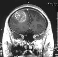
NEW IMAGING TECHNIQUE DISTIGUISHES TUMORS FROM NORMAL TISSUE

Even though the main objective of tumor surgery is to remove tumors as much as possible without disturbing the adjacent normal tissues, the task is very challenging in the operating room as neoplastic tissue is hard to distinguish from the adjacent healthy tissue. Thus, the portion of tumor still remained in the body after surgery causes recurrence, treatment failure, and poor outcome.

Surgery is an important treatment modality for brain tumors. Therefore, distinguishing normal tissue from tumor is extremely important for brain tumor surgery owing to the risk of damaging functional brain structures. Removal of healthy tissue can cause neurologic problems, but leaving tumor tissue behind can allow the cancer to grow and spread again. This is a major problem with glioblastoma multiforme (GBM), the most common form of malignant brain tumor in adults. Glioblastoma tumors grow quickly and are difficult to treat. The tumors infiltrate normal brain tissue and can’t be easily singled out. Therefore, tools designed to safely maximize the removal of tumor tissue are warranted. So far, experimental attempts to tell the difference between tumors and normal tissue during surgery have had limited success until recently, a new study by Ji et al. (2013) reported successful separation of  tumor-infiltrated brain tissue from surrounding healthy tissue in mice using stimulated Raman scattering (SRS) microscopy. Ji and colleagues provided evidence that SRS microscopy can be used to delineate tumor tissue in a human GBM xenograft mouse model, both ex vivo and in vivo, and in human brain tumor surgical specimens.

brain.scan_

Raman spectroscopy is a technique to study the interactions (vibrational, rotational, and other low-frequency modes) between matter and radiation in a system. It is named after the Indian noble laureate Dr. C.V. Raman (1930) who described the effect of light impinges upon a molecule and its interactions with the electron cloud and the bonds of that molecule. Chemical bonds in molecules have their own sets of vibration frequencies, and produce unique patterns of scattered light called Raman spectra. These spectra can be used as fingerprints to identify and differentiate different molecules in a complex environment. Developed on the concept of Raman spectroscopy, SRS is now emerging as an imaging technique to image biological tissues based on the intrinsic vibrational spectroscopy of their molecular components such as lipids, proteins, and DNA. Being free from the drawbacks of the dye-based methods, this label-free imaging technique exhibits high chemical selectivity enabling its use in complex biological applications including brain imaging.

To this end, Ji and colleagues used SRS microscopy to the problem of distinguishing protein-rich glioblastomas from more lipid-rich surrounding tissue and showed that it can be used to detect glioma ex vivo in human GBM xenograft mice, with results that correlated with the interpretation of hematoxylin and eosin (H&E)–stained slides by a surgical pathologist. Most importantly, this study demonstrated that SRS microscopy can detect extensive tumor infiltration in regions that appear grossly normal under standard bright-field conditions. This study suggests that SRS holds promise for improving the accuracy and effectiveness of cancer surgery. However, several challenges remain to be overcome including making a handheld surgical device with motion correction to acquire images from within a surgical cavity.

Reference:

Ji M, Orringer DA, Freudiger CW, Ramkissoon S, Liu X, Lau D, et al. Rapid, label-free detection of brain tumors with stimulated Raman scattering microscopy. Sci Transl Med. 2013;5:201ra119.


New Findings in Cell Based Therapy for GBM

Glioblastoma multiforme (GBM) is the most common and lethalGlioblastoma type of malignant primary brain tumors that account for over 70% of all intracranial cancers.  The current course of GBM treatment consists of surgical resection of the main tumor mass, followed by administration of radiation and chemotherapy. Surgical resection of the primary tumor leads to injury to the surrounding normal tissue, while chemotherapy and radiotherapy cause toxicity to the healthy tissue in the brain.  These undesirable secondary effects of glioma treatments have a major impact on patients’ physical, cognitive and emotional functioning. Nonetheless, despite this aggressive treatment regimen and its harmful side effects, GBM remains virtually incurable, with post-diagnosis median survival persisting less than 14 months.  This dismal prognosis is due to a combination of unique anatomical features of the central nervous system (CNS), in addition to GBMs’ (glioma cells’) exceptional invasive capacity. Glioma cells infiltrate the brain’s highly dense parenchyma, migrating along the corpus callosum and creating new masses within the hemisphere contralateral to the initial tumor mass homing.

Thus, recurrence in postoperative GBM patients is in essence inevitable. Furthermore, GBMs are not only heterogeneous among individual patients, but they are also highly heterogeneous within a single tumor mass. Recent studies have shown compelling evidence of a therapeutically resistant subpopulation of malignant glioma cells that exhibit stem-cell like characteristics, such as multipotency, the ability to self-renew and invade/migrate; these tumor-initiating cells are referred to as glioma stem cells (GSCs) and are believed to be responsible for tumor initiation and recurrence in GBM patients. Furthermore, GSCs have been observed to ensconce within similar niches to neural stem cells (NSCs).

NSCs and mesenchymal stem cells (MSCs) have been shown to have an exceptional migratory ability within brain’s parenchyma and possess a notable inherent tumor tropism.  Thus, several cell-based therapy (CBT) studies and clinical models of malignant tumors have incorporated autonomous tracking of tumor cells by employing NSCs and MSCs to deliver multiple therapeutic genes to specific tumor loci. This target specific drug-delivery model has NSCs or MSCs transduced to express one pro-drug-activating enzyme, which catalyzes the conversion of a particular pro-drug into an active toxic agent, which results in the localization of the chemotherapeutics specifically at the tumor sites.  In addition to its ability to deliver effective cytotoxic damage to the tumor without causing damage to the healthy surrounding tissue, the resulting bystander effect of this system causes cell death not only to the drug-delivery-vehicle cells, but also to the surrounding glioma cells. Although a number of different enzyme/prodrug systems have been utilized in relevant studies, HSV-thymidine kinase (HSV-tk)/ deoxyguanosine analog ganciclovir (GCV) has been the most commonly tested in animal and in vitro models. HSV-tk phosphorylates GCV and produces deoxyguanosine triphosphate which is a polymerase-I inhibitor and DNA chain terminator; cell death occurs upon incorporation of the nucleotide analog within DNA chains.

In recent study published in the journal of Molecular Therapy, Blanco’s group report their findings regarding the interaction between human MSCs (hMSCs) and gliomas and the underlying mechanism for the effectiveness of hAMSC based therapy in GBMs.  In their previous work Blanco’s group showed that the administration of hAMSCs expressing HSV thymidine kinase in glioma tumors significantly promotes tumor growth, whereas induction of cytotoxicity by administration of the prodrug GCV demonstrated a significant antitumor response.

According to this new study, hMSCs differentiate to endothelial lineage (supported by the expression of CD31 marker) within tumors, and integrate in the tumor vascular system where they adopt an endothelial phenotype. Further, Blanco proposed the notion of hMSCs’ ability to home to privileged vascular structures where GSCs also reside, is the underlying characteristic that leads to the effectiveness of cytotoxic hMSCs in regulating the bystander killing of tumor cells.

Although Blanco’s study provides invaluable insights into the GSCs’ niche and its role in malignant brain tumor CBT, hMSCs’ tumor growth promotion is a quality, which makes these cells less than ideal for utilization in human clinical trials in the near future. Future studies on this mechanism using primary patient tumor cells rather than the U87 glioma cell line and human NSCs are needed to further confirm these observations.

Further reading:

Juli R. Bagó, Maria Alieva, Carolina Soler, Núria Rubio, Jerónimo Blanco. Endothelial differentiation of adipose tissue-derived mesenchymal stromal cells in glioma tumors: implications for cell based therapy. Molecular Therapy.