
Whether an individual develops autoimmunity depends on how environmental and genetic factors interact to influence immune function. Genome wide association studies have revealed numerous risk alleles associated with diverse autoimmune diseases and small animal models have helped parse the effects of some of these gene variants on specific components of the immune system (see previous post). These approaches, however, largely ignore two of the more perplexing aspects of autoimmunity: (1) the strong female predominance of many autoimmune diseases and (2) the high incidence of autoimmunity in industrialized areas of the world compared to poorer, rural areas. The issue of sexual dimorphism in autoimmunity has led investigators to surmise that sex hormones are potent modulators of immune function. Indeed, altering estrogen, progesterone, or testosterone levels can influence the progression of autoimmune reactions in animals and people, although the specific mechanisms underpinning these phenomena remain controversial1. The increasing incidence of autoimmunity in the industrialized world has given rise to the “hygiene hypothesis”: exposure to specific microbes early in life is necessary for a fully-functioning immune system. These beneficial exposures are lacking in modernized, clean cities which contributes to immunological derangement and self-reactivity. Evidence supporting the hygiene hypothesis has accumulated in recent years to the point that clinical trials are in progress testing whether purposeful infestation with parasitic worms (thought to be the major microbial exposure missing from a modern upbringing) can control inflammatory bowel disease and multiple sclerosis2.

In a recent paper in Science Magazine, Dr. Jayne Danska’s group at the University of Toronto present an intriguing hypothesis that unifies the roles of early-life microbial exposure and sex hormone levels in driving an autoimmune response3. The group, with lead author Dr. Janet Markle, used a well-established mouse model of type-1 diabetes (T1D)—the non-obese diabetic (NOD) mouse—that is genetically predisposed to develop spontaneous, immune-mediated destruction of beta-islets at around 15 weeks of age, causing diabetes. Similar to several human autoimmune diseases, NOD mice have a 2:1 female-to-male sex bias in progressing to diabetes. In agreement with this, Markle et al. found that male mice housed under standard laboratory conditions were protected from developing diabetes compared to female mice. However, if NOD mice were born and raised in germ-free conditions (i.e. their intestines were never colonized by commensal bacteria) males developed the disease at the same rate as females. In addition, the environment in which the mice were raised impacted their levels of sex hormones: male mice raised in germ-free conditions had lower serum testosterone levels than those raised in standard conditions while germ free female mice had higher testosterone levels than those kept in standard cages. These data supported the conclusion that colonization with commensal bacteria was responsible for the protection of male NOD mice against T1D and that bacterial colonization somehow regulated the production and/or use of testosterone.
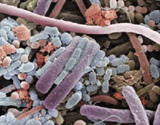
To get a sense of the extent to which gut microbiota influenced general physiology of adult male and female NOD mice, the authors used mass spectrometry to profile almost 200 unique small-molecule metabolites in serum. They found that male and female NOD mice raised in standard conditions had distinct profiles of serum metabolites. In contrast, there were few detectable differences between the metabolite profile of males and females raised in germ free conditions. This data suggested two hypotheses: (1) male and female mice had different physiologic responses to the same commensal bacteria, or (2) male and female mice had different commensal communities influencing their hormone and metabolite levels. The authors then sequenced bacterial 16S ribosomal RNA from the intestines of NOD mice at various stages of maturation (just after weaning, at puberty, and as adults). While male and female NOD mice had indistinguishable gut microbiota after weaning, sex-based differences in commensal bacteria were apparent at puberty and became even more pronounced in adulthood.
Having established that adult male and female NOD mice have distinct bacterial populations in their intestines, Markle et al. showed that transplanting the “male” gut microbiota into pre-pubescent female NOD mice altered the composition of the recipient’s commensal populations for several weeks and resulted in increased serum testosterone levels. Importantly, transplantation of male commensal bacteria protected the female recipients from T1D. Markers of T1D disease activity, such as inflammation of beta-islets and production of auto-antibodies, were reduced in recipients of male gut bacteria compared to unmanipulated females. This effect was abrogated upon treatment with the anti-androgen Flutamide, indicating that testosterone levels were a critical regulator of autoimmune pathogenesis.
Markle et al. have put forth an interesting model in which sexual maturation results in sex-specific programming of intestinal commensal bacteria. These distinct populations have differential effects on host physiology and hormonal balance which, in turn, modulate immune function. The centrality of gut microbiota to immunity raises the intriguing possibility of treating a dysfunctional immune system by altering the make-up of the intestine’s commensal communities. Such studies are already underway for intestinal disorders using “fecal transplants”, but the concept may extend to more systemic autoimmune disorders4. This approach would benefit from knowing the specific effects of different bacterial species on host physiology in order to identify the critical components of effective therapeutic microbial regimens. Similarly, it will be interesting to see which of the changes in metabolite and hormone levels that accompany shifts in commensal populations are most impactful on immune function. The metabolite profiling approach shown in this article could be useful for identifying endogenous compounds that serve as immune modulators.
Reference:
1. Sex differences in spontaneous versus induced animal models of autoimmunity. Lee TP, Chiang BL. Autoimmun Rev. 2012 May;11(6-7):A422-9. doi: 10.1016/j.autrev.2011.11.020. Epub 2011 Dec 4.
2. Vaccine against autoimmune disease: can helminths or their products provide a therapy? Zaccone P, Cooke A. Curr Opin Immunol. 2013 Mar 2. pii: S0952-7915(13)00027-7. doi: 10.1016/j.coi.2013.02.006.
3. Sex differences in the gut microbiome drive hormone-dependent regulation of autoimmunity. Markle JG, Frank DN, Mortin-Toth S, Robertson CE, Feazel LM, Rolle-Kampczyk U, von Bergen M, McCoy KD, Macpherson AJ, Danska JS. Science. 2013 Mar 1;339(6123):1084-8. doi: 10.1126/science.1233521. Epub 2013 Jan 17.
4. Stool transplants: ready for prime time? Weissman JS, Coyle W. Curr Gastroenterol Rep. 2012 Aug;14(4):313-6. doi: 10.1007/s11894-012-0263-7.
