Remodeling of the Tumor Extracellular Matrix Activates YAP in Fibroblasts to Produce Cancer Associated Fibroblasts

When cells undergo transformation and initiate the formation of a solid tumor mass, they cause profound changes on the phenotypes of the cells that surround them1. However, in addition to the changes in cellular phenotype, there is a change in the extracellular matrix that coincides with tumor formation1. It has been demonstrated that the majority of solid tumors have increased stiffness in their extracellular matrix (ECM), which may lead to increased activation of pro-tumor signaling pathways, such as Src, FAK, and RhoA2-4. Recently, it was discovered that increased matrix stiffness may also lead to increased activity of the oncogenic YAP/TAZ complex, which is connected to the Hippo signaling pathways, transcriptional regulators that increase cellular proliferation, decreased cellular contact inhibition, increased cancer stem cell phenotype, and increased metastasis5. However, in a fibroblastrecent edition of Nature Cell Biology Calvo et al. demonstrated that 6.  Not only do the authors demonstrate that YAP/TAZ is active in CAFs, but YAP/TAZ is necessary for CAF development6. They show that CAF activation leads to matrix remodeling towards increased stiffness, via myosin light chain 2 (MYL9/MLC) expression, establishing a feed-forward loop where the ECM plays a vital role6.

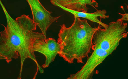
The authors first isolated fibroblasts in different stages towards becoming a CAF and saw that both mechanical-responsive signaling machinery (SMA, FN1, Paxillin, MYL9, MYH10, DIAH1 & F-actin) and mechanical tension were increased in populations containing CAFs. Moreover, tumor cell invasion, and angiogenesis of the tumor microenvironment (shown via endomucin and second-harmonic microscopy) were increased in samples that contained more tumor-associated-like fibroblasts (indicated by vimentin)6.

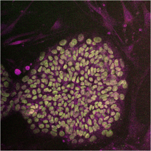
Because of the role of cell-cell and cell-ECM contact in the Hippo signaling pathway, the authors sought to understand whether this pathway is activated in CAFs. They found that YAP, and its co-factor, TAZ to be only upregulated and co-localized in the nucleus of transformed fibroblasts; the target of the activated YAP-TAZ complex6.  Furthermore, upon depletion of YAP, the ability for CAFs to cause matrix stiffness by contraction lessened as well as CAFs ability to form collagen networks and facilitate angiogenesis. Interestingly, when TAZ was inhibited, there was no change in functionality, which may lead to a TAZ-independent function for YAP.

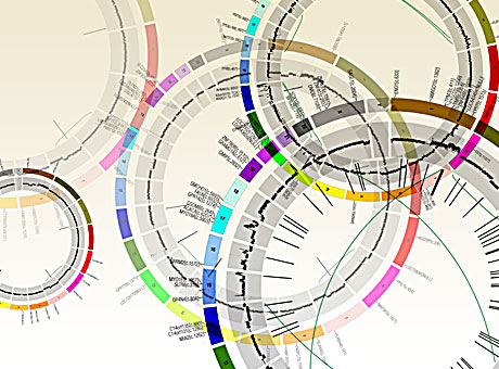
Upon microarray analysis of CAFs treated with siRNA that targets YAP, Calvo et al. found that the expression of many of the genes involved in mechanosensing and motility to be diminished6. Furthermore, when these individual genes were silenced, there was an overall decrease in the amount of cellular invasion of tumors. Many of the YAP-mediated genes, such as ANLN and DIAPH3, were involved in matrix remodeling and cellular invasion. Interestingly, modification of only one protein overexpression resulted in high amounts of matrix-remodeling and invasion: myosin regulatory light polypeptide 9 (MYL9). While not transcriptionally controlled by the YAP/TAZ complex, the authors demonstrate that YAP/TAZ is able to control MYL9 by post-translational modifications, placing YAP as a critical factor in regulating matrix-remodeling and invasion through MYL96.

Calvo et al. next posited that YAP/TAZ activation may not be exclusive to CAFs, but may also occur in normal fibroblasts when placed in a cancerous environment6. They found that fibroblasts placed in culture with tumor conditioned media had higher nuclear translocation of YAP, and higher gel contraction (akin to matrix stiffening) comparable to known promoters of pro-contractile function: L-alpha-lysophosphatidic acid (LPA) and transforming growth factor-beta (TGFβ). However, actomyosin inhibition (by blebbistatin) could not be rescued with LPA and TGFβ. Therefore, while soluble factors may activate matrix contraction, a functional cytoskeleton is essential for matrix contraction. Because of the necessary role of the cytoskeleton, the authors tested whether inhibition of RhoA kinase (ROCK), a kinase involved in regulating translocation and structure of the cell by the cytoskeleton, would affect the nuclear localization of YAP6. Inhibition of ROCK decreased YAP nuclearization and decreased the matrix stiffness. Of note, like ROCK inhibition, inhibition of Src also affected the nuclear localization of YAP as well as complex formation with TEAD1 and TEAD4. However, Src modulation of YAP is downstream of cytoskeletal changes in tension since Src inhibition did not affect stress fibers6.

Since activation of YAP in CAFs  is connected to actomycin-mediated matrix stiffness, and this activation of YAP expresses MYL9, and expression of MYL9 results in matrix-remodeling towards stiffness, the authors posit that this pathway forms a feed-forward loop6. This loop could lead to constitutive activation of YAP pathway in CAFs, causing a robust response and stabilizing the CAF phenotype. However, it is not known what other mechanisms, as well as regulatory mechanisms of YAP, are involved in this process as well as whether the YAP-ECM tension pathway may play a regulatory role in normal fibroblasts.

References:

1. Boudreau, A., van’t Veer, L. J. & Bissell, M. J. An “elite hacker”: breast tumors exploit the normal microenvironment program to instruct their progression and biological diversity. Cell adhesion & migration 6, 236-248, doi:10.4161/cam.20880 (2012).

2. Levental, K. R. et al. Matrix crosslinking forces tumor progression by enhancing integrin signaling. Cell 139, 891-906, doi:10.1016/j.cell.2009.10.027 (2009).

3. Guilluy, C. et al. The Rho GEFs LARG and GEF-H1 regulate the mechanical response to force on integrins. Nature cell biology 13, 722-727, doi:10.1038/ncb2254 (2011).

4. Sawada, Y. et al. Force sensing by mechanical extension of the Src family kinase substrate p130Cas. Cell 127, 1015-1026, doi:10.1016/j.cell.2006.09.044 (2006).

5. Harvey, K. F., Zhang, X. & Thomas, D. M. The Hippo pathway and human cancer. Nature reviews. Cancer 13, 246-257, doi:10.1038/nrc3458 (2013).

6. Calvo, F. et al. Mechanotransduction and YAP-dependent matrix remodelling is required for the generation and maintenance of cancer-associated fibroblasts. Nature cell biology 15, 637-646, doi:10.1038/ncb2756 (2013).


Finding the Right Cancer Culprits Using Mutational Heterogeneity

Imagine this: you are a police officer on patrol and you receive a call that multiple 30-year-old Caucasian males were seen breaking and entering; stealing heirlooms from a nearby neighborhood. The suspects were last seen entering a convention center and, to your dismay, you arrive to find the entire convention center is an antique show containing several 30-year-old Caucasian males carrying heirlooms. What do you do to apprehend your perpetrators?  You could arrest everyone that fits the description and interrogate them. On the other hand, you could scan the crowd for clues that there is a group of people that do not belong, or also radio to the police station for more information to narrow down the crowd. Needless to say, without gaining more contextual information for prudent discernment of the situation, you may arrest the wrong men and let the criminals go free.genomes

This is where cancer genomics is today; the sophistication of sequencing techniques have allowed for datasets that can detect every genomic mutation within cancer cells. Unfortunately, mutation rates are not equal among all genes. While this may seem a non-issue, this could lead scientists to ascertain that a mutated gene is associated with cancer when, in fact, the gene that “matches the description” is more susceptible to mutation, but has no role in oncogenesis. This is exactly what occurred to researchers who found high mutation rates of olfactory genes within lung cancer1. Doubtful of the role of olfactory genes in lung tumorigenesis, these scientists ultimately concluded that the mutation of olfactory genes had no role in the transformation of the lung epithelial cells1.

In Nature, Lawrence et al. further explored this issue, showing that failure to correct for the variability of mutation rates across the genome could lead to false positives for cancer associated genes1. To illustrate the importance of incorporating heterogeneity into the methodologies of data analysis, the authors compared a datasets with similar mutation frequencies to datasets that had different average mutation frequencies and found, when failing to take into account variability of mutations, there was an increase false categorization of cancer associated genes. Furthermore, the authors demonstrate that an analysis of an increasing sample size, as seen in the “big data” datasets of  American Society of Clinical Oncology’s “CancerLinQ™”2 and the Cancer Genome Atlas3, without correcting mutation rates, may exacerbate  the amount of false positives for cancer associated genes by decreasing the threshold needed to reach statistical significance. Lawrence postulated that heterogeneity may affect the detection of appropriate cancers by failing to correct for three contextual events: heterogeneity in mutation rates amongst samples of the same cancer type (patient-specific context), heterogeneity in mutation rates based on nucleotides surrounding a sequence (sequence-specific context), and heterogeneity in mutation rates based on the time that the gene is replicated or transcribed (replication/transcription-specific context).  Using the mutated olfactory genes mentioned above, along 3083 tumor-control pairs spanning 27 different cancer types, the authors demonstrate the importance of these contextually-discerning mutation rates and construct an algorithm for further context-based analysis, called MutSigCV.

Lawrence et al. studied cancer samples of the same cancer type (3,083 tumor-normal pairs across 27 tumor types) with variable average mutation rate. The authors found that, among all pairs and tumor types, there was a 1,000-fold variance in median frequency of mutations within the sample size. In these samples, the lowest variances were amongst hematological and pediatric cancers while the highest were among tumors induced by environmental factors, such as smoking and radiation. Given the importance of having accurate knowledge of the variability of rate of mutation, this underscores the importance in treating different cancer types, as well as patients with the same cancer, with a context-specific treatment protocol.

However, correcting for mutational frequencies attributed to tissue types, and mutations caused by known carcinogens and differences in cancer types, the authors still found that there was high mutational variability within certain samples of the same cancer type. Since mutation variance cannot be wholly accounted for by carcinogens, Lawrence et al. postulated that nucleotide makeup of the gene sequence may play a role in the mutation rate variability. The authors tested mutational heterogeneity in multiple tumors by assaying for 96 possible mutations (taking into account flanking bases) that were simplified into a radial chart for analysis1. Lawrence et al found that certain tumor types clustered into certain mutated sequences with the same flanking nucleotides (for instance lung cancers had a really high C to A mutations) was predominate, but still varied, within a certain cancer type.

While both variance in median mutation rates, and predominance of a specific sequence mutation, within specific cancer types was significant, the most important aspect in mutational heterogeneity seems to be in regional areas across a whole genome of cancer types, attributing to an excess of fivefold differences in median mutation rates1. Lawrence et al. credited this to two factors: the amount a gene is transcribed for the time the DNA section is replicated. The authors discovered that mutation rates are highest in genes with low rates of transcription and late DNA replication events. Comparing falsely-implicated olfactory receptor genes to known cancer associated genes, Lawrence et al. demonstrate different transcription rates and different replication times, with olfactory genes being expressed at cells with lower rates and later replication times. In contrast, cancer associated genes have higher transcription rates and earlier replication times.  In other words, while normal and cancer associated genes are both gaining mutations, the events that lead to these mutations are different. Thus, without parsing out mutational rates compared to replication and transcription, one may falsely assume that similar mutation levels must determine a cancer associated genes.

In the end, the authors surmised that “the rich variation in mutational spectrum across tumours underscores the problems with using an overly simplistic model of the average mutational process for a tumour type and failing to account for heterogeneity within a tumor type.” They state that their new analysis algorithm, MutSigCV, takes into account these context dependent nuances, allowing for cancer genomic analysis of mutations that eliminates these false positives. Using MutSigCV, Lawrence et al. was able to take a list of 450 suspected cancer associated genes in lung carcinoma and narrow the list to 11 suspected genes; genes shown to be linked to cancer1. This underscores the importance of context-specific analysis of big data in terms of cancer genomics. Without such a process, the use of whole genome sequencing for mutation rates for novel drug targets may be inadequate, sending many pharmaceutical and biotech companies toward therapeutic targets that, while look like the right suspect, are just an innocent bystanders that “fit the description”.


References:

1          Lawrence, M. S. et al. Mutational heterogeneity in cancer and the search for new cancer-associated genes. Nature, doi:10.1038/nature12213 (2013).

2          DeMartino, J. K. & Larsen, J. K. Data Needs in Oncology: “Making Sense of The Big Data Soup”. Journal of the National Comprehensive Cancer Network 11, S-1-S-12 (2013).

3          Network, C. G. A. R. Comprehensive genomic characterization of squamous cell lung cancers. Nature 489, 519-525, doi:10.1038/nature11404 (2012).


The Crucial Connection Between Metabolism and the Immune System.

Over the past couple of decades, the field of immunology has been growing at an exponential pace. Today, immunologists that continue to study the intracellular and extracellular components are also creating new therapies that suppress the immune system, increase the immune system, and even fix the dysregulation of the immune system. Likewise, there has been an increase in the study of organism metabolism and intracellular metabolism in various pathologies; such as diabetes and cancer. Interestingly, there is also an increase in studying how immune cells function in terms of their intracellular metabolism, how these metabolic pathways affect the phenotype and activation of immune cells, and how the immune system affects the metabolic functions of its host organism also known as immunometabolism1.

There are multiple metabolic pathways that cells use to make ATP. It has been discovered that some cells may preferentially use the glycolytic pathway to make ATP, even when the components are available for aerobic respiration; an event called the Warburg effect1. On the other hand, some cells may use the glycolytic pathway along with the Krebs cycle and electron transport chain (ETC), known as oxidative phosphorylation (OxPhos), to consume materials to make energy1. However, not all immune cells act alike. For instance, activated neutrophils preferentially use the Warburg effect1.  Interestingly, this metabolic pathway makes the most hydrogen peroxide, which is part of the substance neutrophils use for granulocytic release against pathogens1. Likewise, dendritic cells that have been activated via a toll-like receptor agonist and express inducible nitric oxide synthase (iNOS) are also found to use the Warburg effect and, like neutrophils, the metabolite of iNOS plays a functional role in activated dendritic cells1. Finally, the

macrophage

M1 pro-inflammatory macrophages also use the Warburg pathways to make ATP1. Glycolysis and oxidative phosphorylation are linked when the pyruvate from the glycolytic pathway is used to make acetyl-CoA using the Krebs cycle. Immune cells that use this pathway are activated T-cells, immunosuppressive M2 macrophages, and the pro-inflammatory Th17 T-cells1. Finally, fatty acid oxidation, the process of using lipids to make ATP, is utilized by memory T-cells, regulatory T-cells, and alternatively-activated macrophages1.

Metabolism in the immune system is more than just connected to the cell’s activation. Metabolism is involved in the homeostasis between immune cells as well as between immune cells and their stromal host cells1. Dysregulation of metabolites has been found in many pathologies and the role of metabolite fluctuation caused by host immune cells versus pathogens is an active area of study2. Likewise, metabolism has been shown to be involved in class switching effector T-cells into memory T-cells and is involved in the act of bringing immune cells into quiescence3.

An interesting example of a link between immunity and metabolism can be found in an article published in Nature April 11, 2013 by Tannahill et al. This paper demonstrates that the metabolite succinate, a key component in the Krebs cycle, is integral in lipopolysaccharide-induced macrophage activation4. Furthermore, they demonstrated that the activation of toll-like receptor 4, through LPS stimulation, leads to an increase of intracellular glutamate uptake and upregulation of succinate production under the “gamma-Amminobutyric acid (GABA) shunt” metabolic pathway4. The metabolite succinate then stabilizes hypoxia-inducible factor-1α; a protein that is involved in IL-1β production4. 

The study of metabolism is more than just an academic exercise. Many therapies have been proposed that look at changing the metabolic system for alteration of the dysregulated immune response. One of these is metformin, a drug that is used to help regulate type 2 diabetes2. However, metformin is now actively investigated as an anti-cancer therapeutic, having an effect on changing the tumor immune microenvironment from a pro-tumor phenotype to an anti-tumor phenotype2. As we continue to learn more about the effects of host metabolism on the immune system, the role of the immune system in organism metabolism, and the intracellular metabolic pathways involved in various immune cells during different states of function, we will hopefully be able to develop more therapies that repair immune dysregulation as well as develop immunotherapies for treating various metabolic diseases.

Further Reading

1. Pearce, E. L. & Pearce, E. J. Metabolic pathways in immune cell activation and quiescence. Immunity 38, 633-643, doi:10.1016/j.immuni.2013.04.005 (2013).

2. Mathis, D. & Shoelson, S. E. Immunometabolism: an emerging frontier. Nature reviews. Immunology 11, 81, doi:10.1038/nri2922 (2011).

3. Finlay, D. & Cantrell, D. A. Metabolism, migration and memory in cytotoxic T cells. Nature reviews. Immunology 11, 109-117, doi:10.1038/nri2888 (2011).

4. Tannahill, G. M. et al. Succinate is an inflammatory signal that induces IL-1beta through HIF-1alpha. Nature 496, 238-242, doi:10.1038/nature11986 (2013).

The Hippo-YAP Pathway: New Connection between Cancer and Stem Cells.

First discovered  by laboratories studying  Drosophila development 18 years ago1-3, the Hippo-YAP signaling pathway (also known as the Salvador-Warts-Hippo Pathway) is a novel pathway implicated in organism development, stem cell biology, and cancer biology4. While not much is known about the Hippo-YAP pathway, this signaling mechanism could lead to a promising paradigm in regenerative medicine and treating cancer.

While we are far from fully discovering every aspect Hippo-YAP signaling pathway, some components of this novel pathway have been uncovered. In mammalian cells, the first signal modulator to be stimulated is mammalian STE-20 protein kinase 1 & 2(Mst1/2). This stimulation causes autophosphorylation, which in turn starts a kinase cascade; phosphorylating the proteins Salvador homolog 1 (Sav1), MOB kinase activator 1 (Mob1), and large tumor suppressor 1 & 2 (Lats1/2). Once Lats1/2 is activated, it phosphorylates YAP (Yes-associated protein)4. This phosphorylation of YAP sequesters it outside of the cell and leads to its proteosomal degradation and thus blocking its ability to complex with the protumor TEAD transcription factors, which in turn inhibits proliferation and blocks inhibition of apoptosis4. Interestingly, other alternative mechanisms, such as directly targeting YAP via the WNT pathway, or activation of YAP/TAZ via the SMAD signaling pathway by TGFβ and BMP, have been demonstrated5. While the stimulation of the Hippo pathway is still being revealed, researchers have discovered two mechanisms hippo stimulation: cell-cell contact and activation of G-protein coupled receptors4,5.  Stimulation of G-protein Coupled Receptors (GPCRs), Go with the ligands LPA or S1P and Gs with glucagon and epinephrine, have been shown to activate the Hippo-YAP signaling pathway, causing phosphorylation of Mst1/26. On the other hand, the cell-cell contact method of hippo activation most likely phosphorylates Mst1/2 through the upstream component: Merlin6.  However, while there is phosphorylation of Mst1/2 in both stimulation methods, neither stimulation pathway is known, save one or two components, upstream of Mst1/2. Furthermore, the complexity of this signaling pathway is certain and upstream signals may be redundant6.

The Hippo-YAP signaling pathway plays a crucial role in embryological development. At the middle of this is the Hippo signaling component is transcriptional co-activator with PDZ-binding motif (TAZ, also known as WWTR1). TAZ is able to regulate the signaling mechanisms of the SMAD2/3-4 signaling pathway4; a pathway that regulates the TGF-beta signaling cascade that is important in early embryogenesis7. Furthermore, it has been demonstrated that functional loss of the TAZ protein, and not YAP, will lead to uncontrolled differentiation of human embryonic stem cells (hESCs) as well as loss of self-renewal of hESCs4.  Surprisingly, although YAP is not as important as TAZ to block differentiation, YAP is inactivated during normal hESC differentiation4. In addition to stem cell differentiation, the Hippo-YAP signaling pathway has been shown to be important for polarization of tissues8 in both planar and apicobasal cell polarity5, tissue shape and patterning9, and overall tissue homeostasis9.

While the activation of the Hippo-YAP pathway seems to be important for embryogenesis, the dysregulation of the Hippo-YAP pathway seems to play a striking role in tumorigenesis9. Deletion of the upstream Mst1/2 component of the Hippo-YAP pathway has been shown to cause uncontrolled liver growth. Microscopic analysis of liver biopsies revealed that these tissues were full of hepatocellular carcinoma and cholangiocarcinoma4. Likewise, overactivation of the YAP protein caused uncontrolled, extreme thickening of epidermal layer4. However, the pathway that is thought to be involved in this process of YAP activation is not the canonical Hippo-YAP pathway, but a signaling through alpha catenin. The catenin family and the Hippo-YAP signaling pathway were further shown to interact when overexpression of YAP facilitated the expression of Notch/Wnt signaling pathway indirectly by YAP-driven overexpression of beta catenin4. Because the Notch/Wnt pathways are important for cancer stem cell phenotype10 and cancer metastasis11, further investigation into the roles of the Hippo-YAP signaling pathway could bring a lot of clinical significance.

describe the imageAt the writing of this blog, no proposed drug has been proposed that directly targets the Hippo signaling pathway.  However, many possible targets for therapy are being investigated that would also affect the Hippo signaling pathway5. One of these is targeting the homeodomain-interacting protein kinase 2 (HIPK2) which has been demonstrated to activate YAP 5.In addition, using GPCR antagonists, such as Dobutamine, have been shown to decrease activation levels of YAP5. Also promising, researchers have solved many domains of the YAP structure, which may lead to specific inhibition of this oncogene by potential inhibitors12. Because of the role that the Hippo signaling pathway may play on tumor growth inhibition, it may not be long before candidate drugs targeting this pathway will start to enter the FDA drug pipeline.


Further Reading:

1              Justice, R. W., Zilian, O., Woods, D. F., Noll, M. & Bryant, P. J. The Drosophila tumor suppressor gene warts encodes a homolog of human myotonic dystrophy kinase and is required for the control of cell shape and proliferation. Genes & development 9, 534-546 (1995).

2              Xu, T., Wang, W., Zhang, S., Stewart, R. A. & Yu, W. Identifying tumor suppressors in genetic mosaics: the Drosophila lats gene encodes a putative protein kinase. Development 121, 1053-1063 (1995).

3              Wu, S., Huang, J., Dong, J. & Pan, D. hippo encodes a Ste-20 family protein kinase that restricts cell proliferation and promotes apoptosis in conjunction with salvador and warts. Cell 114, 445-456 (2003).

4              Ramos, A. & Camargo, F. D. The Hippo signaling pathway and stem cell biology. Trends in cell biology 22, 339-346, doi:10.1016/j.tcb.2012.04.006 (2012).

5              Harvey, K. F., Zhang, X. & Thomas, D. M. The Hippo pathway and human cancer. Nature reviews. Cancer 13, 246-257, doi:10.1038/nrc3458 (2013).

6              Yu, F. X. et al. Regulation of the Hippo-YAP pathway by G-protein-coupled receptor signaling. Cell 150, 780-791, doi:10.1016/j.cell.2012.06.037 (2012).

7              Massague, J. TGFbeta signalling in context. Nature reviews. Molecular cell biology 13, 616-630, doi:10.1038/nrm3434 (2012).

8              Yu, F. X. & Guan, K. L. The Hippo pathway: regulators and regulations. Genes & development 27, 355-371, doi:10.1101/gad.210773.112 (2013).

9              Pan, D. The hippo signaling pathway in development and cancer. Developmental cell 19, 491-505, doi:10.1016/j.devcel.2010.09.011 (2010).

10           Takebe, N., Harris, P. J., Warren, R. Q. & Ivy, S. P. Targeting cancer stem cells by inhibiting Wnt, Notch, and Hedgehog pathways. Nature reviews. Clinical oncology 8, 97-106, doi:10.1038/nrclinonc.2010.196 (2011).

11           Fodde, R. & Brabletz, T. Wnt/beta-catenin signaling in cancer stemness and malignant behavior. Current opinion in cell biology 19, 150-158, doi:10.1016/j.ceb.2007.02.007 (2007).

12           Sudol, M., Shields, D. C. & Farooq, A. Structures of YAP protein domains reveal promising targets for development of new cancer drugs. Seminars in cell & developmental biology 23, 827-833, doi:10.1016/j.semcdb.2012.05.002 (2012).

The Shock of Sepsis: The Struggle to Treat SIRS

Since the mid-1880’s, scientists and clinicians have studied the concept of sepsis. Investigations started with physician-scientists such as Ignaz Semmelweiss, Joseph Lister, Hugo Schottmüller and Louis Pasteur, initiating the fields of immunology and diseases related to immunology that continue even to today. Unfortunately, while we increase our knowledge about sepsis (also known as systemic inflammatory response syndrome; SIRS), diagnosis and treatment remain difficult1,2. This is seen as recently as last year, when an infection of a 12-year-old boy , Rory Staunton, lead to fatal septic shock after doctors failed to see the early signs of SIRS3. His death initiated the formation of the Rory Staunton Foundation and subsequent reforms of emergency protocols taken in his home state of New York (labeled as “Rory’s Laws”)3.

describe the imageHowever, hospital policy reforms are just part of the issue; sepsis affects more than 700,000 people in North America every year, with a 30-50% mortality rate4. To date, there are no FDA-approved drugs to fight SIRS4. In fact, after 20 years of intense research into translational medicine for sepsis, none of the proposed treatment approaches have had enough clinical efficacy to be used as treatment paradigms1. Action must be taken to not only change the methodologies of care for sepsis, but to better understand the biological mechanisms of sepsis. From this understanding, we may be able to have more accurate detection of sepsis as well as more effective therapeutics.

Sepsis occurs when an infection causes a systemic inflammatory response5. This disease causes an imbalance of the immune system, leading to changes in the hemodynamics of the host, resulting in coagulation, heart ischemia, and multi-organ failure1.  Originally thought to be caused by overactivation of the innate immune system, many patients did not die from the initial onslaught of inflammation, but from later stages of immune system suppression (also known as “immunoparalysis”) which allowed opportunistic viruses and bacteria to take over the host1. The roles of sepsis may be analyzed by observing the different cell types it affects: the innate system, the adaptive immune system, and non-immune cells.

Since sepsis is due to bacterial and viral infection, the innate immune system is the most well studied aspect of the pathogenesis of sepsis. During SIRS the innate immune cells are pathologically affected: macrophages, dendritic cells and natural killer (NK) cells. During the initial period of infection, increased amounts of pathogen-associated molecular pattern (PAMPs) and damage-associate molecular pattern (DAMP) molecules are found in the host organism. Toll-like receptors (TLRs), such as TLR4 and TLR9, in the innate immune cells recognize these molecules and confer a host inflammatory response.  The inflammation leads to an increased presence of adhesion molecules on both innate and adaptive immune cells4 indicating the desire to enter the site of infection to confer an immune response.  Along the same lines, activation of the complement cascade occurs, leading to increased amounts of C5a protein. The upregulated C5a protein levels cause increased migration of innate immune cells and increased phagocytic ability of macrophages4.

Unfortunately, prolonged exposure of macrophages to C5a leads to dysregulation of macrophage activation and, eventually, apoptosis1. During this apoptotic stage, macrophages release high-mobility group protein B1 (HMGB1) which may lead to increased inflammation5. On the other hand, in neutrophils, sepsis is known to cause an abnormally long proliferation period5 which could lead to organ damage and increased inflammation. Surprisingly, sustained activation of neutrophils may also contribute to the immunoparalysis by sepsis because activated neutrophils increase reactive oxygen species, known to cause immunosuppression, and macrophages that eat apoptotic neutrophils express anti-inflammatory cytokines5. describe the imageProlonged sepsis also decreases the pro-inflammatory ability of dendritic cells; SIRS increases depletion of splenic and myeloid dendritic cells, where the remaining dendritic cells are functionally deficient4.  NK cells, originally shown to play a role in anti-viral immune responses but are also proposed to play a role in bacterial infection responses, have two distinct roles in different phases of sepsis.  In the early phases, NK cells are thought to contribute to the overactive immune responses and systemic inflammation4 however, in later phases of sepsis, NK cells may be compromised which could lead to secondary bacterial or viral infections, thus exacerbating the inflammatory response during SIRS4.

describe the imageIn addition to dysregulation of the innate immune system, the cells that make up the adaptive immune system, T-cells and B-cells, are also affected by sepsis. Sepsis decreases overall T-cell receptor function, and moves Th1 (proinflammatory) T-cells toward a Th2 (immunosuppressive) response1. In addition, CD25+Foxp3+ T-cells, also known as regulatory T-cells, are increased during SIRS5. Interferon-gamma, a proinflammatory cytokine expressed during sepsis, causes an increase in innate response activator (IRA) B Cells, B-cells that recognize PAMPs, impair infection clearance, and accelerate septic shock4,6. During sepsis, the increase of C5a causes T-Cell and B-cell apoptosis4, which leads to immunosuppression4.

Sepsis also has non-immunological effects on the patient. During sepsis, coagulation becomes more pronounced. This increase in coagulation and pre-coagulation states may lead to ischemic injury5.  Sepsis also causes cytopathic hypoxia5 and cardiomyopathy4. Interestingly, SIRS also plays a role on the autonomic nervous system, with increased apoptosis of the adrenal medullary cells, there is a dysregulation of the endocrine system that regulates the autonomic nervous system1.

Currently, there are several studies looking into the pathogenesis of sepsis with the goal being effective treatment. In terms of pathogenesis, recent studies looking at the role of STIM17  and PI3K activation in the initiation of sepsis5. Host deficiencies leading to sepsis are also being studied. Deficiencies, including,  zinc8 and ADAMS-T5 deficiencies, will hopefully elucidate more on the dysregulation found in sepsis. Currently, medical scientists are working with clinicians to study the more nuanced roles of hyper-responsiveness versus system immune suppression4. From these studies, multiple immunotherapies have been proposed: dendritic cell implantation4, regulatory T-cell implantation 4, and the modification of the host immune system by using TLR antagonists4, injections of the proinflammatory cytokines IL-15 and IL-174, and limiting adaptive immune system exhaustion by blocking PD-14. However, several of these studies, such as T-reg implantation, and using TLR antagonists have had limited clinical efficacy and have been prematurely terminated4. This may be due to the lack of an appropriate model to study human sepsis in the pre-clinical setting.

The limitations of current research methodologies for sepsis have shown the need for reform in SIRS research. Recently an article by Seok et al in the Proceedings of the National Academy of Sciences elucidated the limitations of using mouse models to studying human inflammation9. In this article, Seok et al list the differences in temporal responses, gene signatures, and regulated pathways involved inflammatory response following various insults9. The reasons for describe the imagedifferences in the inflammatory response may be described by the evolutionary differences between mouse and human immune systems, the inbred nature of the mice used, and the tendency to focus on a single mechanism in mouse models when there is great overlap of immune responses within a host system. These authors propose studying more of the genetic and epigenetic changes of patient samples during sepsis to find appropriate mouse models for study9. Furthermore, they propose in vitro recapitulation of the inflammation response in diseased tissue9. Unfortunately, the in vitro model is limited and this reductionist approach may miss a key cell, cellular event, or special/temporal arrangement that may be vital to pathogenesis of sepsis.

The use of non-human primates as a sepsis model may be more accurate than either of these, but the ethical and cost-effective issues of using non-human primates remains a deterrent. A final model that may be of use to sepsis researchers in the future is the use of the humanized mouse model; a severely immunocompromised mouse host that has had a recapitulation of the adaptive and/or innate immune systems for in vivo study of human pathologies.  The use of humanized mice for the study of sepsis was proposed by Unsinger et al, where they used two-day-old NOD-scid IL2 receptor-gamma knockout mice and transplanted them with hCD34+ enriched hematopoietic cord blood stem cells.  After establishment of the human immune system, mice were treated with cecal ligation puncture (CLP) model of intra-abdominal peritonitis and assayed for immune response changes10. These mice developed a functional human innate and adaptive immune system with recapitulation of the human immune response to sepsis.  However, while there still remain differences in the humanized mouse model versus the actual human immune response, such as differences in the bacterial flora that is found in the bowels10, this model nonetheless provides a useful tool which, along with more genetic and epigenetic information from patient studies, will lead to more impactful pre-clinical studies and possibly FDA-approved drugs to treat sepsis in the clinics.


References:

1. Rittirsch, D., Flierl, M. A. & Ward, P. A. Harmful molecular mechanisms in sepsis. Nat Rev Immunol 8, 776-787, doi:10.1038/nri2402 (2008).

2. Stearns-Kurosawa, D. J., Osuchowski, M. F., Valentine, C., Kurosawa, S. & Remick, D. G. The pathogenesis of sepsis. Annu Rev Pathol 6, 19-48, doi:10.1146/annurev-pathol-011110-130327 (2011).

3. Dwyer, J. Death of a Boy Prompts New Medical Efforts Nationwide, October 26, 2012).

4. Ward, P. A. & Bosmann, M. A historical perspective on sepsis. Am J Pathol 181, 2-7, doi:10.1016/j.ajpath.2012.05.003 (2012).

5. Cinel, I. & Opal, S. M. Molecular biology of inflammation and sepsis: a primer. Crit Care Med 37, 291-304, doi:10.1097/CCM.0b013e31819267fb (2009).

6. Rauch, P. J. et al. Innate response activator B cells protect against microbial sepsis. Science 335, 597-601, doi:10.1126/science.1215173

7. Gandhirajan, R. K. et al. Blockade of NOX2 and STIM1 signaling limits lipopolysaccharide-induced vascular inflammation. J Clin Invest, doi:10.1172/jci65647 (2013).

8. Liu, M. J. et al. ZIP8 Regulates Host Defense through Zinc-Mediated Inhibition of NF-κB. Cell Rep, doi:10.1016/j.celrep.2013.01.009 (2013).

9. Seok, J. et al. Genomic responses in mouse models poorly mimic human inflammatory diseases. Proc Natl Acad Sci U S A, doi:10.1073/pnas.1222878110 (2013).

10. Unsinger, J., McDonough, J. S., Shultz, L. D., Ferguson, T. A. & Hotchkiss, R. S. Sepsis-induced human lymphocyte apoptosis and cytokine production in “humanized” mice. J Leukoc Biol 86, 219-227, doi:10.1189/jlb.1008615 (2009).

Epithelial to Mesenchymal Transition: the Key to Cancer Metastasis

We have all seen science fiction movies where alien invaders, in search of a fertile planet like earth, start a full scale war against humanity. A common theme in many of these stories, but often overlooked, is the role of the scout. Before any invasion, a solitary member of the invaders, fortified for the harsh journey, is sent on a quest to scout out the new land. As soon as this scout finds a promised land, the colony is formed and the invasion begins. This concept of invasion is seen in nature with viral and bacterial infections, where fortified virus or sporulated bacteria are able to survive harsh conditions and then proliferate in their host system upon arrival. In an ironic display, cancer metastasis follows a similar system; cancer cells are able to leave the primary tumor, travel long distances in harsh conditions, and form colonies in other tissues within an organism. These metastases have grave consequences. In fact in certain cancers, such as melanoma, it is metastasis to vital areas like the brain that makes it life-threatening. One of the biggest areas in cancer biology research is elucidating the mechanisms involved in cancer metastasis, which includes the concept of the epithelial to mesenchymal transition (EMT). It is this process that gives the cancer “scouts” the means to invade vasculature, fortify themselves for a journey to the metastatic site, and resist most therapies at all locations in the tumor-bearing patient. In normal development, the epithelial to mesenchymal transition is a reversible system that is involved in embryonic gastrulation1 and cardiac development1,2. In adulthood, EMT is involved in wound healing3. Unfortunately, EMT is also involved in pathological events such as fibrosis of injured tissue1 and cancer development and progression1-4. Here, we focus on EMT’s role in tumorigenesis, cancer proliferation, treatment resistance, and metastasis.

The epithelial to mesenchymal transition occurs in many different solid tumors of epithelial origin. Many tumors are carcinoma based, which implies that they are usually confined from motility by the basement membrane1 but the process of converting toward a cancer cell that has mesenchymal characteristics allows cancer cells to infiltrate the circulatory and lymphatic systems, causing motility that will later lead to metastasis1,5. As a cancer progresses, epithelial features, such as cellular adhesion marker  E-cadherin3 and intracellular adhesion components such as tight junctions, cytokeratins, and desmosomes, are downregulated5. Coordinating with this downregulation is an upregulation of mesenchymal markers such as N-Cadherin, Vimentin, and Fibronectin3. This phenotypic shift is orchestrated by a coordination of many different mechanisms: epigenetic changes, post-translational modifications of proteins, transcriptional silencing by noncoding-RNAs (ncRNAs), and activation of epithelial to mesenchymal transcription factors (EMT-TFs)3. However, while all mechanisms of EMT development are important, this article will focus on the main orchestrators of EMT: the EMT-TFs.

EMT-TFs are many, but the main transcription factors fall into three families: SNAIL, ZEB, and TWIST. SNAIL has three main transcription factors: SNAIL (Snail1), SLUG (Snail2), and SMUC (Snail3)5. These proteins are zinc finger nucleases that are at the crux of EMT phenotype. In fact, it has been noted that SNAIL, and possibly SLUG, directly repress the expression of E-cadherin by binding to its promoter, CDH13. Furthermore, the SNAIL family has been shown to repress desmoplakin, adherens junctions, occulidins, and cytokeratin upon activation5. While not much is known about SMUC’s role in normal or pathological development, SNAIL, unlike SLUG, is so crucial to cancer metastasis that has been implicated in being an independent prognostic factor in the metastatic potential and severity of cancers5. Interestingly, not only does the SNAIL family propagate the characteristics of EMT transition, they also upregulate other EMT-TFs, such as ZEB1 and ZEB2, to further propagate the mechanism of EMT 5.

The Zinc-finger E-box-binding homeobox (ZEB) family are also a family of transcription factors with zinc-finger nuclease properties3. Like SNAIL, ZEB1 and ZEB2 bind to the E-cadherin promoter5. Moreover, ZEB1 and ZEB2 also downregulate tight/gap junctions, desmosomes and markers of polarity5. In addition, the ZEB family has been implicated in repressing P- and R-cadherins, other markers that inhibit the motility of epithelial cells5. ZEB1 is usually not found on non-cancerous cells but is found highly expressed on many cancer types5. In contrast, ZEB2 is expressed on normal epithelial cells, but is vastly upregulated in cancerous cells5.

describe the imageThe final group is the TWIST family, a basic helix-loop-helix transcription factor involved in different steps in embryonic development1. Interestingly, while TWISTs are vital to embryogenesis, they are absent in normal adult epithelium5. As a cancerous cell progresses , TWIST1 and TWIST2 appear and their reactivity increases in correlation with the tumorigenic progress5. Interestingly, while TWIST1 is involved  in regulating some of SLUG’s EMT effects, and directly drives expression of N-cadherin, it is not directly associated with the downregulation of E-cadherin5.

It is through these EMT-TFs that EMT is involved in tumorigenesis and tumor progression. EMT-TFs are involved in suppressing senescence and increasing cell cycle proliferation5. However, EMT-TFs are not enough to push tumorigenesis on their own, but require another event of tumorigenesis3. Therefore, EMT-TFs may act as a facilitator of tumorigenesis, but not be tumorigenic factors by themselves. EMT is also crucial for tumor invasiveness and metastasis. Besides the aforementioned changes in cellular adhesion molecules, activation of the EMT-TFs causes upregulation of matrix-metalloproteinases (MMP), enzymes involved in degradation of the extracellular matrix and invasion of cancer cells3. It has been demonstrated that the SNAIL family activates the expression of MMP1, MMP2, MMP7, and MT1-MP 5. Furthermore, the upregulation of MMPs additionally activates EMT-TFs, thus forming a feed-forward loop3. EMT events may also go beyond extracellular degradation; TWIST1 is known to be involved in the formation of invadopodia which has been correlated with invasiveness3.

The invasion of metastasis is not just due to intrinsic changes but also to changes in the host, and target microenvironment4. For instance, TGF-β, a cytokine that normally acts as a tumor suppressor, can enhance tumor invasion in later-stage tumors2. Furthermore, TGF-β activates the SNAIL and TWIST families of the EMT-TFs2,5. TGF-β may be produced by myeloid derived suppressor cells and CD11b+/F4/80+ tumor associated macrophages (TAMs) in the primary tumor microenvironment, thus perpetuating the EMT phenotype4. TAMs also express the cytokines fibroblast growth factor (FGF), epidermal growth factor (EGF), and macrophage colony stimulating factor (CSF-1) which are involved in EMT-based invasion as well as recruitment of immune cells toward a metastatic-favored microenvironment4. Moreover, TNF-α, usually correlated with  an anti-tumor phenotype, also stabilizes SNAIL expression, thus implicating it in facilitating EMT3. Besides immune cells, other stromal cells are involved in EMT initiating and metastasis. For instance, mesenchymal stem cells (MSCs) and cancer associated fibroblasts (CAFs) are both involved in EMT initiation and propagation4.

One of the more clinically relevant aspects of EMT in cancer progression is the ability of EMT to confer therapy resistance. Resistance to doxorubicin is breast cancer correlates with higher levels of ZEB15 and both ZEB1 and ZEB2 have been shown to guard against cisplatin therapy5. TWIST1 mediates resistance to paciltaxel and TWIST1 and TWIST2 block daunorubicin by inhibiting degradation of the anti-apoptotic protein, Bcl-2, in bladder, ovarian, and prostate cancer5. Along the same lines, the EMT has been shown to confer upon cells a cancer stem cell-like phenotype2,4, a cancer phenotype also known for its therapy resistance5. TGF-β-driven EMT activates protein involved in stem cell phenotype, such as Sox2, PDGFB, and LIF 2. Furthermore, ZEB1 has been shown to be vital in the formation and maintenance of stem cell phenotype in some cancers5. However, while activation of the EMT pathway may confer a cancer stem cell-like phenotype, it is not necessary to get this phenotype3 as EMT-TFs are not necessarily involved in dedifferentiation3. In fact, some reports shine controversy on EMT-TFs’ role in stem cell development: while colorectal cancer spheroids show higher levels of SNAIL, other studies have shown that overexpression of SNAIL and SLUG in ovarian cancer drives these cancers away from a stem cell phenotype5.

cancer patientBesides the difficulty in treatment that EMT poses, one of the main obstacles that is troubling the cancer metastasis field is difficulty in identification of EMT/MET in cancer in vivo4. Because EMT is an orchestration between tumor and it’s microenvironment, researchers have been unable to definitively demonstrate the role of EMT beyond stromal epithelium4. The field needs better phenotypic markers  to identify tumor epithelial cells from normal epithelial cells as well as a way to trace lineages of human cancers in vivo 4. Another is MET, the reverse of EMT and the end result of metastasis. To date, bona fide evidence for MET is only found in vitro studies and xenograft experiments4. MET explains why the phenotype of metastatic tumors mirror the primary site, but it is not an explanation for the system4. Several methods have been proposed to more appropriately study EMT, such as intravital 2-photon microscopy4. However, the sporadic nature of the EMT event makes observation, even in this system, very difficult4. In such a case, we are left to the spontaneous tumor-forming mouse models or studying xenograft models of immortalized cancer lines that are known to be highly metastatic. In addition, since there are no anatomically distinguishable between mesenchymal and epithelial cells, people have proposed the solutions of creating tumor lines the express reporter genes linked to promoters for epithelial/mesenchymal fates4. If one were able to combine an intravital two-photon system with a xenograft of a highly metastatic cancer line transduced with mesenchymal/epithelial reporter constructs, this would be the most feasible model to study EMT in real-time. As technology advances to detect individual cell populations in real-time, the expectation to solidify the mechanism of epithelial to mesenchymal transition in metastasis will increase the reliably of current EMT findings.


References:

1          Lim, J. & Thiery, J. P. Epithelial-mesenchymal transitions: insights from development. Development 139, 3471-3486, doi:10.1242/dev.071209 (2012).

2          Massagué, J. TGFβ signalling in context. Nat Rev Mol Cell Biol 13, 616-630, doi:10.1038/nrm3434 (2012).

3          Craene, B. D. & Berx, G. Regulatory networks defining EMT during cancer initiation and progression. Nat Rev Cancer 13, 97-110, doi:10.1038/nrc3447 (2012).

4          Gao, D., Vahdat, L. T., Wong, S., Chang, J. C. & Mittal, V. Microenvironmental regulation of epithelial-mesenchymal transitions in cancer. Cancer Res 72, 4883-4889, doi:10.1158/0008-5472.can-12-1223 (2012).

5          Sánchez-Tilló, E. et al. EMT-activating transcription factors in cancer: beyond EMT and tumor invasiveness. Cell Mol Life Sci 69, 3429-3456, doi:10.1007/s00018-012-1122-2 (2012).


Cancer Stem Cell Hypothesis: Proceed with Caution

CSC picture

In 1937, the cancer stem cell hypothesis was proposed to explain the concept of tumor heterogeneity (Clevers, 2011). In the mid 1990s, alongside the boom of stem cell biology, the theory that subpopulations of leukemia with stem cell-like properties was reintroduced with seminal work from John Dick (Clevers, 2011). These subpopulations were named “cancer stem cells” (although many today prefer the term “tumor-initiating cells”) due to their tumorigenicity and apparent self-renewal, thus mimicking the adult stem cell properties of multipotency and self-renewal. Today, cancer stem cell populations have been identified for cancers of the brain, pancreas, ovary, colon, liver, as well as leukemia (Magee et al, 2012). However, while confirmation of tumor-initiating cells in all tumors cannot be proven (Magee et al, 2012), the study of cancer stem cells remains important due to its possible impact on current cancer therapy.

In many tumors, cancer cell subpopulations are believed by many to be resistant to chemotherapy and radiation therapy (Clevers, 2011) (Magee et al, 2012). The importance becomes clearer when one looks at the possible outcome of not taking into account targeting cancer stem cells when developing cancer therapy. Computer simulations have shown that use of therapy that only targets non-tumorigenic cancer cells would enrich for the tumorigenic tumor-initiating cells, exacerbating the malignancy of many cancers (Vermeulen et al, 2012). This would explain why many cancers are more malignant after treatment with current therapy. In addition, the current intricacy of dealing with heterogeneity of the tumor is an issue since many different cancerous cell types respond differently to current therapies (Vermeulen et al, 2012) thus making ideal therapy difficult.

The cancer stem cell hypothesis is not the only theory to be brought forth to explain tumor heterogeneity.  One belief is in the stochastic model of tumor heterogeneity, where variances in genetics and epigenetics cause the heterogeneity of tumor. Due to selection of more robust subpopulations, clonal evolution causes cell populations to proliferative non-uniformly in a tumor (Magee et al, 2012). Another proposed theory of tumor heterogeneity is the belief in the variation of extrinsic factors caused by the changes in the tumor microenvironment (Magee et al, 2012). In this model, cells that are closer to areas, such as the vasculature, form a niche that change the properties of tumor cells in a temporary, or permanent, manner (Magee et al, 2012). The cancer stem cell model states that a distinct subset of a tumor is tumorigenic and has the ability to self-renew (form more tumorigenic cells) or differentiate into the bulk of the non-tumorigenic cells of the tumor (Magee et al, 2012).  Within the tumor-initiating cell community, there has been increasing support for a non-mutually exclusive model which has a combination of the hypotheses listed above that may contribute to tumor heterogeneity (Clevers, 2011)(Magee et al, 2012). One must account for all of these factors as possibilities when studying populations that may be tumorigenic inside a tumor model. For instance, attempting to study a certain population of ovarian cancer stem cells in vitro does not recapitulate the microenvironment and may negatively affect observation outcomes.

There have been many proposed mechanisms for identifying and studying cancer stem cells. These include the isolation of specific surface marker phenotypes, the use of cultures that are thought to favor the clonogenicity of the cancer stem cell population (such as the sphere forming cultures), serial transplantations of certain populations into immunocompromised mice to check tumorigenicity, and microscopic analysis of tumor heterogeneity through markers. However, there are limitations and caveats that one must consider when using these techniques to study cancer stem cell biology. First, studies have indicated that the cancer stem cell phenotype may be a context-specific event, showing up only in certain patient samples at certain ages (Magee et al, 2012). Furthermore, it is still unknown whether non-tumorigenic cells may become (through spontaneous formation or de-differentiation) tumorigenic cancer stem cells.  This phenotypic plasticity calls into question the isolation of certain populations and the validity of ex-post facto tumor heterogeneity since confirmatory data to the initial isolation of a cancer stem cell population and subsequent studies would be lacking.

Therefore, if one were to study cancer stem cells, the key is to not rely too heavily on one assay, but to interweave all of the assays for bona fide tumor-initiating cell experimentation.  For instance, one should study different populations of brain tumors, keeping in mind the limitations of their results, and be able to recapitulate their findings in a properly formed sphere formation assay as well as in an in vivo limited dilution model of tumorgenecity.  Even to this end, the expression of cancer stem cells, both in number and property, may be extremely patient specific and rigorous testing of individual cases must be performed before any basic-science concepts are used for treatment.

Things to keep in mind when studying cancer stem cells:

  • Although controversial, Cell surface markers have been correlated with a cancer stem cell phenotype, these include:
  • Glioma: CD133, SSEA1, CD49f, Musashi-1, and Nestin
  • Breast: BMI-1, CD44, CD24, CD49f, ALDHA1, and EpCAM
  • Lung: ALDHA1, CD90, CD117, and EpCAM
  • Upregulation of certain stem cell associated genes, such as Nestin, Oct4, Sox2, Nanog, Mushashi1, Notch1, and Notch4 have also been traditionally used to identify cancer stem cell subpopulations
  • Multiple primary tumors tend to be better specimens to study compared to immortalized cancer cell lines, which have undergone many mutations through passages that may affect the representative phenotype of tumor-initiating cells.
  • Many labs studying cancer stem cells agree that lineage tracing and side-by-side fate mapping of tumor subpopulations is essential for proper tumor-initiating cell studies.
  • Single-cell, serial transplantation into immunocompromised mice, if feasible in your system, is an adequate assay to test for cancer stem cell phenotype. However, there are still possible issues of minor immunoediting in immunocompromised mice. If one is dealing with murine specimens, the use of a syngeneic mouse line may limit this issue.
  • The possibility of quiescent cancer stem cells must be taken into account
    • This can be studied looking at the cell population of interest and performing a western blot analysis on stem cell associated proteins (such as Sox2, Nestin, Oct 4, Nanog, etc.) with cell proliferation markers (increase of cell cycle regulators, such as p21, Cyclin D2, TP53 and a downregulation of cell cyclin proliferative markers such as Cyclin B1, cdc20, and Myc)(Moore and Lyle, 2011).
    • Use label-retention/chase experiments, such as tritiated thymidine (3H-TdR) or 5-bromo-2-deoxy-uridine (BrdU), on your cell of interest is also a good technique and an in vivo alternative a (Moore and Lyle, 2011).
  • Genetically engineered mouse models that spontaneously form tumors are tools that allow for the study of tumor-initiating cells while controlling for most artificial biases seen in engraftment of xenospecies cells and/or high-passage cancer cells. There is some concern whether artificial plastic culture conditions may affect the in vitro study of cell populations due to the lack of mechanical sensitivity found in the actual tumor microenvironment.  This may be controlled for by assaying clonal analysis on a 3D scaffold system that is representative of the primary location of the tumor (Pastrana et al, 2011).
    • Sphere formation assays may select against tumor initiating cells that do not form spheres (Read  and Wechsler-Reya,  2012).

Suggested Reading/ References:

The cancer stem cell: premises, promises, and challenges. Clevers H. Nature Medicine 2011 Mar 7:17(3).

Cancer Stem Cells: Impact, Heterogeneity, and Uncertainty. Magee JA, Piskounova E, Morrison SJ. Cancer Cell 2012 Mar 20:(21).

The developing cancer stem-cell model: clinical challenges and opportunities. Vermeulen L, De Sousa e Melo F, Richel DJ, Medema JP. Lancet Oncology Feb: (13):e83-89.

Quiescent, Slow –Cycling Stem Cell Populations in Cancer: A Review of Evidence and Discussion of Significance. Moore N and Lyle S. Journal of Oncology, 2011.

Spheres without Influence: Dissociationg In Vitro Self-Renewal from Tumorigenic Potential in Glioma. Read TA, Wechsler-Reya RJ. Cancer Cell, 2012 Jan 17: (21).

Eyes Wide Open: A Critical Review of Sphere-Formation as an Assay for Stem Cells. Pastrana E, Silva-Vargas V, Doetsch F. Cell Stem Cell May6:(8).