Upcoming Immunology Conferences: July – September, 2013

I previously posted on  Immunology Conferences: March- June, 2013 and 2013 Conferences in Tumor Immunology and Cancer Immunotherapy

This listing will include upcoming Immunology-related conferences from July – September, 2013.


July:

Frontiers in Immunology Researchdescribe the image

July 1 – 4, 2013.

Monte Carlo, Monaco


14th International TNF Conference

July 7 – 10, 2013.

Loews Le Concorde, Quebec, Canada.

Travel grant application deadline: April 5, 2013.

Early Registration deadline: April 5, 2013.


British Society of Allergy & Clinical Immunology Annual Meeting: Allergy Across the Ages

July 8 – 10, 2013.

The International Centre, Telford, UK.

Abstract submission deadline: April 8, 2013.

BSACI membership deadline: June 10, 2013.

Travel fellowship deadline: May 20, 2013.

Early Registration deadline: May 26, 2013.


FASEB Conference: Autoimmunity

July 7 – 12, 2013

Saxtons River, Vermont, USA.

Early Registration deadline: June 3, 2013.


AAI Introductory Course in Immunology

July 13 – 18, 2013.

University of Pennsylvania, Philadelphia, PA, USA.

An intensive introductory immunology course.

Registration deadline: June 28, 2013.


FASEB Conference: Molecular Mechanisms of Lymphocyte Development and Function

July 14 – 19, 2013.

Steamboat Springs, Colorado, USA.

Early Registration deadline: June 3, 2013.


16th International Congress of Mucosal Immunology (ICMI 2013)

July 17 – 20, 2013.

Westin Bayshore Vancouver, Vancouver, Canada.

Late Breaking Abstract Submission Deadline: April 15, 2013.


The American Society for Virology 32nd Annual Scientific Meeting

July 20 – 24, 2013.

Pennsylvania State University, State College, PA, USA.

Early Registration Deadline: May 31, 2013.

T Follicular Helper Cells: Basic Discoveries and Clinical Applications

July 21 – 26, 2013.

The Chinese University of Hong Kong, Hong Kong, China.

Short-talk abstracts deadline: May 15, 2013.

Application deadline: June 23, 2013.

AAI Advanced Course in Immunology
July 28 – August 2, 2013.

Seaport World Trade Center, Boston, MA, USA.

Registration deadline: July 12, 2013.


August:

FASEB Conference: Gastrointestinal Tract XV: Epithelia, Microbes, Inflammation and Cancer

August 11–15, 2013.

Steamboat Springs, Colorado, USA

Registration deadline: July 3, 2013.


14th European Meeting on Complement in Human Disease (EMCHD)

August 17-21, 2013.

Jena, Germany.

Early registration deadline: June 21, 2013.


15th International Congress of Immunology

August 22-27, 2013.

MiCo – Milano Congressi, Milan, Italy.

Late Abstract Deadline: June 30, 2013.

Early Registration Deadline: April 15, 2013.


Immune-related Pathologies: Understanding Leukocyte Signalling and Emerging therapies (IMPULSE 2013)

August 31– September 3, 2013.

Mátraháza, Hungary

Abstract Submission Deadline: June 15, 2013.

Early Registration Deadline: June 15, 2013.


September:

ESF-EMBO Symposium: B Cells From Bedside To Bench And Back Again

September 2–7, 2013.

Pultusk, Poland.

Application Deadline: June 3, 2013.


7th Leukocyte signal Transduction Conference

September 8 – 13, 2013.

Grecotel Kos Imperial Hotel, Kos, Greece.

Early Registration Deadline: June 15, 2013.

Abstract Submission Deadline: June 15, 2013.

Travel Award Application Deadline: June 30, 2013.

2nd International Conference on ImmunoMetabolism: Molecular and Cellular Immunology of Metabolism

September 15–20, 2013.

Sheraton Conference Center, Rhodes, Greece.

Abstract Submission Deadline: June 15, 2013.

Early Registration Deadline: June 15, 2013.

Travel Award Application Deadline: June 30, 2013.


Cytokines 2013: From Molecular Mechanisms to Human Disease

September 29 – October 3, 2013

Hyatt Regency San Francisco, San Francisco, CA, USA.

Early Registration Deadline: May 7, 2013.

Abstract Submission Deadline: May 7, 2013.

A free iPhone/iPad App for the meeting is now available!

The Android version will be out very soon.


3rd International Lymphoid Tissue Meeting

September 15–17, 2013.

Rotterdam, The Netherlands.

Abstract Submission Deadline: July 1, 2013.

Early Registration Deadline: July 15, 2013.


Websites that list upcoming Conferences & Events in Immunology, Tumor Immunology, and Cancer Immunotherapy:

The American Association of Immunologists (AAI) Meetings and Events Calendar

Nature Reviews Immunology’s list of conferences

Cancer Immunity Journal’s List of Conferences

FASEB Scientific Research Conferences Calendar 


When can B cells make IL-17?

IL-17 cytokines are best known for their roles in describe the imageimmune defense against bacterial and fungal infections and in the pathogenesis of inflammatory autoimmune diseases including rheumatoid arthritis(RA), multiple sclerosis(MS), and psoriasis.   Cells that are known to be producers of IL-17 include CD4+ TH17 T cells, CD8+ TC17 T cells, type 3 innate lymphoid cells (ILC3/lymphoid tissue-inducer cells), gd T cells, and NKT cells.  However, in a recent article in Nature Immunology, Bermejo et. al demonstrate that B cells are primary producers of IL-17 during infection with the protozoan parasite,Trypanosoma cruzi.  Furthermore, B cells activated IL-17 production through an entirely novel pathway.

IL-17 comprises a family of six related homo- or hetero-dimeric functioning cytokines (IL-17A – IL-17F) that signal through a complex family of multimeric IL-17 receptors (IL-17RA – IL-17RE).  IL-17 cytokines activate a unique signaling pathway through the adaptor protein Act1 which leads to induction of several transcription factors including NF-kB, IκBζ, C/EBPδ, C/EBPβ, MAPK, and PI3K.

In the study by Bermejo et. al, the authors sought to identify all of the cell types producing IL-17 during in vivo infection of mice with T. Cruzi.  Interestingly, the majority of splenic cells producing IL-17 at 10 and 19 days post-infection were CD3 and instead expressed CD19 and B220, markers of B cells.  Further characterization of these cells revealed expression of the plasmablast marker CD138, but not germinal center B cell markers.  IL-17+B220+ cells were absent in T. Cruzi infected B cell-deficient μMT mice and these mice were less able to control the infection and had higher levels of IFN-gamma and TNF.  Thus, B cells were not only the major IL-17 producers during T. Cruzi infection, but played a major role in protective immune responses and limiting immune pathology.

In mice, the generation of IL-17 producing TH17 cells is driven by IL-6, IL-23, and the transcription factors RORgammaT, ROR-alpha, and Ahr.  However, IL-17 producing B cells were still generated in T. Cruzi infected mice or B cells lacking IL-6, IL-23R, RORgammaT, Ahr, or treated with inhibitors of ROR-alpha.  In vitro exposure of purified B cells to T. Cruzi induced production of IL-17; however this was not mimicked by incubation of B cells with TLR2, TLR4, or TLR9 ligands, further indicating the lack of a role for inflammatory cytokines upregulated by TLRs in inducing IL-17 in B cells.  T cells did not produce IL-17 in response to T. Cruzi.  Thus the known regulators of IL-17 producing cells were not involved in the unique induction of IL-17 in B cells by T. Cruzi.

The authors focused on T. Cruzi trans-sialidase, a surface GPI-anchored enzyme, as being the potential IL-17 inducing signal, due to its previously described activity as a B cell mitogen.  Treatment of B cells with recombinant enzymatically active trans-sialidase recapitulated IL-17 production and a blocking antibody against the enzymatic site of trans-sialidase completely inhibited the induction of IL-17 during T. Cruzi exposure.  Through a series of experiments, the authors determined that trans-sialidase-mediated sialylation of the surface marker CD45 was required for B cell IL-17 production.  The signaling pathway downstream of CD45 leading to IL-17 induction was found to involve Src kinases, Btk, and Tec.

Finally, the authors examined if this phenomenon occurs in human B cells as well.  CD19+ B cells were isolated from tonsils and the production of IL-17 was also seen in response to T. Cruzi exposure in a CD45 and Btk dependant fashion.

In conclusion, this was an exciting study that demonstrated not only that B cells are major IL-17 producers during parasitic infections, but also identified a unique signaling pathway that mediates this effect in both mice and humans.  Many questions remain about how this unique signal transduction pathway operates only in plasmablast B cells but not other cell types, despite the widespread expression of CD45 by hematopoietic cells.  Furthermore, the role of IL-17 production by B cells in immune responses to other pathogens that express trans-sialidases as well as the role for these B cells in IL-17-driven immune pathologies remains to be explored.

Further Reading:

Trypanosoma cruzi trans-sialidase initiates a program independent of the transcription factors RORγt and Ahr that leads to IL-17 production by activated B cells.  Bermejo DA, Jackson SW, Gorosito-Serran M, Acosta-Rodriguez EV, Amezcua-Vesely MC, Sather BD, Singh AK, Khim S, Mucci J, Liggitt D, Campetella O, Oukka M, Gruppi A, Rawlings DJ. Nat Immunol. 2013 May;14(5):514-22. doi: 10.1038/ni.2569.

IL-17-producing B cells combat parasites.  León B, Lund FE. Nat Immunol. 2013 May;14(5):419-21. doi: 10.1038/ni.2593.

Recent advances in the IL-17 cytokine family.  Gaffen SL. Curr Opin Immunol. 2011 Oct;23(5):613-9. doi: 10.1016/j.coi.2011.07.006.

Development and evolution of RORγt+ cells in a microbe’s world.  Eberl G. Immunol Rev. 2012 Jan;245(1):177-88. doi: 10.1111/j.1600-065X.2011.01071.x.


Tissue Resident CD8+ Memory T cells: A front line defense?

Memory CD8+ T cells continually Tcell cytolysis
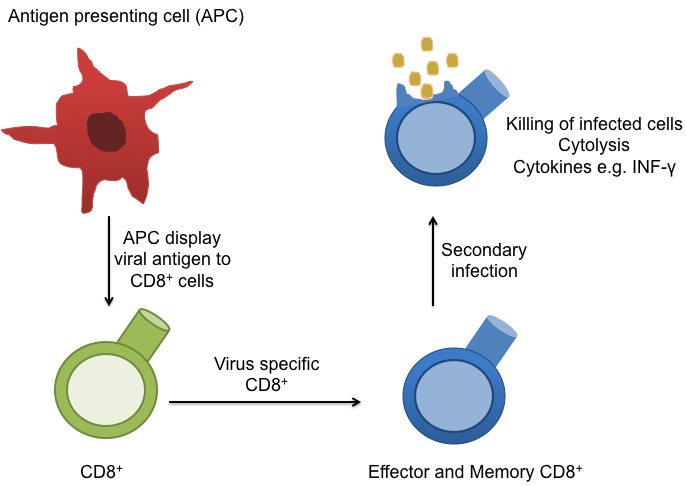
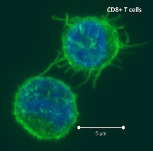
circulate in the blood, lymph, and secondary lymphatic organs as they patrol for the presence of secondary infections.  Recently, a class of effector memory CD8+ T cells has also been shown to migrate to and reside long-term in non-lymphoid tissues. These tissue resident memory T cells can quickly respond to tissue infections with their cognate pathogen.  CD8+ T cells mainly function in killing of infected cells though cytolysis and by producing effector cytokines, including IFN-gamma to activate other immune cells.  In a recent article in Nature Immunology, Schenkel et. al demonstrate an additional function for these tissue resident memory CD8+ T cells: as major producers of chemokines that recruit circulating memory T cell forces to the site of infection.

In this study, Lymphocytic choriomeningitis virus (LCMV) was injected intraperitoneally into female mice that had been adoptively transferred with naïve TCR transgenic CD8+ T cells specific for the gp33 epitope expressed by LCMV.  Two months later, mice were transcervically infected with a Vaccinia virus strain engineered to express gp33 (VV-gp33) and the kinetics of the CD8 T cell recall response in the mucosal tissues of the female reproductive tract were assessed.  Gp33-specific tissue resident memory CD8+ T cells were required for the rapid (within 2 days) recruitment of additional circulating gp33-specific T cells into the tissues.  However, if the secondary infection was with a Vaccinia virus strain expressing a different antigen (OVA), then relatively few memory T cells were seen accumulating in the reproductive tract tissue.  This specificity was also seen when gp33 versus OVA peptides were transcervically injected into the tissues instead of infection with VV.

To derive the mechanisms underlying the recruitment of T cells to infected tissue sites, chemokine expression was assessed.  CXCL9 as well as multiple other chemokines were found to be rapidly induced in various cells residing in the reproductive tissues including endothelial cells, tissue dendritic cells, and memory CD8+ T cells.  Production of IFN-gamma by the tissue resident memory CD8+ T cells was required for both recruitment of additional memory CD8+ T cells to the site as well as CXCL9 production by endothelial cells.

Antigen presenting cells as well as activated T cells produce copious amounts of IFN-gamma and it is expected that IFN-gamma production would elicit CXCL9 expression as the alternate name of CXCL9 is monokine induced by IFN-gamma (MIG).  In this study however, it was production of IFN-gamma by the antigen-specific (gp33) tissue resident memory CD8+ T cells that was critical in the expression of chemokines such as CXCL9 and further recruitment of additional memory CD8+ T cells into the infected tissues.  Antigen presenting cells alone were not as productive in this process because when the secondary infection was instead performed with an OVA-antigen expressing vaccinia virus, significant numbers of OVA-specific T cells were not recruited to the infected tissues.

Thus, this study demonstrated that antigen-specific CD8+ memory T cells that reside in tissues function to significantly amplify innate immune alarms to secondary infections by recruiting additional circulating CD8+ memory T cells to the infected site.

Further Reading:

Sensing and alarm function of resident memory CD8(+) T cells.  Schenkel JM, Fraser KA, Vezys V, Masopust D. Nat Immunol. 2013 May;14(5):509-13. doi: 10.1038/ni.2568. Epub 2013 Mar 31.

Hidden memories: frontline memory T cells and early pathogen interception.  Masopust D, Picker LJ. J Immunol. 2012 Jun 15;188(12):5811-7. doi: 10.4049/jimmunol.1102695.

Chemokine monokine induced by IFN-gamma/CXC chemokine ligand 9 stimulates T lymphocyte proliferation and effector cytokine production.  Whiting D, Hsieh G, Yun JJ, Banerji A, Yao W, Fishbein MC, Belperio J, Strieter RM, Bonavida B, Ardehali A. J Immunol. 2004 Jun 15;172(12):7417-24.

The Crucial Connection Between Metabolism and the Immune System.

Over the past couple of decades, the field of immunology has been growing at an exponential pace. Today, immunologists that continue to study the intracellular and extracellular components are also creating new therapies that suppress the immune system, increase the immune system, and even fix the dysregulation of the immune system. Likewise, there has been an increase in the study of organism metabolism and intracellular metabolism in various pathologies; such as diabetes and cancer. Interestingly, there is also an increase in studying how immune cells function in terms of their intracellular metabolism, how these metabolic pathways affect the phenotype and activation of immune cells, and how the immune system affects the metabolic functions of its host organism also known as immunometabolism1.

There are multiple metabolic pathways that cells use to make ATP. It has been discovered that some cells may preferentially use the glycolytic pathway to make ATP, even when the components are available for aerobic respiration; an event called the Warburg effect1. On the other hand, some cells may use the glycolytic pathway along with the Krebs cycle and electron transport chain (ETC), known as oxidative phosphorylation (OxPhos), to consume materials to make energy1. However, not all immune cells act alike. For instance, activated neutrophils preferentially use the Warburg effect1.  Interestingly, this metabolic pathway makes the most hydrogen peroxide, which is part of the substance neutrophils use for granulocytic release against pathogens1. Likewise, dendritic cells that have been activated via a toll-like receptor agonist and express inducible nitric oxide synthase (iNOS) are also found to use the Warburg effect and, like neutrophils, the metabolite of iNOS plays a functional role in activated dendritic cells1. Finally, the

macrophage

M1 pro-inflammatory macrophages also use the Warburg pathways to make ATP1. Glycolysis and oxidative phosphorylation are linked when the pyruvate from the glycolytic pathway is used to make acetyl-CoA using the Krebs cycle. Immune cells that use this pathway are activated T-cells, immunosuppressive M2 macrophages, and the pro-inflammatory Th17 T-cells1. Finally, fatty acid oxidation, the process of using lipids to make ATP, is utilized by memory T-cells, regulatory T-cells, and alternatively-activated macrophages1.

Metabolism in the immune system is more than just connected to the cell’s activation. Metabolism is involved in the homeostasis between immune cells as well as between immune cells and their stromal host cells1. Dysregulation of metabolites has been found in many pathologies and the role of metabolite fluctuation caused by host immune cells versus pathogens is an active area of study2. Likewise, metabolism has been shown to be involved in class switching effector T-cells into memory T-cells and is involved in the act of bringing immune cells into quiescence3.

An interesting example of a link between immunity and metabolism can be found in an article published in Nature April 11, 2013 by Tannahill et al. This paper demonstrates that the metabolite succinate, a key component in the Krebs cycle, is integral in lipopolysaccharide-induced macrophage activation4. Furthermore, they demonstrated that the activation of toll-like receptor 4, through LPS stimulation, leads to an increase of intracellular glutamate uptake and upregulation of succinate production under the “gamma-Amminobutyric acid (GABA) shunt” metabolic pathway4. The metabolite succinate then stabilizes hypoxia-inducible factor-1α; a protein that is involved in IL-1β production4. 

The study of metabolism is more than just an academic exercise. Many therapies have been proposed that look at changing the metabolic system for alteration of the dysregulated immune response. One of these is metformin, a drug that is used to help regulate type 2 diabetes2. However, metformin is now actively investigated as an anti-cancer therapeutic, having an effect on changing the tumor immune microenvironment from a pro-tumor phenotype to an anti-tumor phenotype2. As we continue to learn more about the effects of host metabolism on the immune system, the role of the immune system in organism metabolism, and the intracellular metabolic pathways involved in various immune cells during different states of function, we will hopefully be able to develop more therapies that repair immune dysregulation as well as develop immunotherapies for treating various metabolic diseases.

Further Reading

1. Pearce, E. L. & Pearce, E. J. Metabolic pathways in immune cell activation and quiescence. Immunity 38, 633-643, doi:10.1016/j.immuni.2013.04.005 (2013).

2. Mathis, D. & Shoelson, S. E. Immunometabolism: an emerging frontier. Nature reviews. Immunology 11, 81, doi:10.1038/nri2922 (2011).

3. Finlay, D. & Cantrell, D. A. Metabolism, migration and memory in cytotoxic T cells. Nature reviews. Immunology 11, 109-117, doi:10.1038/nri2888 (2011).

4. Tannahill, G. M. et al. Succinate is an inflammatory signal that induces IL-1beta through HIF-1alpha. Nature 496, 238-242, doi:10.1038/nature11986 (2013).

Identification of a Novel Eomesodermin Expressing T cell Subset

41BB (CD137) is a costimulatory receptor transiently upregulated on T cells following activation.  41BB is activated by its ligand 41BBL (TNFSF9), a TNF receptor superfamily member expressed by activated antigen presenting cells and anti-41BB agonistic antibodies are in clinical trials for cancer immunotherapy.  In a recent article in The Journal of Experimental Medicine, Curran et. al demonstrate that 41BB activation of T cells leads to the generation of a novel subset of CD4+ and CD8+ T cells dependant on the master transcription factor Eomesodermin (Eomes).

describe the imageActivation of 41BB on T cells leads to enhanced T cell survival.  Anti-41BB-agonistic antibodies have demonstrated significant anti-tumor activity in mice by enhancing anti-tumor cytotoxic T cell responses.  Thus, there are currently several clinical trials underway exploring the efficacy of anti-41BB-agonist antibodies in several types of cancers, including melanoma, renal carcinoma, ovarian cancer, and lymphoma.  In a previous study by the same group (Curran et. al, PLoS One, 2011), an observation was made that a unique subset of T cells infiltrated B16 melanoma tumors in mice after anti-41BB-agonistic antibody treatment.  These T cells expressed the inhibitory receptor KLRG1, and elicited strong anti-tumor activity.  Thus, in the current study, the authors sought to further characterize this T cell subset in mice.

To define the phenotype and functions of tumor-associated KLRG1+ versus KLRG1T cells types, T cells were isolated from B16 tumors established in mice, following treatment with anti-41BB antibodies plus irradiated Flt3-ligand–expressing B16 cells (FVAX) or FVAX alone. The addition of FVAX further enhanced the tumor-infiltrating frequency of KLRG1+ T cells elicited by anti-41BB antibodies.  Gene expression analysis revealed that KLRG1+ CD4+ and CD8+ T cells expressed significantly higher levels of cytoxicity genes: multiple granzymes, perforin, and FasL, than KLRG1T cells.  In vitro cytotoxicity assays with B16 melanoma cell targets demonstrated enhanced killing capacity of KLRG1+ compared with KLRG1 CD4+ and CD8+ T cells.

Superior cytotoxic functions are generally associated with CD4+ TH1 and CD8+ TC1 T cell subsets, dependant on the transcription factor T-bet (TBX21).  However, analysis of expression of the known master transcription factors governing different T cell subsets, found that expression of Eomes but not T-bet was elevated in KLRG1+ T cells.  Runx3 expression was also slightly elevated in KLRG1+ versus KLRG1T cells.  Furthermore, transgenic mice lacking Eomes expression in CD4+ cells (CD4-CRE/Eomesflox/flox) did not develop tumor-infiltrating KLRG1+ T cells after anti-41BB antibody treatment, demonstrating the necessity of Eomes for development of these cells, even when Eomes expression is only absent in the CD4+ T cell compartment.  Thus, these novel subsets of KLRG1+ T cells were termed CD4+ THEO and CD8+ TCEO T cells.

Interestingly, KLRG1+ T cells play a role not only in anti-tumor immunity, but were induced and found at significant levels in spleens and livers from mice infected with Listeria Monocytogenes or LCMV.

As this is a newly described T cell subset, many questions remain.  However, most relevant is whether equivalents of these cells exist in humans, and the roles they play in human diseases.

Further Reading:

Systemic 4-1BB activation induces a novel T cell phenotype driven by high expression of Eomesodermin.  Curran MA, Geiger TL, Montalvo W, Kim M, Reiner SL, Al-Shamkhani A, Sun JC, Allison JP. J Exp Med. 2013 Apr 8;210(4):743-55.

Combination CTLA-4 blockade and 4-1BB activation enhances tumor rejection by increasing T-cell infiltration, proliferation, and cytokine production.  Curran MA, Kim M, Montalvo W, Al-Shamkhani A, Allison JP. PLoS One. 2011 Apr 29;6(4):e19499.

Immunotherapy of cancer with 4-1BB.  Vinay DS, Kwon BS. Mol Cancer Ther. 2012 May;11(5):1062-70. doi: 10.1158/1535-7163.MCT-11-0677. Epub 2012 Apr 24.

Immune regulation by 4-1BB and 4-1BBL: complexities and challenges.  Wang C, Lin GH, McPherson AJ, Watts TH. Immunol Rev. 2009 May;229(1):192-215.

Can shutting down the IRF3 kinases be the magic pill to treat obesity?

amlexanox
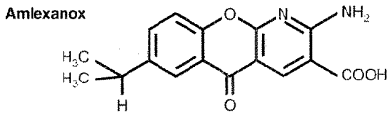
Can amlexanox, a small molecule drug that has been approved for asthma, allergic rhinitis and aphthous ulcers be an effective treatment for obesity? Many are excited about this prospective presented in a recent article in Nature Medicine by Reilly et. al. as simply Googling “amlexanox” and “obesity” resulted in a plethora of news articles on this report.  Interestingly, the two highly related proteins allegedly inhibited by amlexanox that led to this result in mice are the IRF3-kinases, TBK1 (TANK-binding Kinase-1, T2K, NAK) and IKK-ε (IκB kinase-epsilon, IKK-i), whose major known functions are during pathogen infections: the activation of IRF-3 (interferon regulatory kinase-3), the major transcription factor regulating expression of interferon-β (IFNβ).

Inflammation is considered a key link between obesity and insulin resistance.  The canonical NF-ĸB signaling pathway has been shown to playing a major role in this linkage.  IKK-ε and TBK1 are two IKK-β-related kinases with an unclear role in NF-ĸB activation, and thus their role in obesity and insulin resistance was explored in this and a previous study by the same group (Chiang et. al).

In this study, the authors show that expression and kinase activity of IKK-ε and TBK1 were increased in mice fed a high-fat diet and in response to TNFα in an in vitro adipocyte inflammation model. In the previous study, IKK-ε deficient mice were partially resistant to development of obesity and insulin resistance when fed a high fat diet.  Thus, the authors sought to determine if inhibiting these kinases would have a therapeutic effect on obesity.  A screen for small molecule inhibitors to block IKK-ε and TBK1 identified amlexanox, a drug currently used to treat asthma, allergic rhinitis and aphthous ulcers.  Amlexanox treatment inhibited the in vitro kinase activity of both IKK-ε and TBK1 at a much lower concentration than it did the related canonical NF-ĸB pathway kinase IKK-β.

To determine the effect of in vivo inhibition of these kinases on obesity indexes, mice fed a high fat diet were treated with amlexanox.  Amlexanox-treated mice gained significantly less weight than non-treated mice, and treatment of mice with pre-established diet-induced obesity led to a significant, but reversible weight loss accompanied by a decrease in adipose tissue, without a lower food intake.  Additionally, ob/ob mice which are genetically disposed to overt obesity due to deficient leptin expression, also lost adipose tissue mass when treated with amlexanox.

Interestingly, compared with mice fed a normal diet, mice on a high fat diet have a decreased core body temperature. Amlexanox-treated mice had body temperatures raised to normal levels along with increased oxygen consumption, indicating that an increase in energy expenditure of these mice may contribute to their weight loss.  Other indexes of obesity and glucose intolerance also returned to normal levels following amlexanox treatments.

Despite the promising results achieved with this drug in decreasing obesity and insulin resistance, the mechanisms by which inhibition of these kinases lead to this effect remains unclear.  Thus, future studies need to clarify the effects of amlexanox on IKK-ε and TBK1 regulation of the NF-ĸB pathway and obesity-associated inflammation, as well as address effects on the IRF3-interferon pathway.  Finally, it will be important to determine if other molecules are targeted by this drug.


Further Reading:

An inhibitor of the protein kinases TBK1 and IKK-ɛ improves obesity-related metabolic dysfunctions in mice.  Reilly SM, Chiang SH, Decker SJ, Chang L, Uhm M, Larsen MJ, Rubin JR, Mowers J, White NM, Hochberg I, Downes M, Yu RT, Liddle C, Evans RM, Oh D, Li P, Olefsky JM, Saltiel AR. Nat Med. 2013 Mar;19(3):313-21.

The protein kinase IKKepsilon regulates energy balance in obese mice.  Chiang SH, Bazuine M, Lumeng CN, Geletka LM, Mowers J, White NM, Ma JT, Zhou J, Qi N, Westcott D, Delproposto JB, Blackwell TS, Yull FE, Saltiel AR. Cell. 2009 Sep 4;138(5):961-75.

Turning off the inflammatory, but not the metabolic, flames.  Calay ES, Hotamisligil GS. Nat Med. 2013 Mar 6;19(3):265-7.

Deficiency of T2K leads to apoptotic liver degeneration and impaired NF-kappaB-dependent gene transcription.  Bonnard M, Mirtsos C, Suzuki S, Graham K, Huang J, Ng M, Itié A, Wakeham A, Shahinian A, Henzel WJ, Elia AJ, Shillinglaw W, Mak TW, Cao Z, Yeh WC. EMBO J. 2000 Sep 15;19(18):4976-85.

Heterozygous TBK1 mutations impair TLR3 immunity and underlie herpes simplex encephalitis of childhood.  Herman M, Ciancanelli M, Ou YH, Lorenzo L, Klaudel-Dreszler M, Pauwels E, Sancho-Shimizu V, Pérez de Diego R, Abhyankar A, Israelsson E, Guo Y, Cardon A, Rozenberg F, Lebon P, Tardieu M, Heropolitanska-Pliszka E, Chaussabel D, White MA, Abel L, Zhang SY, Casanova JL. J Exp Med. 2012 Aug 27;209(9):1567-82.

TANK-binding kinase 1 (TBK1) controls cell survival through PAI-2/serpinB2 and transglutaminase 2.  Delhase M, Kim SY, Lee H, Naiki-Ito A, Chen Y, Ahn ER, Murata K, Kim SJ, Lautsch N, Kobayashi KS, Shirai T, Karin M, Nakanishi M. Proc Natl Acad Sci U S A. 2012 Jan 24;109(4):E177-86.

*Image courtesy of RXlist.com*

The Immunoscore: bringing immunological parameters to the clinic for cancer patient prognosis

Cancer stages
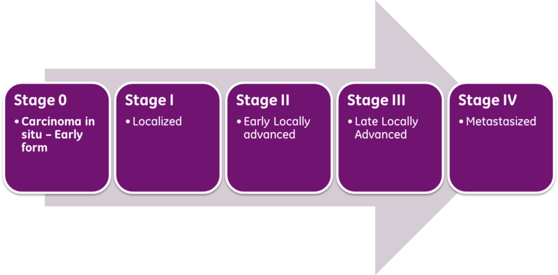
Classical prognosis of cancer patients utilizes the AJCC/UICC (American Joint Committee on Cancer / International Union Against Cancer) “TNM” classification system, in which T (Tumor) is indicative of primary tumor size and invasion properties, N (Nodes) indicates the extent of tumor invasion into draining and regional lymph nodes, and M (Metastasis), describes the presence and extent of metastatic lesions at diagnosis.  The combinations of these parameters are then used to assess a patient’s stage at diagnosis and predict patient outcome.  The exact parameter definitions vary for each cancer type.

However, it has long been known that while the TNM system provides a fairly good estimate for patient populations overall, there is still significant heterogeneity within each stage as to tumor recurrence and ultimate outcome.  This is unsurprising given this staging system evaluates only tumor characteristics and fails to account for other patient parameters. In particular, the integrity of the patient’s immune system, our inherent natural protection against tumor development, has been shown to have a significant impact on disease progression and patient outcome.

In colorectal cancer as well as other cancer types, much progress has been made in ascertaining the prognostic significance of cytotoxic CD8+ T cell infiltration into the tumor microenvironment.  The densities of CD8+ T cell presence in the invasive margin (IM) and the center of the tumor (CT) have been shown to have significant prognostic value.  In a study by Pages et. al. (J Clin Oncol. 2009), assessment of CD8+ T cells and CD45RO+ memory T cell densities in CT/IM tumor regions in stage I and II colorectal cancer patients significantly predicted recurrence and overall survival, showing that application of this system is particularly relevant in early stage patients to better direct treatment strategies.  Multiple cytotoxic CD8+ T cell and TH1 phenotyping markers have shown prognostic significance in human cancer patients, including CD8, CD3, CD45RO, and Granzyme B expression.  However, as discussed by Dr. Jerome Galon in the Oct 3, 2012 J Transl Med. article, CD8 and CD3 represent the most robust markers for adoption into routine clinical practice as CD45RO, and Granzyme B expression are intensity-dependant evaluations and thus much more technically difficult to standardize.

Thus, the proposed immunoscore relies on immunohistochemistry staining for CD8+ and CD3+ T cells in CT/IM tumor regions using standardized antibodies and protocols.  Quantitative assessment of their densities is then determined and scored on whole tissue slides using specified slide scanning and staining analysis software.

In 2012, an international task force, led by Dr. Galon was established to promote the routine usage of this classification system in clinical diagnosis of cancer patients.  The goals of this taskforce include feasibility and standardization of the quantitative immunohistochemistry protocol used to derive the score, worldwide validation of the immunoscore for colorectal cancer patient prognosis, as well as the application of this classification system for other cancer types.  Thus, the immunoscore may soon become a standard clinical practice and aid in better prognostic stratification of patients and therapeutic guidance.


Further Reading:

http://www.immunescore.org/

Website: American Joint Committee on Cancer Staging

Cancer classification using the Immunoscore: a worldwide task forceGalon J, Pagès F, Marincola FM, Angell HK, Thurin M, Lugli A, Zlobec I, Berger A, Bifulco C, Botti G, Tatangelo F, Britten CM, Kreiter S, Chouchane L, Delrio P, Arndt H, Asslaber M, Maio M, Masucci GV, Mihm M, Vidal-Vanaclocha F, Allison JP, Gnjatic S, Hakansson L, Huber C, Singh-Jasuja H, Ottensmeier C, Zwierzina H, Laghi L, Grizzi F, Ohashi PS, Shaw PA, Clarke BA, Wouters BG, Kawakami Y, Hazama S, Okuno K, Wang E, O’Donnell-Tormey J, Lagorce C, Pawelec G, Nishimura MI, Hawkins R, Lapointe R, Lundqvist A, Khleif SN, Ogino S, Gibbs P, Waring P, Sato N, Torigoe T, Itoh K, Patel PS, Shukla SN, Palmqvist R, Nagtegaal ID, Wang Y, D’Arrigo C, Kopetz S, Sinicrope FA, Trinchieri G, Gajewski TF, Ascierto PA, Fox BA.  J Transl Med. 2012 Oct 3;10:205.

The immune score as a new possible approach for the classification of cancer.  Galon J, Pagès F, Marincola FM, Thurin M, Trinchieri G, Fox BA, Gajewski TF, Ascierto PA. J Transl Med. 2012 Jan 3;10:1.

The immune contexture in human tumours: impact on clinical outcome. Fridman WH, Pages F, Sautes-Fridman C, Galon J.  Nat Rev Cancer 2012, 12:298-306.

In situ cytotoxic and memory T cells predict outcome in patients with early-stage colorectal cancerPagès F, Kirilovsky A, Mlecnik B, Asslaber M, Tosolini M, Bindea G, Lagorce C, Wind P, Marliot F, Bruneval P, Zatloukal K, Trajanoski Z, Berger A, Fridman WH, Galon J. J Clin Oncol. 2009 Dec 10;27(35):5944-51.

Image courtesy of simplyanon on Wikipedia

Induction of Tumor cell senescence by TH1 cytokines IFN-g and TNF

interferon betaInterferons, including type I (IFNα/β) and type II (IFNγ) are known to be critical for mediating multiple aspects of tumor immunity, by targeting both immune cells for activation, and cancer cells for expression of MHC and genes associated with growth arrest and apoptosis.  Thus, expression of cytokines such as IFNγ by immune cells has been shown to be critical in anti-tumor immune responses.  IFNγ is one of the major effector cytokines of CD4+ TH1 cells and cytotoxic CD8+ T cells.  However, the full effects of IFNγ as well as other TH1 cytokines in mediating anti-tumor effects have not been fully elucidated.

Previous observations were made that tumor-specific CD4+ TH1 cells, IFNγ, and TNF were required for controlling tumor growth.  However, tumor growth arrest induced by IFNγ and TNF occurred without either significant T cell infiltration into the tumor, or appreciable tumor cell destruction (Müller-Hermelink et. al, 2008).  Thus, the anti-tumor mechanisms of the combined action of IFNγ and TNF produced by TH1 cells remained to be defined.  In an interesting follow-up study by the same group published in the February 21, 2013 issue of Nature, Braumüller et. al, further explore the mechanisms by which IFNγ and TNF produced by TH1 cells induce direct tumor cell growth arrest.

Culturing of pancreatic β-cancer cells with IFNγ plus TNF directly induced tumor cell growth arrest in the G1/G0 phase.  Interestingly, even following removal of IFNγ and TNF, tumor cells remained growth arrested for at least two weeks in vitro, indicating IFNγ and TNF induced cellular senescence.  Both IFNγ and TNF were required to induce senescence as either alone was not sufficient.

Induction of the p16INK4a gene by the combined actions of IFNγ-STAT1 and TNF-TNFR1 pathways was found to mediate this effect via consequential hypo-phosphorylation of the p16–retinoblastoma protein (Rb), thus maintaining its activated state.   Rb mediates senescence growth arrest by suppressing E2F, a transcription factor that promotes expression of cell cycle progression genes.  The role for these pathways was further validated by short hairpin (sh)-RNA knockdown of p16INK4a and p19 which inhibited tumor cell senescence by IFNγ and TNF.  The authors further demonstrated this phenomenon of TH1 cell – IFNγ and TNF induced tumor cell senescence in multiple cancer cell types as well as in an in vivo pancreatic cancer model.  Tumor cells rendered senescent by IFNγ and TNF in vivo remained arrested, even in the absence of T cells, B cells, and NK cells, following implantation into NOD–SCID/IL2rγ−/− mice.

Thus these studies define a mechanism of tumor-growth inhibition by the direct actions of the CD4+ TH1 cytokines IFNγ and TNF in mediating tumor cell senescence through activation of the Rb pathway.

The role of IFNγ in mediating tumor-immune responses is increasingly complex. IFNγ production has been negatively correlated with effective anti-tumor CD8+ T cell responses in some models (Gattinoni et. al) and also has been shown to induce expression of the immune inhibitory receptor PD-L1 (Lyford-Pike et. al).  Thus, these studies highlight that the contextual roles of immune cell effector cytokines are critical in their functions for regulation of tumor immunity and direct effects in tumor cells themselves.

Further Reading:

T-helper-1-cell cytokines drive cancer into senescence.  Braumüller H, Wieder T, Brenner E, Aßmann S, Hahn M, Alkhaled M, Schilbach K, Essmann F, Kneilling M, Griessinger C, Ranta F, Ullrich S, Mocikat R, Braungart K, Mehra T, Fehrenbacher B, Berdel J, Niessner H, Meier F, van den Broek M, Häring HU, Handgretinger R, Quintanilla-Martinez L, Fend F, Pesic M, Bauer J, Zender L, Schaller M, Schulze-Osthoff K, Röcken M. Nature. 2013 Feb 21;494(7437):361-5. doi: 10.1038/nature11824.

TNFR1 signaling and IFN-gamma signaling determine whether T cells induce tumor dormancy or promote multistage carcinogenesis.  Müller-Hermelink N, Braumüller H, Pichler B, Wieder T, Mailhammer R, Schaak K, Ghoreschi K, Yazdi A, Haubner R, Sander CA, Mocikat R, Schwaiger M, Förster I, Huss R, Weber WA, Kneilling M, Röcken M. Cancer Cell. 2008 Jun;13(6):507-18.

Cellular senescence: when bad things happen to good cells. Campisi, J. & d’Adda di Fagagna, F. Nature Rev. Mol. Cell Biol. 8, 729–740 (2007).

Acquisition of full effector function in vitro paradoxically impairs the in vivo antitumor efficacy of adoptively transferred CD8+ T cells.  Gattinoni L, Klebanoff CA, Palmer DC, Wrzesinski C, Kerstann K, Yu Z, Finkelstein SE, Theoret MR, Rosenberg SA, Restifo NP. J Clin Invest. 2005 Jun;115(6):1616-26.

Evidence for a role of the PD-1:PD-L1 pathway in immune resistance of HPV-associated head and neck squamous cell carcinoma.  Lyford-Pike S, Peng S, Young GD, Taube JM, Westra WH, Akpeng B, Bruno TC, Richmon JD, Wang H, Bishop JA, Chen L, Drake CG, Topalian SL, Pardoll DM, Pai SI. Cancer Res. 2013 Jan 3.

photo credit: AJC1 via photopin cc

Development of Anti-viral CD4+ T cells in unexposed individuals

HIVIn a study published in the February 2013 issue of Immunity, Su. et. al., characterized the CD4+ T cell repertoire from adult human peripheral blood mononuclear cells (PBMC) and made a fascinating observation: that memory T cells can develop against pathogens the host has never been exposed to, including HIV-1, cytomegalovirus (CMV), and herpes simplex virus (HSV).

To determine the baseline frequencies of pathogen and self-antigen-specific CD4+ T cell populations in naive individuals by flow cytometry, PBMCs were stained with major histocompatibility complex (MHC)-peptide tetramers for HLA-DR4 -restricted epitopes.  The MHC-peptides assessed included peptides from viral pathogens including HIV-1 (gag p24), CMV (pp65), and HSV (VP16), as well as self-peptides: the melanoma-associated antigen gp100, the arthritis-associated antigen fibrinogen, and the diabetes-associated antigen preproinsulin. For both self and pathogen-associated antigens, the frequency of tetramer-positive cells was found to be one to ten cells per million CD4+ T cells, in individuals previously unexposed to these viruses.  Prior exposure to viral pathogens was determined by highly sensitive serological tests.

Interestingly, on average across individuals, greater than 50% of these antigen-specific CD4+ T cells were found to be memory T cells based on CD45RO+ staining and expression of CD4+ memory T cell-associated genes.  Furthermore, viral antigen-specific CD45RO+ memory T cells secreted IFNg when stimulated with phorbol myristate acetate (PMA) plus ionomycin.  Sequencing of the TCR-beta chain from naïve and memory HIV-1-specific CD4+ T cells indicated that the antigen-specific memory phenotype but not naive CD4+ T cell populations had arisen from clonal expansion.  Thus the memory phenotype of these antigen-specific T cells indicates prior antigen experience and subsequent clonal expansion.

The question remained though, as to how individuals had developed CD4+ memory T cell responses against pathogens they had not apparently been exposed to.  One hypothesis for the existence of these CD4+ memory T cell populations is development via cross-reactivity to other antigens.  BLAST analysis of the HIV-1 peptide sequence identified several similar sequences present in gut and soil-resident bacteria, marine algae, and plants.  HIV-1-specific CD4+ memory T cell clones from various individuals showed cross-reactivity to several of these environmental microbial antigens as measured by production of IFNg and IL-2, and proliferation when stimulated with MHC-peptides.  Interestingly, antigen-specific CD4+ memory T cells elicited by vaccination of individuals with an H1N1 influenza virus vaccine were reciprocally cross reactive to several other microorganisms.

Overall, this study shows that pathogen-specific CD4+ memory T cells commonly arise in individuals through exposure to other environmental microbes containing similar peptide sequences.  The question remains however, as to whether these cross-reactive antigen-specific T cells are able to exhibit protective immune responses.


Further Reading:

Virus-Specific CD4 (+) Memory-Phenotype T Cells Are Abundant in Unexposed Adults.  Su LF, Kidd BA, Han A, Kotzin JJ, Davis MM. Immunity. 2013 Feb 5. pii: S1074-7613(13)00052-6.

Derivation and maintenance of virtual memory CD8 T cells.  A.D. Akue, J.Y. Lee, S.C. Jameson.  J. Immunol., 188 (2012), pp. 2516 – 2523

Nonrandom attrition of the naive CD8+ T-cell pool with aging governed by T-cell receptor:pMHC interactions. B.D. Rudd, V. Venturi, G. Li, P. Samadder, J.M. Ertelt, S.S. Way, M.P. Davenport, J. Nikolich-Žugich.  Proc. Natl. Acad. Sci. USA, 108 (2011), pp. 13694–13699.

Maintenance of peripheral naive T cells is sustained by thymus output in mice but not humans.  I. den Braber, T. Mugwagwa, N. Vrisekoop, L. Westera, R. Mögling, A.B. de Boer, N. Willems, E.H. Schrijver, G. Spierenburg, K. Gaiser et al.  Immunity, 36 (2012), pp. 288–297

Photo credit: Microbe World / Foter.com / CC BY-NC-SA

The Shock of Sepsis: The Struggle to Treat SIRS

Since the mid-1880’s, scientists and clinicians have studied the concept of sepsis. Investigations started with physician-scientists such as Ignaz Semmelweiss, Joseph Lister, Hugo Schottmüller and Louis Pasteur, initiating the fields of immunology and diseases related to immunology that continue even to today. Unfortunately, while we increase our knowledge about sepsis (also known as systemic inflammatory response syndrome; SIRS), diagnosis and treatment remain difficult1,2. This is seen as recently as last year, when an infection of a 12-year-old boy , Rory Staunton, lead to fatal septic shock after doctors failed to see the early signs of SIRS3. His death initiated the formation of the Rory Staunton Foundation and subsequent reforms of emergency protocols taken in his home state of New York (labeled as “Rory’s Laws”)3.

describe the imageHowever, hospital policy reforms are just part of the issue; sepsis affects more than 700,000 people in North America every year, with a 30-50% mortality rate4. To date, there are no FDA-approved drugs to fight SIRS4. In fact, after 20 years of intense research into translational medicine for sepsis, none of the proposed treatment approaches have had enough clinical efficacy to be used as treatment paradigms1. Action must be taken to not only change the methodologies of care for sepsis, but to better understand the biological mechanisms of sepsis. From this understanding, we may be able to have more accurate detection of sepsis as well as more effective therapeutics.

Sepsis occurs when an infection causes a systemic inflammatory response5. This disease causes an imbalance of the immune system, leading to changes in the hemodynamics of the host, resulting in coagulation, heart ischemia, and multi-organ failure1.  Originally thought to be caused by overactivation of the innate immune system, many patients did not die from the initial onslaught of inflammation, but from later stages of immune system suppression (also known as “immunoparalysis”) which allowed opportunistic viruses and bacteria to take over the host1. The roles of sepsis may be analyzed by observing the different cell types it affects: the innate system, the adaptive immune system, and non-immune cells.

Since sepsis is due to bacterial and viral infection, the innate immune system is the most well studied aspect of the pathogenesis of sepsis. During SIRS the innate immune cells are pathologically affected: macrophages, dendritic cells and natural killer (NK) cells. During the initial period of infection, increased amounts of pathogen-associated molecular pattern (PAMPs) and damage-associate molecular pattern (DAMP) molecules are found in the host organism. Toll-like receptors (TLRs), such as TLR4 and TLR9, in the innate immune cells recognize these molecules and confer a host inflammatory response.  The inflammation leads to an increased presence of adhesion molecules on both innate and adaptive immune cells4 indicating the desire to enter the site of infection to confer an immune response.  Along the same lines, activation of the complement cascade occurs, leading to increased amounts of C5a protein. The upregulated C5a protein levels cause increased migration of innate immune cells and increased phagocytic ability of macrophages4.

Unfortunately, prolonged exposure of macrophages to C5a leads to dysregulation of macrophage activation and, eventually, apoptosis1. During this apoptotic stage, macrophages release high-mobility group protein B1 (HMGB1) which may lead to increased inflammation5. On the other hand, in neutrophils, sepsis is known to cause an abnormally long proliferation period5 which could lead to organ damage and increased inflammation. Surprisingly, sustained activation of neutrophils may also contribute to the immunoparalysis by sepsis because activated neutrophils increase reactive oxygen species, known to cause immunosuppression, and macrophages that eat apoptotic neutrophils express anti-inflammatory cytokines5. describe the imageProlonged sepsis also decreases the pro-inflammatory ability of dendritic cells; SIRS increases depletion of splenic and myeloid dendritic cells, where the remaining dendritic cells are functionally deficient4.  NK cells, originally shown to play a role in anti-viral immune responses but are also proposed to play a role in bacterial infection responses, have two distinct roles in different phases of sepsis.  In the early phases, NK cells are thought to contribute to the overactive immune responses and systemic inflammation4 however, in later phases of sepsis, NK cells may be compromised which could lead to secondary bacterial or viral infections, thus exacerbating the inflammatory response during SIRS4.

describe the imageIn addition to dysregulation of the innate immune system, the cells that make up the adaptive immune system, T-cells and B-cells, are also affected by sepsis. Sepsis decreases overall T-cell receptor function, and moves Th1 (proinflammatory) T-cells toward a Th2 (immunosuppressive) response1. In addition, CD25+Foxp3+ T-cells, also known as regulatory T-cells, are increased during SIRS5. Interferon-gamma, a proinflammatory cytokine expressed during sepsis, causes an increase in innate response activator (IRA) B Cells, B-cells that recognize PAMPs, impair infection clearance, and accelerate septic shock4,6. During sepsis, the increase of C5a causes T-Cell and B-cell apoptosis4, which leads to immunosuppression4.

Sepsis also has non-immunological effects on the patient. During sepsis, coagulation becomes more pronounced. This increase in coagulation and pre-coagulation states may lead to ischemic injury5.  Sepsis also causes cytopathic hypoxia5 and cardiomyopathy4. Interestingly, SIRS also plays a role on the autonomic nervous system, with increased apoptosis of the adrenal medullary cells, there is a dysregulation of the endocrine system that regulates the autonomic nervous system1.

Currently, there are several studies looking into the pathogenesis of sepsis with the goal being effective treatment. In terms of pathogenesis, recent studies looking at the role of STIM17  and PI3K activation in the initiation of sepsis5. Host deficiencies leading to sepsis are also being studied. Deficiencies, including,  zinc8 and ADAMS-T5 deficiencies, will hopefully elucidate more on the dysregulation found in sepsis. Currently, medical scientists are working with clinicians to study the more nuanced roles of hyper-responsiveness versus system immune suppression4. From these studies, multiple immunotherapies have been proposed: dendritic cell implantation4, regulatory T-cell implantation 4, and the modification of the host immune system by using TLR antagonists4, injections of the proinflammatory cytokines IL-15 and IL-174, and limiting adaptive immune system exhaustion by blocking PD-14. However, several of these studies, such as T-reg implantation, and using TLR antagonists have had limited clinical efficacy and have been prematurely terminated4. This may be due to the lack of an appropriate model to study human sepsis in the pre-clinical setting.

The limitations of current research methodologies for sepsis have shown the need for reform in SIRS research. Recently an article by Seok et al in the Proceedings of the National Academy of Sciences elucidated the limitations of using mouse models to studying human inflammation9. In this article, Seok et al list the differences in temporal responses, gene signatures, and regulated pathways involved inflammatory response following various insults9. The reasons for describe the imagedifferences in the inflammatory response may be described by the evolutionary differences between mouse and human immune systems, the inbred nature of the mice used, and the tendency to focus on a single mechanism in mouse models when there is great overlap of immune responses within a host system. These authors propose studying more of the genetic and epigenetic changes of patient samples during sepsis to find appropriate mouse models for study9. Furthermore, they propose in vitro recapitulation of the inflammation response in diseased tissue9. Unfortunately, the in vitro model is limited and this reductionist approach may miss a key cell, cellular event, or special/temporal arrangement that may be vital to pathogenesis of sepsis.

The use of non-human primates as a sepsis model may be more accurate than either of these, but the ethical and cost-effective issues of using non-human primates remains a deterrent. A final model that may be of use to sepsis researchers in the future is the use of the humanized mouse model; a severely immunocompromised mouse host that has had a recapitulation of the adaptive and/or innate immune systems for in vivo study of human pathologies.  The use of humanized mice for the study of sepsis was proposed by Unsinger et al, where they used two-day-old NOD-scid IL2 receptor-gamma knockout mice and transplanted them with hCD34+ enriched hematopoietic cord blood stem cells.  After establishment of the human immune system, mice were treated with cecal ligation puncture (CLP) model of intra-abdominal peritonitis and assayed for immune response changes10. These mice developed a functional human innate and adaptive immune system with recapitulation of the human immune response to sepsis.  However, while there still remain differences in the humanized mouse model versus the actual human immune response, such as differences in the bacterial flora that is found in the bowels10, this model nonetheless provides a useful tool which, along with more genetic and epigenetic information from patient studies, will lead to more impactful pre-clinical studies and possibly FDA-approved drugs to treat sepsis in the clinics.


References:

1. Rittirsch, D., Flierl, M. A. & Ward, P. A. Harmful molecular mechanisms in sepsis. Nat Rev Immunol 8, 776-787, doi:10.1038/nri2402 (2008).

2. Stearns-Kurosawa, D. J., Osuchowski, M. F., Valentine, C., Kurosawa, S. & Remick, D. G. The pathogenesis of sepsis. Annu Rev Pathol 6, 19-48, doi:10.1146/annurev-pathol-011110-130327 (2011).

3. Dwyer, J. Death of a Boy Prompts New Medical Efforts Nationwide, October 26, 2012).

4. Ward, P. A. & Bosmann, M. A historical perspective on sepsis. Am J Pathol 181, 2-7, doi:10.1016/j.ajpath.2012.05.003 (2012).

5. Cinel, I. & Opal, S. M. Molecular biology of inflammation and sepsis: a primer. Crit Care Med 37, 291-304, doi:10.1097/CCM.0b013e31819267fb (2009).

6. Rauch, P. J. et al. Innate response activator B cells protect against microbial sepsis. Science 335, 597-601, doi:10.1126/science.1215173

7. Gandhirajan, R. K. et al. Blockade of NOX2 and STIM1 signaling limits lipopolysaccharide-induced vascular inflammation. J Clin Invest, doi:10.1172/jci65647 (2013).

8. Liu, M. J. et al. ZIP8 Regulates Host Defense through Zinc-Mediated Inhibition of NF-κB. Cell Rep, doi:10.1016/j.celrep.2013.01.009 (2013).

9. Seok, J. et al. Genomic responses in mouse models poorly mimic human inflammatory diseases. Proc Natl Acad Sci U S A, doi:10.1073/pnas.1222878110 (2013).

10. Unsinger, J., McDonough, J. S., Shultz, L. D., Ferguson, T. A. & Hotchkiss, R. S. Sepsis-induced human lymphocyte apoptosis and cytokine production in “humanized” mice. J Leukoc Biol 86, 219-227, doi:10.1189/jlb.1008615 (2009).