Unique anti-tumor functions of IFNg vs. IL-17 producing CD8 cells

interferon gammaThe anti-tumor effectiveness of IFNγ-producing CD4+ TH1 cells and CD8+ TC1 cells is well accepted.  However, the role of IL-17 producing CD4+ (TH17) and CD8+ T cells (TC17) in promoting or inhibiting tumor growth remains unclear, as various studies have shown both tumor-inhibiting and tumor-promoting functions of these cell types.  Thus, context is a key determinant factor in the role of IL-17 producing T cell subsets in tumor immune responses.

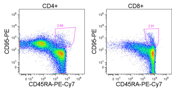
In the current February 15, 2013 issue of The Journal of Immunology, Yu. et. al. compared the role of adoptively transferred tumor-specific TC1 and TC17 cells in controlling tumor growth.  In this murine model, anti-gp100 T cells from Pmel-1 TCR transgenic mice were polarized ex-vivo into TC1 or TC17 phenotypes, and adoptively transferred into luciferase-expressing B16F10 melanoma lung metastatic tumor-bearing mice that had undergone total body irradiation.

The efficacy of anti-tumor TC1 or TC17 cells in mediating tumor regression was monitored by measuring tumor burden by luciferase luminescence and overall survival.  By these measures, both TC1 and TC17 cells exhibited anti-tumor activity, however TC1 cells were superior.  TC1 cells completely inhibited tumor growth, while TC17 cells delayed tumor growth but were ultimately unable to control the tumor.

Adoptively transferred TC1 cells were found to produce only IFNγ but not IL-17, while TC17 cells expressed high levels of IL-17 and could also differentiate into IFNγ-producing cells, as is known for these cell subsets.  In a fascinating observation, the authors found that eliminating IFNγ-responsiveness in tumor cells completely reversed the relative efficacy of TC1 and TC17 cells. IFNγ-responsiveness in tumor cells was required for TC1 mediated anti-tumor activity, indicating a critical role for IFNγ-responsive genes in promoting tumor cell recognition by TC1 cells and/or growth inhibition or sensitivity to apoptosis. However, when tumor cells could no longer respond to IFNγ, TC17 cells were now able to induce complete tumor regression. In cytokine-neutralization assays, in vivo IFNγ was required however, for the anti-tumor effectiveness of TC17 cells while IL-17 was not.  This indicates that in the context of TC17 cell therapy, IFNγ is required for modulation of non-tumor cells in the tumor microenvironment, while IFNγ signaling in the tumor cells themselves is puzzlingly detrimental.

Within the first few days of adoptive transfer, TC1 cells proliferated faster in vivo than TC17 cells.  However, after two and four weeks, in vivo levels of TC17 cells were higher or similar in the spleen and lungs compared with TC1 cells in mice bearing wild-type as well as IFNγ-nonresponsive tumors.  Thus, the superior in vivo persistence of TC17 cells may be a factor in TC17 cell-elicited anti-tumor responses.  IFNγ elicits its effects through activation of the STAT1 transcription factor, while IL-17 signals through an alternate ACT1 pathway to activate NF-ĸBIL-22, also released by TC17 cells activates STAT3.  Thus an interaction between these pathways in tumor cells may mediate the differential requirement for IFNγ-responsiveness in TC1 vs. TC17 mediated anti-tumor effects.

While it remains unclear why TC17 cells in this model were able to effectively control IFNγ-nonresponsive tumor cells but not wild-type tumor cells, and TC1 cells exhibited the opposite propensity, these observations have important implications for future enactment of adoptive cell transfer for tumor therapeutics.  Because of their inherent stem-like propensity for long term in vivo persistence and demonstrations of highly effective anti-tumor functions, TH17 cells have been proposed to be a superior CD4+ cellular subset for adoptive anti-tumor T cell therapy in combinations with CD8+ T cells.  However, the observations in this study suggest that different cytokine-producing CD8+ TC and CD4+ TH subsets will vary in their effectiveness depending on factors such as the tumor’s ability to respond to cytokines including IFNγ.


Further Reading:

Adoptive Transfer of Tc1 or Tc17 Cells Elicits Antitumor Immunity against Established Melanoma through Distinct Mechanisms.  Yu Y, Cho HI, Wang D, Kaosaard K, Anasetti C, Celis E, Yu XZ. J Immunol. 2013 Feb 15;190(4):1873-81.

Tumor-specific Th17-polarized cells eradicate large established melanoma.  Muranski P, Boni A, Antony PA, Cassard L, Irvine KR, Kaiser A, Paulos CM, Palmer DC, Touloukian CE, Ptak K, Gattinoni L, Wrzesinski C, Hinrichs CS, Kerstann KW, Feigenbaum L, Chan CC, Restifo NP. Blood. 2008 Jul 15;112(2):362-73.

Phenotype, distribution, generation, and functional and clinical relevance of Th17 cells in the human tumor environments.  Kryczek I, Banerjee M, Cheng P, Vatan L, Szeliga W, Wei S, Huang E, Finlayson E, Simeone D, Welling TH, Chang A, Coukos G, Liu R, Zou W. Blood. 2009 Aug 6;114(6):1141-9.

Structure and signalling in the IL-17 receptor superfamily.  Sarah L. Gaffen.  Nat Rev Immunol. 2009 August; 9(8): 556.

Human TH17 cells are long-lived effector memory cells.  Kryczek I, Zhao E, Liu Y, Wang Y, Vatan L, Szeliga W, Moyer J, Klimczak A, Lange A, Zou W. Sci Transl Med. 2011 Oct 12;3(104):104ra100.

Photo credit: AJC1 via photopin cc

T cell dysfunction in cancer: Anergy, Exhaustion, or Senescence?

Immune responses against cancer have been shown to be effective in eliminating tumors.  However anti-tumor immunity is limited by dysfunctional T cells which have been described in cancer patients.  Understanding how dysfunctional T cells arise in cancer and the potential mechanisms for restoring functionality are critical for developing effective immunotherapeutics.  In the current issue of Current Opinion in Immunology, Dr. Weiping Zou’s group, (Crespo. et. al.,) review the various types of T cell dysfunction that occur in the tumor microenvironment.

Anergy, exhaustion, and senescence are three different mechanisms underlying T cell hyporesponsiveness, which share distinguishing phenotypic features but arise by different mechanisms and under different experimental settings.  However, what is the difference between these three types of T cell dysfunction and which contribute to impaired T cell responses in the setting of cancer?

Anergy: T cell anergy generally refers to a hyporesponsive state in T cells induced by triggering the TCR either without adequate concomitant co-stimulation through CD28 or in the presence of high co-inhibitory molecule signaling.  Without both TCR and CD28 signals, IL-2 is not effectively transcribed and instead, anergy-associated genes such as GRAIL are expressed which contribute to impaired TCR signaling via negative feedback.

In the tumor microenvironment cancer, altered expression of B7 family members by APCs leads to an enhanced expression of B7 family co-inhibitory molecules including PD-L1 and a reduction in the B7 family co-stimulatory molecules CD80 and CD86.  Thus, T cell activation in this environment could lead to induction of anergy.

Exhaustion: T cell exhaustion occurs as a result of chronic over-stimulation, such as occurs in the settings of chronic viral infections including hepatitis C virus (HCV) and HIV, autoimmunity, and cancer.  Exhausted T cells progressively lose the ability to express effector cytokines including IL-2, IFNg, and TNFα.  They also express multiple inhibitory receptors including PD-1 and LAG-3, lose cytotoxic and proliferative potential, and may ultimately be driven to apoptosis.

Because anti-tumor T cells are persistently exposed to antigen in the tumor microenvironment, exhaustion is a likely mechanism contributing to T cell dysfunction in cancer patients.  As such, exhausted T cells have been described in patients with melanoma, ovarian cancer and hepatocellular carcinoma.

Senescence:  Senescence is thought to occur due to the natural life span, or aging of cells.  However, senescent T cells have been observed in the settings of chronic inflammation and persistent infection in young individuals, indicating other factors beyond a person’s age, such as DNA damage, drive acquisition of this state.  Senescent T cells are marked by deficient CD28 expression, telomere shortening, expression of regulatory receptors such as TIM-3 and KLRG-1, and inability to progress through the cell cycle.

Cells with features of senescence have been described in patients with lung cancer, head and neck cancer, hepatocellular carcinoma, melanoma and lymphoma.  Thus, senescence may also contribute to T cell dysfunction in cancer patients.

In conclusion, there is evidence that anergy, exhaustion, and senescence may all be contributing toward T cell dysfunction in cancer.  In the review, Crespo. et. al., make the point that distinguishing cells in these states may be complicated as they overlap phenotypically and in expression of various markers.  Furthermore, the mechanisms mediating establishment of these three states is not well defined.  However, it is important to clarify the mechanisms by which T cells gain and maintain dysfunction in cancer in order to best develop effective immunotherapeutics.


Further Reading:

 T cell anergy, exhaustion, senescence, and stemness in the tumor microenvironment.  Crespo J, Sun H, Welling TH, Tian Z, Zou W.  Curr Opin Immunol. 2013 Jan 5.

The three main stumbling blocks for anticancer T cells.  Baitsch L, Fuertes-Marraco SA, Legat A, Meyer C, Speiser DE. Trends Immunol. 2012 Jul;33(7):364-72.

Induction of T cell anergy: integration of environmental cues and infectious tolerance. P. Chappert, R.H. Schwartz.  Curr. Opin. Immunol., 22 (2010), pp. 552–559.

T cell exhaustion.  E.J. Wherry.  Nat Immunol, 12 (2011), pp. 492–499.

T-cell senescence: a culprit of immune abnormalities in chronic inflammation and persistent infection.  A.N. Vallejo, C.M. Weyand, J.J. Goronzy.  Trends Mol Med, 10 (2004), pp. 119–124.

Upcoming Immunology Conferences: March – June, 2013

antibodiesI previously posted about 2013 Conferences in Tumor Immunology and Cancer ImmunotherapyThis listing will include other upcoming Immunology-related conferences.


Keystone Symposium: Understanding Dendritic Cell Biology to Improve Human Disease

March 3 – 8, 2013.

Keystone, Colorado, USA.

Registration Deadline: March 3, 2013.


Gordon Research Conference: Cell Biology of Megakaryocytes & Platelets

March 10 – 15, 2013.

Galveston, Texas, USA

Application Deadline: February 10, 2013.


World Immune Regulation Meeting (WIRM) VII: Innate and Adaptive Immune Response and Role of Tissues in Immune Regulation

March 13 – 16, 2013.

Congress Center, Davos, Switzerland.

Registration is still open online.


Keystone Symposium: Host Response in Tuberculosis

This is a joint meeting with the Keystone meeting on Tuberculosis: Understanding the Enemy

March 13 – 18, 2013.

Whistler, British Columbia, Canada.

Registration Deadline: March 3, 2013.


Keystone Symposium: Tuberculosis: Understanding the Enemy

This is a joint meeting with the Keystone meeting on Host Response in Tuberculosis.

March 13 – 18, 2013.

Whistler, British Columbia, Canada.

Registration Deadline: March 3, 2013.


Keystone Symposium: Immune Activation in HIV Infection: Basic Mechanisms and Clinical Implications

April 3 – 8, 2013.

Breckenridge, Colorado, USA.

Registration Deadline: April 3, 2013.


Canadian Society for Immunology 26th Annual Spring Meeting.

April 5 – 8, 2013.

TELUS Whistler Conference Centre, Whistler, British Columbia, Canada.

Registration is open online.  Early registration ends March 1, 2013.

Abstract Submission Deadline: March 1, 2013.


Keystone Symposium: Immunopathology of Type 1 Diabetes

April 4 – 9, 2013.

Whistler, British Columbia, Canada.

Registration Deadline: April 4, 2013.


Keystone Symposium: Advances in the Knowledge and Treatment of Autoimmunity

April l4 – 9, 2013.

Whistler, British Columbia, Canada.

Registration Deadline: April 4, 2013.


Molecular Pattern Recognition Receptors

April 11 – 13, 2013.

Boston, Massachusetts, USA.

Early Registration Deadline: March 1, 2013.


Clinical Immunology Society Annual Meeting: Regulation and Dysregulation of Immunity

April 25 – 28, 2013.

Miami, Florida, USA.

Pre-Registration Deadline: April 3, 2013.


T cell Function and Modulation Meeting

April 28 – May 1, 2013.

Makena Beach & Golf Resort, Maui (Makena), HI.

Registration can be submitted online and is limited to the first 125 attendees.


IMMUNOLOGY 2013, AAI Annual Meeting and Centennial Celebration

May 3 – 7, 2013.

Hawaii Convention Center, Honolulu, Hawaii, USA.

Registration is open online.  Early registration ends March 18, 2013.

Abstract Submission Deadline: February 13, 2013.


Keystone Symposium: The Innate Immune Response in the Pathogenesis of Infectious Disease

May 5 – 10, 2013.

A Universidade Federal de Ouro Preto, Ouro Preto, Brazil.

Early Registration Deadline: March 5, 2013.


Cell Symposia: Microbiome and Host Health

May 12 – 14, 2013.

Lisbon, Portugal.

Abstract Submission Deadline: February 8, 2013.

Early Registration Deadline: March 9, 2013.


30 Years of HIV Science: Imagine the Future

May 21 – 23, 2013.

The International Research Centre in Paris, the Institut Pasteur Conference Centre, Paris, France.

Abstract Submission Deadline: March 1, 2013.

Early Registration Deadline: March 20, 2013.


Abcam: Allergy & Asthma 2013

May 23 – 24, 2013.

Bruges, Belgium.

Oral Abstract Submission Deadline: February 22, 2013.

Poster Abstract Submission Deadline: March 25, 2013.

Early Registration Deadline: March 25, 2013.


ISIR 2013 – Building Bridges in Reproductive Immunology.

May 28 – June 1, 2013.

Boston Park Plaza Hotel, Boston, Massachusetts, USA.

Registration is open online.

Abstract Submission Deadline: February 15, 2013.


78th Cold Spring Harbor Symposium on Quantitative Biology: Immunity & Tolerance

May 29 – June 3, 2013.

Cold Spring Harbor Laboratory, New York, USA

Abstract and Registration Deadline: March 15, 2013.


6th International Singapore Symposium of Immunology.

June 5 – 6, 2013.

Matrix Level 2 Auditorium, Biopolis, Singapore.

Registration is open online.

Abstract Submission Deadline: April 5, 2013.


Cell Symposium: Immunometabolism: From Mechanisms to Therapy

June 9 – 11, 2013.

The Sheraton Centre Toronto Hotel, Toronto, Canada.

Abstract Submission Deadline: February 22, 2013.

Early Registration Deadline: April 5, 2013.


Gordon Research Conference: Mucosal Health & Disease

June 9 – 14, 2013.

Stonehill College, Easton, Massachusetts, USA.

Application Deadline: May 12, 2013.


Gordon Research Conference: Phagocytes

June 9 – 14, 2013.

Waterville Valley, New Hampshire, USA.

Application Deadline: May 12, 2013.


European Academy of Allergy & Clinical Immunology and World Allergy Organization: World Allergy & Asthma Congress

June 22 – 26, 2013.

Milan, Italy.

Early Registration Deadline: February 20, 2013.


Aegean Conference: 10th International Conference on Innate Immunity

June 23 – 28, 2013.

Kos, Greece.

Early Registration and Abstract Submission Deadline: March 15, 2013.

Gordon Research Conference: Apoptotic Cell Recognition & Clearance

June 23 – 28, 2013.

University of New England, Biddeford, Maine, USA.

Application Deadline: May 26, 2013.


Abcam: Inflammasomes in Health and Disease

June 24 – 25, 2013.

Boston, Massachusetts, USA.

Oral Abstract Submission Deadline: April 26, 2013.

Poster Abstract Submission Deadline: May 17, 2013.

Early Registration Deadline: April 26, 2013.


FOCIS 2013

June 27 – 30, 2013.

Boston, Massachusetts, USA.

Late-breaking Abstract Submission Deadline: April, 2, 2013.

Registration Deadline for Poster Presenters:  March 28, 2013.

Websites that list upcoming Conferences & Events in Immunology, Tumor Immunology, and Cancer Immunotherapy:

The American Association of Immunologists (AAI) Meetings and Events Calendar

Nature Reviews Immunology’s list of conferences

Cancer Immunity Journal’s List of Conferences

New role for p53 in regulating the inflammatory tumor microenvironment

small 3316209998p53 (TP53), a transcription factor activated by various types of cellular stress, is one of the most well studied tumor suppressor proteins.  Stress signals such as various types of DNA damage lead to p53 activation and subsequent induction of genes involved in cell cycle arrest, DNA repair, and apoptosis.

In many types of cancers including colorectal cancer, loss of function mutations in p53 commonly occur, particularly in the DNA-binding domain.  Thus, transcriptional activity of p53 is altered or lost, and cells with damaged genomes maintain viability.  In colorectal cancer, p53 mutations commonly occur at early stages and are thought to allow for accumulation of DNA damage and survival of mutated cells during tumorigenesis.

In the January issue of Cancer Cell, Schwitalla et. al, demonstrate that p53 plays a role in not only its classically associated functions as a critical DNA-damage checkpoint in early stages of tumorigenesis, but during the progression stage, p53 plays an additional significant role in regulating the inflammatory tumor microenvironment.

To study the role of p53 during various stages of carcinogenesis, the authors established a colon tumor model in which azoxymethane (AOM), a promoter of Wnt-driven tumorigenesis that induces stabilizing mutations in β-catenin, was administered to mice with a p53-deficiency in intestinal epithelial cells.  A gene expression analysis of AOM-derived tumors from wild-type versus p53-deficient mice, found that p53 deficiency led to upregulation of genes involved in inflammation and epithelial-mesenchymal transition (EMT).  A large number of genes upregulated in p53-deficient tumors were found to be regulated by the inflammatory transcription factor NF-ĸB, and phosphorylated p65, a major transcriptional co-factor in the classical NF-ĸB pathway, was enhanced in p53-deficient tumors.  NF-ĸB regulated genes included the EMT gene Twist as well as myeloid cell chemokines CXCL1, CXCL2, and CCL2, indicating that loss of p53 may lead to EMT and recruitment of inflammatory myeloid cells via upregulation of the NF-ĸB pathway.

Reciprocal regulation of the NF-ĸB and p53 pathways has long been known, and these pathways play opposing roles in cell fate in response to stress. However, in this study, the authors demonstrated that p53-deficient intestinal epithelial cells were impaired in their epithelial barrier function: leakage of intestinal bacteria and LPS resulted in the observed NF-ĸB activation and NF-ĸB-regulated gene expression.  Interestingly, NF-ĸB signaling was required in both tumor cells and the myeloid cells for maximal tumor progression.  NF-ĸB signaling in intestinal epithelial cells was required for recruitment of myeloid cells to the tumor microenvironment and EMT induction. NF-ĸB signaling in hematopoietic cells was required for tumor cell proliferation, survival, and metastasis, by inducing expression of cytokines that activate STAT3 in the tumor cells.

Thus, in this model, p53 plays an additional role during tumor progression by regulating inflammation in the tumor microenvironment, via its effects on intestinal barrier function.  In my last blog post, I discussed another recent study demonstrating links between the TNFα/NF-ĸB, IL-6/STAT3, and SphK1/S1P pathways in chronic inflammation driven colorectal cancer.  Thus, this study adds another layer of links to consider in the complicated interactions between inflammatory cells and tumor cells in the tumor microenvironment that drive colorectal cancer progression: p53, NF-ĸB, and STAT3.


Further Reading:

Loss of p53 in Enterocytes Generates an Inflammatory Microenvironment Enabling Invasion and Lymph Node Metastasis of Carcinogen-Induced Colorectal Tumors.  Schwitalla S, Ziegler PK, Horst D, Becker V, Kerle I, Begus-Nahrmann Y, Lechel A, Rudolph KL, Langer R, Slotta-Huspenina J, Bader FG, Prazeres da Costa O, Neurath MF, Meining A, Kirchner T, Greten FR. Cancer Cell. 2013 Jan 14;23(1):93-106.

p53 and NF-κB: different strategies for responding to stress lead to a functional antagonism.  Ak P, Levine AJ. FASEB J. 2010 Oct;24(10):3643-52.

Alterations of the TP53 gene in gastric and esophageal carcinogenesis.  Bellini MF, Cadamuro AC, Succi M, Proença MA, Silva AE. J Biomed Biotechnol. 2012;2012:891961.

Transcriptional control of human p53-regulated genes. T. Riley, E. Sontag, P. Chen, A. Levine.  Nat. Rev. Mol. Cell Biol., 9 (2008), pp. 402–412.

Immunity, inflammation, and cancer.  S.I. Grivennikov, F.R. Greten, M. Karin.  Cell, 140 (2010), pp. 883–899.

photo credit: sc63 via photopin cc

Signal amplification loops in chronic inflammation and Colon Cancer

The inflammatory gastrointestinal diseases Crohn’s disease, inflammatory bowel diseases (IBD), and ulcerative colitis (UC), are all known to be key risk factors in the development of colorectal cancer.  TNFα and IL-6 are two pro-inflammatory cytokines that signal to activate the NF-ĸB and STAT3 transcription factors, respectively.  Both pathways are known to play a role in the persistent inflammation that drives development of colitis-associated colorectal cancer.

Sphingosine-1-phosphate (S1P) is a lysophospholipid that is metabolized from phosphorylation of sphingosine by the kinases, SphK1 and SphK2, and signals through the G-protein coupled receptor sphingosine-1-phosphate receptor-1 (S1PR1).  The S1P-S1PR1 signaling pathway has been shown to activate STAT3 and induce expression of both IL-6 and S1PR1, driving a positive feedback loop leading to persistent STAT3 activation in tumor cells (Lee et. al., 2010).  S1P and SphK1 have also been shown to play a critical role in the NF-ĸB signaling pathway.  S1P was required for TRAF2 E3 ubiquitin ligase activity and activation of NF-ĸB by TNFα independently from the S1PR1 receptor pathway (Alvarez et. al., 2010).  SphK1 has also been linked to development of colorectal cancer.  SphK1 was highly expressed in human colons cancers as compared with normal mucosa, and SphK1-/- mice were less susceptible to azoxymethane induction of colon cancer (Kawamori et.al., 2009).

In a study by Liang et al. in this month’s issue of Cancer Cell, the authors close these links between the TNFα/NF-ĸB, IL-6/STAT3, and SphK1/S1P pathways in chronic inflammation driven colorectal cancer.  In the dextran sodium sulfate (DSS) murine model of colitis driven tumorigenesis, deficiency of SphK2 led to a compensatory upregulation of SphK1 and S1P production, and exacerbated DSS driven colitis and subsequent tumorigenesis.  S1P-driven NF-ĸB activation was enhanced, as was production of TNFα, IL-6, and STAT3 activation in SphK2-/- mice, and the SphK1/S1P pathway primarily exerted its activities in colonic mucosa infiltrating immune cells.  FTY720, an antagonist of S1PR1 and SphK1, was able to inhibit the S1P positive-feedback loop and block persistent activation of STAT3 and NF-ĸB and production of IL-6.  Additionally, FTY720 inhibited the development of tumors in this colitis driven tumorigenesis murine model.

In summary, these studies show that SphK1 drives a feed-forward signal amplification loop that exacerbates inflammation and promotes tumorigenesis by production of S1P.  S1P then drives S1PR1 activation of STAT3 leading to further production of S1PR1, as well as activates NF-ĸB and induction of TNFα and IL-6 transcription, which further feed back on the NF-ĸB and STAT3 pathways, respectively.  Blocking this amplification loop by inhibiting S1PR1 and SphK1 may thus be a promising treatment strategy for colitis-associated colorectal cancer.


Further Reading:

Sphingosine-1-Phosphate Links Persistent STAT3 Activation, Chronic Intestinal Inflammation, and Development of Colitis-Associated Cancer.  Liang J, Nagahashi M, Kim EY, Harikumar KB, Yamada A, Huang WC, Hait NC, Allegood JC, Price MM, Avni D, Takabe K, Kordula T, Milstien S, Spiegel S. Cancer Cell. 2013 Jan 14;23(1):107-20.

Sphingosine 1-Phosphate Is a Missing Link between Chronic Inflammation and Colon Cancer.  Pyne NJ, Pyne S. Cancer Cell. 2013 Jan 14;23(1):5-7.

STAT3-induced S1PR1 expression is crucial for persistent STAT3 activation in tumors.  H. Lee H, Deng J, Kujawski M, Yang C, Liu Y, Herrmann A, Kortylewski M, Horne D, Somlo G, Forman S, Jove R, Yu H.. Nat. Med., 16 (2010), pp. 1421–1428.

Sphingosine-1-phosphate is a missing cofactor for the E3 ubiquitin ligase TRAF2.  Alvarez SE, Harikumar KB, Hait NC, Allegood J, Strub GM, Kim EY, Maceyka M, Jiang H, Luo C, Kordula T, Milstien S, Spiegel S. Nature. 2010 Jun 24;465(7301):1084-8.

Role for sphingosine kinase 1 in colon carcinogenesis. T. Kawamori, T. Kaneshiro, M. Okumura, S. Maalouf, A. Uflacker, J. Bielawski, Y.A. Hannun, L.M. Obeid.  FASEB J., 23 (2009), pp. 405–414.

Sphingosine 1-phosphate and its receptors: an autocrine and paracrine network.  Rosen H. and Goetzl EJ.   Nature Reviews Immunology 5, 560-570, July 2005.

 Immunity, inflammation, and cancer.  S.I. Grivennikov, F.R. Greten, M. Karin.  Cell, 140 (2010), pp. 883–899.

Intestinal inflammation and cancer.  T.A. Ullman, S.H. Itzkowitz.  Gastroenterology, 140 (2011), pp. 1807–1816.

Antagonism of Tumor-Immunity by Chemotherapeutics

dendritic cellSignificant steps forward are being made in immunotherapeutic approaches for treatment of cancer.  Over the past few years, two cancer immunotherapeutics were FDA approved.  In 2010, Dendreon Corporation’s Provenge, an autologous cellular vaccine, was approved for hormone refractory metastatic prostate cancer.  In 2011, Bristol-Myers Squibb’s anti-CTLA4 antibody Ipilimumab, was FDA approved for late stage melanoma.  Recent promising clinical trial results indicate several additional immune modulating therapies are likely to join this prestigious list in the coming years.  In addition, combinations of immune therapies with chemotherapeutics are being tested in clinical trials.  In light of this, it is important to know how chemotherapies interact with the immune system, in order to best generate synergistic effects.

There are multiple ways that chemotherapies may modulate anti-tumor immunity.  Some therapeutics such as anthracyclines induce an immunogenic cell death characterized by release of endogenous danger signals such as HMGB1, that activate antigen presenting cells (APCs) to elicit anti-tumor T cell responses.  Standard apoptosis elicited by a range of other therapeutics however, is often non-immunogenic.  Chemotherapies can induce lymphopaenia which may have both negative and positive effects on anti-tumor immunity, including loss of anti-tumor effector T cells as well as negative regulatory T cells and myeloid derived suppressor cells (MDSC).

In the recent January issue of Nature Medicine, Bruchard et. al., demonstrate that the widely prescribed chemotherapeutic agents, gemcitabine and 5-fluorouracil (5-FU) activated the NLRP3 (NOD-like receptor family, pyrin domain containing-3) inflammasome complex in MDSCs, leading to perturbed anti-tumor immunity and reduced therapeutic effect of these drugs.

The NLRP3 inflammasome is activated in response to damage associated molecular patterns (DAMPs) released during infection with a plethora of pathogens.  NLRP3 activation leads to formation of the multi-protein inflammasome complex that activates caspase-1.  IL-1b is a pro-inflammatory cytokine that is transcribed as an inactive pro-peptide and requires processing by caspase-1 into its active secreted form, and is a major inflammasome effector molecule.

In the study by Bruchard et. al., gemcitabine and 5-FU activated the NLRP3 inflammasome complex in MDSCs, and led to characteristic activation of caspase-1 and IL-1b.   In contrast, the chemotherapeutics Deticene, taxol, oxaliplatin, mitomycin C, and doxorubicin did not activate this pathway.  Cathepsin B release from damaged lysosomes into the cytosol was shown as the trigger of NLRP3 activation by gemcitabine and 5-FU.  Importantly, increased serum concentrations of IL-1b, and enhanced caspase-1 and cathepsin B activity in circulating MDSCs were found colorectal cancer patients one day after 5-FU treatment, validating the relevance of these observations.

Studies on the subset of CD4+ T helper cells that produce IL-17 (TH17) have demonstrated both positive and negative roles for these cells in cancer pathogenesis, and the contexts by which TH17 cells can play opposing roles is unclear.  In this study, IL-1b released by MDSCs treated with 5-FU promoted differentiation of CD4+ T cells into TH17 cells.  5-FU treatment promoted IL-17 production in PBMCs from colorectal cancer human patients as well.  Mice lacking inflammasome components or IL-17 demonstrated enhanced survival when treated with 5-FU.  Thus, in mice, 5-FU elicited TH17 cells play a pro-tumorigenic role.  Whether or not 5-FU elicited TH17 cells also promote tumor growth in human patients is a critical question to be addressed.

Finally, treatment of mice with the soluble form of IL-1Ra blocked the effects of IL-1b and promoted the anti-tumor effects of 5-FU.  Therefore, IL-1b blockade represents a rational immunotherapeutic strategy to enhance the effects of 5-FU and gemcitabine chemotherapies.

In conclusion, this study identified key mechanisms of immune modulation by gemcitabine and 5-fluorouracil in murine models and human cancer patients.  Chemotherapeutics elicit cellular damage by many different mechanisms, and understanding how each drug interacts with the immune system will be important for promoting critical synergy between chemotherapies and anti-tumor immunity.

Further Reading:

Chemotherapy-triggered cathepsin B release in myeloid-derived suppressor cells activates the Nlrp3 inflammasome and promotes tumor growth.  Bruchard M, Mignot G, Derangère V, Chalmin F, Chevriaux A, Végran F, Boireau W, Simon B, Ryffel B, Connat JL, Kanellopoulos J, Martin F, Rébé C, Apetoh L, Ghiringhelli F. Nat Med. 2013 Jan;19(1):57-64.

Immunological aspects of cancer chemotherapy. Zitvogel, L., Apetoh, L., Ghiringhelli, F. & Kroemer, G. Nat. Rev. Immunol. 8, 59–73 (2008).

Dual role of immunomodulation by anticancer chemotherapy.  Shurin MR.  Nat Med. 2013 Jan;19(1):20-2.

Inflammasomes and their roles in health and disease.  Lamkanfi M, Dixit VM. Annu Rev Cell Dev Biol. 2012;28:137-61.

5-Fluorouracil selectively kills tumor-associated myeloid-derived suppressor cells resulting in enhanced T cell–dependent antitumor immunity. Vincent, J. et al. Cancer Res. 70, 3052–3061 (2010).

Gemcitabine selectively eliminates splenic Gr-1+/CD11b+ myeloid suppressor cells in tumor-bearing animals and enhances antitumor immune activity. Suzuki, E., Kapoor, V., Jassar, A.S., Kaiser, L.R. & Albelda, S.M. Clin. Cancer Res. 11, 6713–6721 (2005).

Restoration of antitumor immunity through selective inhibition of myeloid derived suppressor cells by anticancer therapies. Apetoh, L., Vegran, F., Ladoire, S. & Ghiringhelli, F. Curr. Mol. Med. 11, 365–372 (2011).

2013 Conferences in Tumor Immunology and Cancer Immunotherapy


Tumor Invasion and Metastasis

January 20 – 23, 2013

Omni San Diego Hotel, San Diego, CA

Online registration is closed, but attendees may register onsite on at the conference registration desk on a first-come, first-served basis beginning from Sunday, January 20 at 4:00 p.m.


Keystone Symposia: Cancer Immunology and Immunotherapy

January 27 – February 1, 2013.

Fairmont Hotel Vancouver, Vancouver, British Columbia, Canada.

Registration is still open until January 27, 2013.


Keystone Symposia: Antibodies as Drugs

January 27 – February 1, 2013.

Fairmont Hotel Vancouver, Vancouver, British Columbia, Canada.

Abstract submission is closed. Registered attendees can bring a poster onsite.

Registration is still open online.


Global Technology Community: The 2nd Novel Immunotherapeutics Summit

January 30 – February 1, 2013.

The Westin Gaslamp Quarter, San Diego, California, USA.

The summit includes a pre-summit workshop and 4 concurrent tracks.

Abstracts can be submitted online, and registration is available for Workshop alone, the Workshop + the Immunotherapetics & Immunomonitoring Track, or the entire Summit.

**Purchase an All Conference Pass for attendance to ALL Global Technology Community Conferences for One Year for $3,995 ($1,990 for Acad/Govt)**

Workshop: Immune Responses in Tumor Microenvironment

January 30, 2013.  This is a half day workshop.

Track: 5th Immunotherapeutics & Immunomonitoring

Track: 11th Cytokines & Inflammation

Track: 2nd Allergy & Respiratory Drug Discovery

Track: Immunogenicity & Immunotoxicity


2013 Tumor Immunology Lab Symposium: Tumor vs Immune System: A Cytokine Battle!

February 6, 2013.

Radboud University Nijmegen Medical Centre, Nijmegen, Netherlands.

Registration is Free, however limited to 150 attendees!


First Symposium of the Cancer Research Center of Lyon

February 13 – 15, 2013.

Convention Center, Lyon, France.

Online registration deadline: February 3, 2013.


Global Technology Community: 2nd Novel Cancer Therapeutics Summit

February 25 – 26, 2013.

Palms Casino Resort / Palms Place, Las Vegas, Nevada, USA.

Abstracts and registration can be submitted online.


IMMUNO 2013: 10th International Conference on New Trends in Immunosuppression and Immunotherapy

March 11 – 12, 2013.

Hotel Fira Palace, Barcelona, Spain.

Registration is still open online.


Arrowhead’s 2nd Annual Cancer Immunotherapy Conference: Stem Cells and Cancer Immunotherapy

April 4-5, 2013

The Washington Post Conference Center, Washington, D.C., USA.

Abstract submissions are due by March 10, 2013 to: poster@arrowheadpublishers.com

Registration and application for Speaking Opportunities can be done online.


Global Technology Community: Cancer Immunotherapy and Immunomonitoring Conference

April 22 – 25, 2013.

Hilton Garden Inn, Krakow, Poland.

Abstracts and registration can be submitted online.


Cancer Immunotherapy Consortium 2013 Scientific Colloquium: Entering the Era of Combination Therapies: Practical Implementation

April 25-27, 201.

Willard Intercontinental, Washington, DC, USA.

Early Registration Deadline: March 15, 2013.


Roche – Nature Medicine Imunology Symposium 2013: Host Immunity to Cancer and Chronic Viral Infections

April 28–30, 2013.

Roche Forum, Buonas, Switzerland.

This is a closed symposium.  Only 50 attendees will be selected to participate in addition to the invited speakers.  Applications for attendance and abstracts can be submitted online.

Application & Abstract Submission Deadline: February 21, 2013.


T cell Function and Modulation Meeting

April 28 – May 1, 2013.

Makena Beach & Golf Resort, Maui (Makena), HI.

Registration can be submitted online and is limited to the first 125 attendees.


CIMT Annual Meeting

May 14 – 16, 2013.

Rheingoldhalle Congress Center, Mainz, Germany

The Association for Cancer Immunotherapy (CIMT) Annual Meeting is the largest meeting in Europe focused on research and development in cancer immunotherapy.

Abstract submission deadline: March 15, 2013.

Early Registration deadline: March 15, 2013.


Cold Spring Harbor Asia Conference: Tumour Immunology and Immunotherapy

October 28 – November 1, 2013.

Suzhou Dushu Lake Conference Center, Suzhou, China.

Abstract submission deadline: August 16, 2013.

Early Registration deadline: August 16, 2013.


Websites that list upcoming Conferences & Events in Immunology, Tumor Immunology, and Cancer Immunotherapy:


The American Association of Immunologists (AAI) Meetings and Events Calendar

Nature Reviews Immunology’s list of conferences

Cancer Immunity Journal’s List of Conferences


Identification of human stem cell-like memory T cells in PBMC

Memory T cell populations are heterogeneous in phenotype and function and many questions remain as to the mechanisms mediating their long term persistence.  Recent research by several groups have described populations of antigen-experienced T cells within human peripheral blood mononuclear cells (PBMC) that exhibit stem cell-like characteristics: increased self-renewal capacity and the ability to derive the more differentiated central and effector memory and effector populations in vitro and in vivo, and may thus be the cell type mediating memory T cell persistence.

In 2011, Gattinoni et. al. identified a population of stem cell-like memory T cells (TSCM) with surface markers characteristic of naive T cells in human PBMCs.   TSCM cells were CD45RO, CCR7+, CD45RA+, CD62L+, CD27+, CD28+ and IL-7Ra+.  The TSCM population comprised 2-3% of CD8+ and CD4+ T cells in healthy donors.  These TSCM cells could be differentiated from naïve T cells by high expression of CD95 and IL-2Rb, markers which are also expressed by memory T cells.  Furthermore, the TSCM population exhibited a gene expression profile that was intermediate between naïve (TN) and central memory (TCM) cells.

Like memory T cells, these TSCM cells were antigen-experienced and exhibited rapid effector activity upon T cell receptor (TCR) stimulation.  Importantly, they also exhibited the stem-like property of self-renewal in the presence of homeostatic IL-15 signals.  Following TCR stimulation, TSCM cells could differentiate into TCM and effector memory (TEM) T cell subsets, and the authors demonstrated a progressive differentiation pattern of TN à TSCM à TCM à TEM, where no differentiation in the opposite direction was observed following TCR stimulation of sorted TSCM, TCM, and TEM populations.  Human TSCM cells also survived significantly longer and produced more progeny in vivo then either TCM or TEM populations in a NOD.Cg-PrkdcscidIl2rgtm1Wjl/SzJ (NSG) mouse xenograft adoptive transfer model.

One of the most exciting clinical implications of this work however, was the demonstration that TSCM cells exhibited profoundly superior anti-tumor activity than either TCM or TEM  populations in a xenograft mouse tumor model where mesothelin-specific human T cell populations were transferred into NSG mice bearing human mesothelioma M108 tumors.  Thus, TSCM cells may be the most effective cellular subset for use in adoptive T cell therapy in cancer patients.

Subsequent to this finding, a paper was published by Lugli et. al., describing a protocol for identifying and isolating human TSCM cells from PBMCs as well as their in vitro expansion.  The flow cytometry staining panel proposed for TSCM cell identification includes antibodies targeting CD3, CD8, CD4, CD45RO, CCR7, either CD62L, CD27, CD28 or CD45RA, and CD95CD58 and CD122 (IL-2Rb) were also proposed as additional markers for better differentiation of TSCM cells from naïve populations which express these at lower levels.

Identification of human stem cell-like memory T cells in PBMC


Interestingly, the naïve-like TSCM population described by Gattinoni et. al. is not the only memory T cell population demonstrated to have “stem-like” characteristics.

In a quest to understand the mechanism by which patients who have undergone multiple rounds of cytotoxic chemotherapy induced lymphopenia maintain resistance to viral infections, Turtle et. al. described a population of human PBMC CD8+ T cells within the central and effector memory populations that were distinguished by expression of high levels of IL-18Rα and the natural killer (NK) cell receptor CD161.  These cells exhibited a hematopoietic stem cell-like capacity to efflux chemotherapeutic agents mediated by expression of ABCB1, survive chemotherapy, and replenish the virus-specific memory T cell pool in acute myeloid leukemia (AML) patients.

Human memory TH17 cells also have stem cell-like characteristics.  Despite their effector memory-like surface marker phenotype being CD45RO+ CCR7CD62L, compared with TH1 and TH2 subsets, TH17 cells were shown to have increased capacities for proliferation, in vivo persistence, resistance to apoptosis, and higher expression levels of stem-cell associated genes HIF1a, Notch, Bcl2, OCT4, and Nanog.  TH17 cells were able to differentiate into TH1 and TREG subsets.  TH17 cells also express CD161 and thus may overlap with Turtle et. al.’s CD161+ABCB1+ stem-like memory cells.

However, conflicting evidence has been presented as to the identity of CD161+ IL-17 expressing cells and whether or not these cells are in fact Vα7.2+ mucosal associated invariant T cells (MAITs) which are selected by nonpolymorphic MHC class Ib molecules.  MAIT cells however are not known to be virus-specific whereas the CD161+ABCB1+ stem-like memory population identified by Turtle et. al. included influenza and EBV-specific populations.  Thus much remains to be clarified regarding the overlap between CD161+ABCB1+ stem-like memory populations, IL-17 expressing CD4+ (TH17) and CD8+ (TC17) cells which also express CD161, and CD161+ IL-17 expressing MAIT cell populations.

In summary, the ability to identify various stem-like memory CD4 and CD8 human T cell populations in human PBMC using flow cytometry allows for many questions to be addressed about the phenotype, functions, and clinical applications of these cells.

.

Further Reading:

A human memory T cell subset with stem cell-like properties.  Gattinoni L, Lugli E, Ji Y, Pos Z, Paulos CM, Quigley MF, Almeida JR, Gostick E, Yu Z, Carpenito C, Wang E, Douek DC, Price DA, June CH, Marincola FM, Roederer M, Restifo NP. Nat Med. 2011 Sep 18;17(10):1290-7.

Identification, isolation and in vitro expansion of human and nonhuman primate T stem cell memory cells.  Lugli E, Gattinoni L, Roberto A, Mavilio D, Price DA, Restifo NP, Roederer M. Nat Protoc. 2012 Dec 6;8(1):33-42.

A distinct subset of self-renewing human memory CD8+ T cells survives cytotoxic chemotherapy.  Turtle CJ, Swanson HM, Fujii N, Estey EH, Riddell SR. Immunity. 2009 Nov 20;31(5):834-44.

Human TH17 cells are long-lived effector memory cells.  Kryczek I, Zhao E, Liu Y, Wang Y, Vatan L, Szeliga W, Moyer J, Klimczak A, Lange A, Zou W. Sci Transl Med. 2011 Oct 12;3(104):104ra100.

Human MAIT cells are xenobiotic-resistant, tissue-targeted, CD161hi IL-17-secreting T cells.  Dusseaux M, Martin E, Serriari N, Péguillet I, Premel V, Louis D, Milder M, Le Bourhis L, Soudais C, Treiner E, Lantz O. Blood. 2011 Jan 27;117(4):1250-9. doi: 10.1182/blood-2010-08-303339. Epub 2010 Nov 17.

CD161-expressing human T cells.  Fergusson JR, Fleming VM, Klenerman P. Front Immunol. 2011;2:36. doi: 10.3389/fimmu.2011.00036.