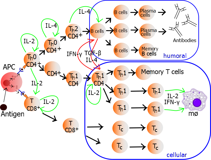
CD4+ T helper type 1 (TH1) cells are the effector T cell population that governs cell mediated immune responses against intracellular pathogens including viruses and intracellular bacteria. TH1 cells mediate their effect by secreting cytokines such as interferon-gamma (IFNγ) and IL-2, and express cell surface markers including CXCR3 and CCR5 and the characteristic TH1 master transcription factor T-bet (TBX21) which can also be used for detection of TH1cells by flow cytometry, as discussed in a previous blog post.
Differentiation of naïve human CD4+ T cells down the TH1 pathway involves cytokines such as IL-12 which activates STAT4, and induces expression of IFNγ and T-bet. As such, in vitro protocols differentiating peripheral blood mononuclear cells (PBMC)-derived naïve CD4+ T cells into TH1 cells involves incubation with IL-12 in the context of T cell activation through the T cell receptor (TCR) complex.
In my experience, TH1 cells are by far the easiest CD4+ helper T cell population to generate in vitro. In order to generate TH1 cells from human PBMC, naïve CD4+ T cells must first be isolated. Multiple methods of naïve CD4+ T cell isolation can be utilized, and magnetic bead- based methods are common and easy methods. Companies such as Miltenyi Biotech and Stem Cell Technologies offer kits for isolation of untouched naïve CD4+ T cells from PBMC by negative isolation methodologies.
Following isolation, naïve CD4+ T cells are activated through the TCR complex. Tissue culture plates can be coated with anti-CD3 (OKT1) and anti-CD28 antibodies in PBS prior to culture. Alternatively, naïve CD4+ T cells can be cultured with Dynal CD3/CD28 T Cell Expander Dynabeads (Life Technologies) at a 1 bead per cell ratio. A third alternative involves coating tissue culture plates with anti-CD3 alone and obtaining CD28 co-stimulation by the addition of autologous monocytes isolated from PBMCs into the culture.
To generate TH1 cells, recombinant human IL-12 is added alone, or at a lower dose in combination with anti-IL-4 blocking antibodies to inhibit the counteractive effects of IL-4 and TH2 pathways on TH1 cell polarization. Finally recombinant human IL-2 is added to promote T cell proliferation. Media and cytokines/blocking antibodies are refreshed every two to three days depending on the cell density, and as the cells expand the time to refresh the media shortens.
 TH1 cells can be generated and assayed for functions including IFNγ expression in as few as three days. If long term or clonal T cells assays are of interest, cells can be expanded in the presence of IL-2 for 2-3 weeks following single cell cloning. As previously discussed, TH1 cells can be identified by IFNγ expression following a 4-6 hour incubation with TCR activation by plate bound anti-CD3 plus anti-CD28, CD3/CD28 Dynabeads, or PMA/ionomycin in the presence of brefeldin-A. Cells are then fixed, permeabilized, and stained for cell surface markers and intracellular IFNγ.
Finally, as a comparison, tandem experiments can be run in which naïve CD4+ T cells are maintained under non-polarizing (TH0) conditions. For this, often no cytokines aside from IL-2 are added. However the addition of anti-IL-12 and anti-IL-4 may be necessary to inhibit any cells from differentiating down TH1 or TH2 pathways by production of these cytokines by the T cells themselves.
In conclusion, generation of CD4+ TH1cells from human PBMC is a relatively simple and straightforward protocol, and very high percentages of TH1cells can be obtained through optimized protocols.
Further Reading:
Differentiation of effector CD4 T cell populations (*). Zhu J, Yamane H, Paul WE. Annu Rev Immunol. 2010;28:445-89.
Memory and flexibility of cytokine gene expression as separable properties of human T(H)1 and T(H)2 lymphocytes. Messi M, Giacchetto I, Nagata K, Lanzavecchia A, Natoli G, Sallusto F. Nat Immunol. 2003 Jan;4(1):78-86.
A critical function for transforming growth factor-beta, interleukin 23 and proinflammatory cytokines in driving and modulating human T(H)-17 responses. Volpe E, Servant N, Zollinger R, Bogiatzi SI, Hupé P, Barillot E, Soumelis V. Nat Immunol. 2008 Jun;9(6):650-7.

