 CXCR5 is a chemokine receptor expressed by and used to identity human CD4+ follicular helper T cells (TFH). TFH cells, as their name implies, promote the differentiation and survival of memory and plasma B cells in the B cell follicular and germinal center regions of secondary lymphoid organs. CXCR5+ TFH-like central memory CD4+ T cells (CD4+ TCM) also circulate in peripheral blood and can be detected among human peripheral blood mononuclear cells (PBMC). CXCR5+ cells comprise 20-25% of CD4+ TCM cells in human PBMC. However, what are the identifying markers and functional differences of CXCR5+ vs. CXCR5– CD4+ T cells from human PBMC and the prototypical CXCR5+ TFH cells in secondary lymphoid organs?
I have previously discussed markers that can be used for identification of human CD4+ TH1, TH2, and TH17 T cell helper subsets as well as CD4+ FoxP3+ regulatory T cells. CXCR5 can be upregulated transiently on activated T cells, however a subset of PBMC T cells constitutively express CXCR5, indicating this is a uniquely functioning subset identifiable by this marker using flow cytometry or other methods. PBMC CXCR5+ CD4+ TCM cells exhibit many but not all features of TFH cells present in secondary lymphoid organs, and thus may be the circulating memory counterpart of TFH cells.
CXCL13, the chemokine ligand for CXCR5, is highly expressed in B cell follicles and likely plays an important role in recruitment of CXCR5+ TFH cells to B cell zones. Expression of ICOS by TFH cells has been demonstrated to be essential for their function in B cell help. Additionally, TFH cells are higher expressers of CXCL13, as well as IL-21, IL-10, Bcl-6, and PD-1 than other helper T cell subsets.
Studies by Chevalier et al, and Morita et. al. compared the functional properties of CXCR5– and CXCR5+ CD4+ TCM cells from human PBMC. PBMC CXCR5+ CD4+ cells are resting central memory cells in phenotype, being CD45RA–, CCR7+ and CD62L+, but not expressing activated TFH markers such as ICOS and CD69. Upon stimulation, CXCR5+ CD4+ TCM promote significantly higher B cell plasmablast differentiation and Ig secretion than CXCR5– CD4+ TCM cells, attributable to enhanced expression of ICOS and IL-10. However, Bcl-6 expression was not found to be different between these PBMC subsets, and conclusions for expression levels and role of IL-21 were contradictory between these studies.
The question of whether PBMC CXCR5+ CD4+ TCM are distinct from TH1, TH2, and TH17 T cells was addressed by these studies as well. While Chevalier et al. found that CXCR5+ TCM cells were more non-polarized and secreted comparatively lower levels of cytokines associated with TH1, TH2, and TH17 T cells, Morita et. al, identified TH1, TH2, and TH17 T cells within CXCR5+ compartment, albeit at somewhat different frequencies than CXCR5– cells. Thus PBMC CXCR5+ CD4+ TCM are a heterogenous subset with features of both TFH cells and the various TH1, TH2, and TH17 subsets. Further interrogation of the functions of these populations are needed.
PBMC CXCR5+ CD4+ cells have been identified as a highly relevant population to study in the context of vaccination and human disease. Patients with systemic lupus erythematosus (SLE) have higher percentages of circulating CD4+CXCR5+ ICOS+ cells. Patients with autoimmune juvenile dermatomyositis (JDM) were found to have an altered CXCR5+ compartment where the overall frequency of CXCR5+ cells was not different, but the ratio of TH2 and TH17 to TH1 cells within the CXCR5+ population was enhanced and associated with disease activity.
In the vaccine setting, emergence of a population of circulating ICOS+CXCR3+CXCR5+CD4+ T cells was found in individuals 7 days after influenza vaccination, and correlated with increased antibody titers and B cell plasmablasts. These cells could also induce plasma cell differentiation in vitro and thus are important in the development of vaccine elicited protective antibody responses.
In conclusion, PBMC CXCR5+ CD4+ T cells are an important cellular subset to study in the context of human disease. These cells are likely the circulating memory component of TFH cells, and alterations in frequencies and functions are associated with various human diseases and protective antibody responses following vaccination.
Further Reading:
Human blood CXCR5(+)CD4(+) T cells are counterparts of T follicular cells and contain specific subsets that differentially support antibody secretion. Morita R, Schmitt N, Bentebibel SE, Ranganathan R, Bourdery L, Zurawski G, Foucat E, Dullaers M, Oh S, Sabzghabaei N, Lavecchio EM, Punaro M, Pascual V, Banchereau J, Ueno H. Immunity. 2011 Jan 28;34(1):108-21.
CXCR5 expressing human central memory CD4 T cells and their relevance for humoral immune responses. Chevalier N, Jarrossay D, Ho E, Avery DT, Ma CS, Yu D, Sallusto F, Tangye SG, Mackay CR. J Immunol. 2011 May 15;186(10):5556-68.
Expansion of circulating T cells resembling follicular helper T cells is a fixed phenotype that identifies a subset of severe systemic lupus erythematosus. N. Simpson, P.A. Gatenby, A. Wilson, S. Malik, D.A. Fulcher, S.G. Tangye, H. Manku, T.J. Vyse, G. Roncador, G.A. Huttley et al. Arthritis Rheum., 62 (2010), pp. 234–244.
Induction of ICOS+CXCR3+CXCR5+ TH Cells Correlates with Antibody Responses to Influenza Vaccination. Bentebibel S. E. et al. Sci Transl Med. 13 March 2013: Vol. 5, Issue 176, p. 176ra32.
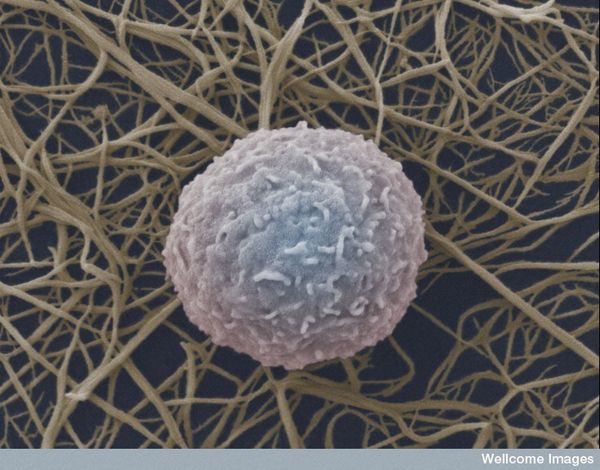
Photo credit: wellcome images / Foter.com / CC BY-NC-ND


