ISSCR 2013 Meeting Updates: Can Alzheimer’s Disease be modeled in a dish?

Human pluripotent stem cells (hPSCs) can differentiate into all cell types of the body, and can thereby serve as great models to examine the pathological mechanisms of various human diseases.  At the International Society for Stem Cell Research (ISSCR) 11th Annual Meeting, various stem cell experts highlighted the current human stem cell models for Alzheimer’s disease, and discussed the potential future directions of the field.

Alzheimer’s disease (AD) is the most common neurodegenerative dementia, affecting ~30 million people worldwide.  AD occurs in two main forms:  early-onset, familial AD (FAD) and late-onset, sporadic AD (SAD).  Both 40004_webare characterized by extensive neuronal loss and the aggregation of two proteins in the brain: amyloid β peptide (Aβ) and tau.  Aβ peptide is derived from the amyloid precursor protein (APP) via cleavage by two proteases, β-secretase and γ-secretase.  According to the amyloid cascade hypothesis, elevated levels of Aβ are necessary and sufficient to trigger disease 1.  Tau is synthesized in neurons and normally functions in binding to tubulin and stabilization of microtubules.  However, in AD, tau is hyper-phosphorylated, resulting in dissociation from microtubules, aggregation, and formation of neurofibrillary tangles (NFTs).  Although the pathological hallmarks of AD consist of these amyloid plaques and NFTs, how the two are related to each other and how they contribute to clinical onset and progression of AD is still under investigation.  By the time a patient manifests symptoms of a mild dementia, there is already significant neuronal loss and substantial accumulation of plaques and tangles.  One major limitation to our understanding of AD has been the lack of live, patient-specific neurons to examine disease progression.

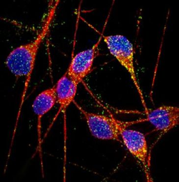
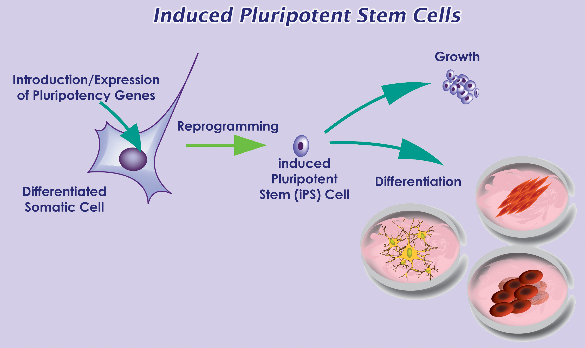
With recent advances in reprogramming technology, scientists can now generate induced pluripotent stem cells (iPSCs), and thereby use live, patient-specific models to examine disease phenotypes in a dish.  At the ISSCR meeting, Larry Goldstein presented his lab’s recent work on using hiPSC models to study AD.  They generated iPSCs from two patients with FAD caused by a duplication of the APP gene, two patients with SAD, and two control individuals.  Next, neurons were generated from the iPSC lines by directed differentiation and fluorescence-activated cell sorting (FACS) purification 2.  Neurons from one SAD and two FAD patients demonstrated significantly higher levels of secreted Aβ and phosphorylated tau (p-tau) 3.  To determine whether there is an association between APP processing and elevated p-tau levels, they treated iPSC-derived neurons with γ-secretase and β-secretase inhibitors.  Interestingly, pharmacologic inhibition of β-secretase resulted in a significant reduction in the levels of Aβ and p-tau.  Treatment with the γ-secretase inhibitor only reduced Aβ levels, but not p-tau levels.  This suggests that products of APP processing other than Aβ might contribute to elevated p-tau levels, highlighting a potential weakness with the amyloid cascade hypothesis.

Other groups have proposed alternative hypotheses to explain AD pathogenesis.  Haruhisa Inoue presented his group’s work on using human iPSC models to examine how intracellular Aβ oligomers contribute to AD.  They generated iPSCs from one patient with FAD caused by the APP-E693Δ mutation, two patients with SAD, and three control individuals.  Corticol neurons were derived using small molecule inhibitors of bone morphogenic protein (BMP) and activin/nodal signaling as previously described 4.  Aβ oligomers accumulated in neurons derived from the FAD patient and one SAD patient, but not in the control neurons 5.  Specifically, the Aβ oligomers accumulated in the endoplasmic reticulum (ER), and triggered ER and oxidative stress in the neurons.  In addition, treatment with docosahexaenoic acid (DHA) alleviated the stress responses.  Although the drug has previously failed in some clinical trials of AD treatment, Inoue’s work suggests that DHA might be effective for a subset of patients.

In summary, Goldstein and Inoue presented convincing evidence that human iPSC models can be used to study early AD pathogenesis and patient-specific drug responses.  Although it can take decades for symptoms to manifest in patients, disease phenotypes can be observed using iPSC models.  However, the fact that only one out of two SAD patients generated a disease phenotype highlights the need of future iPSC studies to examine larger numbers of patients to account for the observed heterogeneity in AD pathogenesis.

References:

1          Hardy, J. & Selkoe, D. J. The amyloid hypothesis of Alzheimer’s disease: progress and problems on the road to therapeutics. Science 297, 353-356, doi:10.1126/science.1072994 (2002).

2          Yuan, S. H. et al. Cell-surface marker signatures for the isolation of neural stem cells, glia and neurons derived from human pluripotent stem cells. PLoS One 6, e17540, doi:10.1371/journal.pone.0017540 (2011).

3          Israel, M. A. et al. Probing sporadic and familial Alzheimer’s disease using induced pluripotent stem cells. Nature 482, 216-220, doi:10.1038/nature10821 (2012).

4          Morizane, A., Doi, D., Kikuchi, T., Nishimura, K. & Takahashi, J. Small-molecule inhibitors of bone morphogenic protein and activin/nodal signals promote highly efficient neural induction from human pluripotent stem cells. J Neurosci Res 89, 117-126, doi:10.1002/jnr.22547 (2011).

5          Kondo, T. et al. Modeling Alzheimer’s disease with iPSCs reveals stress phenotypes associated with intracellular Abeta and differential drug responsiveness. Cell Stem Cell 12, 487-496, doi:10.1016/j.stem.2013.01.009 (2013).


ISSCR 2013 Meeting Updates: Is a Cure in Sight for Type 1 Diabetes?

Last week, the International Society for Stem Cell Research (ISSCR) held its 11th Annual Meeting in Boston, MA.  Over 3,000 stem cell researchers from around the world gathered to hear the leading experts share their research and perspectives in this fast-paced field.  One interesting topic was recent advances in cell-replacement therapy for treatment of type 1 diabetes.

Diabetes mellitus is a metabolic disease that results from a failure in glucose regulation, often leading to severe hyperglycemia and tissue/organ damage.  Pancreatic β-cells respond to high blood glucose levels by secreting insulin, which acts on other tissues to promote glucose uptake from the blood.  Type 1 diabetes (T1D) results from autoimmune destruction of insulin-producing β-cells of the pancreas.  The lack of insulin leads to increased blood and urine glucose.  T1D is often fatal unless treated with exogenous administration of insulin daily and regular blood glucose monitoring for the patient’s entire life.  However, this treatment does not match the effect of having endogenous β-cells.  Thus, scientists in the field of regenerative medicine have focused on strategies for generation of β-cells for cell-replacement therapy.

Douglas Melton presented his lab’s describe the imagecurrent progress on generation of functional β-cells from human embryonic stem cells (hESCs).  Since 2006, D’Amour et al. demonstrated that they had developed a robust differentiation protocol to produce hESC-derived pancreatic endocrine cells capable of synthesizing insulin 1.  However, the derived cells failed to secrete insulin appropriately in response to the addition of glucose, a required function of true β-cells.  Thus, one focus of Melton’s group has been to identify the signals to generate functional β-cells.  Pancreatic islets are complex structures consisting of multiple cell types, including the insulin-producing β-cells as well as endothelial cells in the surrounding blood vessels.  Thus, endothelial signals are known to promote pancreatic development 2.  Melton’s group found that co-culture of β-cells with endothelial cells promotes functional maturation of the hESC-derived β-cells.  In addition, they recently identified another hormone, betatrophin, which is secreted by the liver and functions in promoting β-cell replication 3.  Thus, increasing the levels of this hormone may generate more β-cells.  Although various groups have demonstrated that large amounts of glucose-responsive, insulin-secreting β-cells can be generated in vitro, one concern that remains to be addressed is how the cells will be protected from an autoimmune attack once delivered to the patient.  One potential strategy involves encapsulation of the β-cells into an immunoprotective device prior to delivery.

Other mature cells have also iPSDerivationbeen proposed as a source of new β-cells.  Sarah Ferber presented her lab’s work on inducing liver cells to transdifferentiate into β-cells for autologous cell-replacement therapy.  Transdifferentiation is the process by which one type of adult cell is directly converted into another type of cell.  They used the transcription factor, pancreatic and duodenal homeobox gene 1 (PDX-1), and soluble factors to induce the developmental shift of adult human liver cells into functional insulin-producing cells 4.  Not only did the transdifferentiated liver cells produce insulin, but they also released in a glucose-regulated manner.  When transplanted into diabetic, immunodeficient mice, the cells ameliorated hyperglycemia over a 60-day period.  Thus, PDX-1-induced transdifferentiated liver cells offer the potential to replace β-cells’ function in vivo.  Furthermore, transplantation of autologous β-cells would circumvent a host versus graft immune response, as well as allow the patient to be the donor of his or her own insulin-producing cells.

In summary, Melton’s and Ferber’s presentations demonstrated the tremendous progress that has been made in the last decade to generate functional β-cells for use in treatment of T1D.

References

1          D’Amour, K. A. et al. Production of pancreatic hormone-expressing endocrine cells from human embryonic stem cells. Nat Biotechnol 24, 1392-1401, doi:10.1038/nbt1259 (2006).

2          Nikolova, G. et al. The vascular basement membrane: a niche for insulin gene expression and Beta cell proliferation. Dev Cell 10, 397-405, doi:10.1016/j.devcel.2006.01.015 (2006).

3          Yi, P., Park, J. S. & Melton, D. A. Betatrophin: A Hormone that Controls Pancreatic beta Cell Proliferation. Cell 153, 747-758, doi:10.1016/j.cell.2013.04.008 (2013).

4          Sapir, T. et al. Cell-replacement therapy for diabetes: Generating functional insulin-producing tissue from adult human liver cells. Proc Natl Acad Sci U S A 102, 7964-7969, doi:10.1073/pnas.0405277102 (2005).

Identification of New Potential Drug Targets for Treatment of Lupus

autoantibodies Systemic lupus erythematosus (SLE) is a complex autoimmune disease that afflicts tens of millions of people worldwide.  The most prominent feature is generation of “autoantibodies” to self-proteins and nucleic acids, resulting in immune complex (IC) formation and organ inflammation.  Affected patients may demonstrate rashes, joint pain, anemia, or kidney damage, and untreated complications can often be fatal.  In addition, most SLE patients demonstrate continuously elevated levels of interferon (IFN) α, which is naturally produced by activated plasmacytoid dendritic cells (pDCs) 1.  pDCs are a rare subset of DCs found in the blood and peripheral lymphoid organs that function in host defense by secreting proinflammatory cytokines to initiate the innate immune response.  pDCs are activated following engagement of Toll-like receptors (TLRs), which recognize molecular signatures of bacteria and viruses.  Studies have shown that the frequency of circulating pDCs is significantly reduced in SLE patients, due to increased migration to inflammatory sites in affected organs 2.  Although pDCs have been implicated in contributing to autoimmunity via continuous type I IFN production, their exact role in lupus pathogenesis has not been clearly elucidated.

Recently, in PNAS, Baccala et al. provided direct evidence that in the absence of pDCs, the disease manifestations of Lupus were significantly decreased 3.  Since IRF8 is a hematopoietic cell-specific transcription factor known to be essential for pDC development 4, the authors knocked out IRF8 in NZB mice, a widely used mouse model for SLE.  Appropriately, pDCs were absent in IRF8-deficientNZB mice, and type I IFNs were undetectable even after injection with CpG DNA, a standard method of inducing the interferon pathway.  Interestingly, autoantibody production was almost completely abrogated and kidney disease was drastically improved compared to wild-type NZB mice.  Taken together, their results suggest that without pDCs, SLE disease manifestations are significantly reduced.

Next, the authors sought to examine specifically how pDCs promote systemic autoimmunity.  They used another mouse model with a mutation in Slc15a4, which is characterized by normal development of pDCs but an absence of type I IFN production by pDCs.  It is still unclear how a mutation in Slc15a4 leads to a disruption in proinflammatory cytokine production in pDCs, but since Slc15a4 is a peptide/histidine transporter, others hypothesize that it transports free histidine from the endosome to the cytosol to enable cathepsin-mediated cleavage of endosomal TLRs required for subsequent signaling 5.  Similar to the IRF8-deficient NZB mice, Slc15a4 mice had significantly reduced autoantibodies, decreased kidney disease, and extended survival.  This finding rules out the possibility that pDCs contribute to disease through other functions outside of type I IFN production.

In summary, Baccala et al. provide direct evidence that pDCs contribute to the abnormal manifestations of SLE via hyperproduction of type I IFNs.  Thus, IRF8 and Slc15a4 serve as new potential drug targets for treatment of SLE.  Current therapies involve broad immunosuppressive drugs, which suppress multiple arms of the immune system, increasing a patient’s risk for various infections and cancer.  Specific pharmacologic inhibition of IRF8 or Slc15a4 could prevent Lupus-specific flare-ups, as well as manifestations of other autoimmune diseases.

References

1          Gilliet, M., Cao, W. & Liu, Y. J. Plasmacytoid dendritic cells: sensing nucleic acids in viral infection and autoimmune diseases. Nat Rev Immunol 8, 594-606, doi:10.1038/nri2358 (2008).

2          Ronnblom, L. The type I interferon system in the etiopathogenesis of autoimmune diseases. Ups J Med Sci 116, 227-237, doi:10.3109/03009734.2011.624649 (2011).

3          Baccala, R. et al. Essential requirement for IRF8 and SLC15A4 implicates plasmacytoid dendritic cells in the pathogenesis of lupus. Proc Natl Acad Sci U S A 110, 2940-2945, doi:10.1073/pnas.1222798110 (2013).

4          Tsujimura, H., Tamura, T. & Ozato, K. Cutting edge: IFN consensus sequence binding protein/IFN regulatory factor 8 drives the development of type I IFN-producing plasmacytoid dendritic cells. J Immunol 170, 1131-1135 (2003).

5          Park, B. et al. Proteolytic cleavage in an endolysosomal compartment is required for activation of Toll-like receptor 9. Nat Immunol 9, 1407-1414, doi:10.1038/ni.1669 (2008).

Identification of a new HSC viral transduction enhancer, Vectofusin-1

HSC gene therapy is an emerging therapeutic option for several disorders of the blood and immune system.  Ex vivo cell therapies are based on the ability to isolate CD34+ cells from a patient or a normal donor, expansion ex vivo with genetic modification, and systemic administration into the patient following myeloablative treatment.  An efficient method for gene transfer into HSCs is required for successful gene therapy.  Lentiviral vectors (LVs) have emerged as a robust and versatile tool for ex vivo and in vivo gene delivery into multiple cell types, including HSCs.  LVs can either be pseudotyped with viral envelope glycoproteins that confer a broad tropism, such as the vesicular stomatitis virus G (VSV-G) protein, or those that confer a specific HSC tropism, including gibbon ape leukemia virus (GALVTR), feline endogenous retrovirus RD114 (RD114TR), or amphotropic murine leukemia virus (MLV-A) proteins.  However, viral envelopes vary in transduction efficiency.  Thus, transduction protocols often involve the addition of factors to enhance viral entry, including cationic polymers (polybrene) 1 or fibronectin fragments (Retronectin) 2.

gene therapy

Recently, in Molecular Therapy-Nucleic Acids, Fenard et al identified another viral entry enhancer, Vectofusin-1 3.  Vectofusin-1 is a synthetic, histidine-rich cationic amphipathic peptide derived from the LAH4 peptide family.  LAH4 peptides and their derivatives are known to be efficient DNA transfection agents 4.  In this study, the authors examined whether Vectofusin-1 would also enhance gene transfer of LVs into CD34+ cells derived from human umbilical cord blood.  Indeed, Vectofusin-1 significantly increased the transduction efficiency of LVs pseudotyped with various envelopes (VSV-G, GALVTR, RD114TR, MLV), with transduction levels ranging from 50-80% compared to undetectable transduction levels in its absence.  In addition, the increased transduction efficiency was not cytotoxic.  Addition of Vectofusin-1 during transduction of CD34+ cells did not negatively affect subsequent myeloerythroid differentiation in colony-forming cell (CFC) assays in vitro, or hematopoietic reconstitution in immunodeficient BALB-Rag/γC mice in vivo.  The mechanism for the increased transduction efficiency was attributed to insertion of the peptide in the viral and cellular membranes, resulting in an enhancement in both adhesion and fusion of the viral particles with the cell’s plasma membrane.

In short, the authors demonstrated that Vectofusin-1 is a promising LV entry enhancer that can be potentially used in ex vivo transduction of HSCs for subsequent use in clinical applications.  Addition of Vectofusin-1 to the transduction medium had similar effects as the commonly used Retronectin, although the latter is used to coat plates, suggesting a different mechanism of action.  Future experiments will determine whether Vectofusin-1 and Retronectin can be used together to synergistically enhance HSC transduction.


References:

1          Davis, H. E., Morgan, J. R. & Yarmush, M. L. Polybrene increases retrovirus gene transfer efficiency by enhancing receptor-independent virus adsorption on target cell membranes. Biophys Chem 97, 159-172 (2002).

2          Pollok, K. E. & Williams, D. A. Facilitation of retrovirus-mediated gene transfer into hematopoietic stem and progenitor cells and peripheral blood T-lymphocytes utilizing recombinant fibronectin fragments. Curr Opin Mol Ther 1, 595-604 (1999).

3          Fenard, D. et al. Vectofusin-1, a new viral entry enhancer, strongly promotes lentiviral transduction of human hematopoietic stem cells. Mol Ther Nucleic Acids 2, e90, doi:10.1038/mtna.2013.17 (2013).

4          Kichler, A., Leborgne, C., Marz, J., Danos, O. & Bechinger, B. Histidine-rich amphipathic peptide antibiotics promote efficient delivery of DNA into mammalian cells. Proc Natl Acad Sci U S A 100, 1564-1568, doi:10.1073/pnas.0337677100 (2003).

Reprogramming of old HSCs reverses functional defects associated with aging

HSCs must continuously self-renew to replenish the pool of mature blood cells throughout the life an adult.  One requirement for extensive self-renewal is high telomerase activity to prevent telomere shortening.  HSCs isolated from adult bone marrow have shorter telomeres than cells from fetal liver or umbilical cord blood 1, suggesting that proliferative potential may decrease with age.  Also, HSC aging is associated with decreased lymphoid potential, as well as an up-regulation of genes involved in leukemic transformation 2.  Consequently, “aging” HSCs may have functional defects that might be detrimental for therapeutic strategies involving genetic manipulation and transplantation of HSCs for the treatment of various blood disorders.

describe the image

Previous studies have demonstrated that during reprogramming, developing iPSCs express epigenetic features of ESCs, and lose those of the starting somatic cell 3.  In addition, a previous study reported a significant elongation of telomeres in derived iPSCs compared to the starting somatic cell 4.  Thus, it may be possible that reprogramming reverses functional defects associated with HSC aging.

Recently, in Blood, Wahlestedt et al examined whether characteristics of aging HSCs are reversible 5.  First, they derived iPSCs from young and aged murine HSCs.  To examine their differentiation potential, they injected the derived iPSCs into murine blastocysts and analyzed the engraftment of the donor cells in the developing chimeric embryos.  Overall, iPSCs derived from aged HSCs demonstrated similar differentiation potential compared to that of younger HSCs.  The engraftment of bone marrow mononuclear cells from primary chimeric mice in a competitive transfer experiment was comparable to that of young HSCs.  Aged HSCs, on the other hand, demonstrated a significant reduction in repopulation capacity.  Interestingly, aged iPSC-derived HSCs also generated naïve T cells at similar levels as young HSCs.

Next, the authors examined telomere length following re-differentiation of the young and aged iPSCs.  Telomeres in aged HSCs were ~11% shorter compared to young HSCs.  However, telomeres of the HSCs derived from the aged iPSCs demonstrated a 2-fold elongation compared to blastocyst control HSCs.  This 2-fold elongation was maintained even after transplantation.  Overall, these results indicate that iPSC induction from HSCs results in elongation of telomeres.

In short, Wahlestedt et al demonstrated that reprogramming does indeed reverse some of the functional defects associated with chronologically aged HSCs, including decreased differentiation potential and shortened telomeres.  However, the study did not address whether the iPSCs derived from aged HSCs had an increased DNA mutation frequency, since HSC aging is also associated with a higher mutation rate.  It would also be interesting to determine whether the above phenomena are also observed in reprogramming of aged human HSCs.  If iPSC induction does indeed result in an “epigenetic reset,” then HSCs derived from iPSCs may have unique characteristics favorable for use in clinical settings.


References

1          Vaziri, H. et al. Evidence for a mitotic clock in human hematopoietic stem cells: loss of telomeric DNA with age. Proc Natl Acad Sci U S A 91, 9857-9860 (1994).

2          Rossi, D. J. et al. Cell intrinsic alterations underlie hematopoietic stem cell aging. Proc Natl Acad Sci U S A 102, 9194-9199, doi:10.1073/pnas.0503280102 (2005).

3          Maherali, N. et al. Directly reprogrammed fibroblasts show global epigenetic remodeling and widespread tissue contribution. Cell Stem Cell 1, 55-70, doi:10.1016/j.stem.2007.05.014 (2007).

4          Marion, R. M. et al. Telomeres acquire embryonic stem cell characteristics in induced pluripotent stem cells. Cell Stem Cell 4, 141-154, doi:10.1016/j.stem.2008.12.010 (2009).

5          Wahlestedt, M. et al. An epigenetic component of hematopoietic stem cell aging amenable to reprogramming into a young state. Blood, doi:10.1182/blood-2012-11-469080 (2013).

Efficient generation of iPSCs from human cord blood and peripheral blood

The initial finding that pluripotency could be induced in human somatic cells revolutionized the field of regenerative medicine, since patient-specific stem cells can now be generated to further examine the causes and mechanisms of various human diseases.  Since the discovery of human iPSCs in 2007 1, various studies have focused on improving the reprogramming methods in order to increase the induction efficiency, as well as to further simplify the protocol.  iPSCs are commonly generated from dermal fibroblasts.  However, skin biopsies are required to isolate fibroblast cells, highlighting the necessity to identify an alternative source of cells for reprogramming that would involve less invasive surgical procedures for isolation.

Cord blood cells and peripheral blood mononuclear cells (PMNCs) are attractive sources for the generation of iPSCs due to the low invasiveness of their collection, as well as the abundance of blood banks for potential donors.  iPSCs were first derived from human peripheral blood in 2009 2.  CD34+ cells were mobilized from peripheral blood and subsequently transduced with retroviruses delivering OCT4, SOX2, KLF4, and MYC vectors.   Although the reprogramming was successful, use of retroviral vectors requires genomic integration of transgenes that may increase the risk of tumor formation during clinical applications.  Thus, recent studies have focused on generation of “integration-free” iPSCs.  Yet, development of integration-free methods often means compromising the reprogramming efficiency.  iPSC induction in CD34+ cells using non-integrating episomal plasmids resulted in ~0.03% reprogramming efficiency 3.

hESC H9p40 resized 600 resized 600
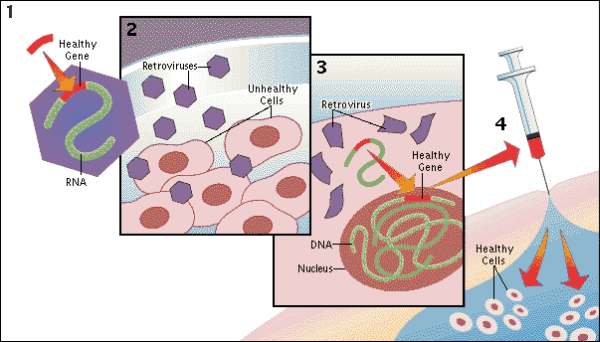
Recently, in Stem Cells, Yamanaka’s group reported a protocol that increased the efficiency of iPSC induction from CD34+ cord blood and peripheral blood 4.  They previously identified an efficient combination of episomal plasmids for reprogramming of adult fibroblasts, termed the “Y4” combination, consisting of plasmids encoding OCT3/4, SOX2, KLF4, L-MYC, LIN28, and an shRNA for TP53 5.  Transfection of CD34+ cells from human cord blood with the Y4 combination resulted in up to 0.1% reprogramming efficiency across two donors.  iPSC induction efficiency of PMNCs isolated from peripheral blood with the Y4 mixture, on the other hand, was inconsistent across donors.  To further increase the reproducibility of iPSC induction from multiple donors, the authors added a vector encoding EBNA1, which is required for episomal plasmid replication and should thereby increase expression of the episomal plasmids.  Addition of the EBNA1 vector to the Y4 mixture resulted in 0.1% reprogramming efficiency in PMNCs across seven donors.  Both, the CD34+– and PMNC-derived iPSCs were molecularly and functionally identical to hESCs.

In summary, Okita et al identified a new protocol allowing efficient generation of integration-free iPSCs from blood.  Previous studies reported ~0.02%- 0.03% induction efficiency from peripheral blood 2 and isolated CD34+ cells 3.  Here, the authors reported a reprogramming efficiency of ~0.06%, with a maximum of 0.1%.  In addition, the new protocol could induce iPSCsfrom frozen PMNCs as efficiently as from freshly isolated cells.  Thus, with the increasing number of potential donors available at cord blood banks, iPSCs can be now efficiently generated for use in autologous or allogeneic stem cell therapy.


References

1          Takahashi, K. et al. Induction of pluripotent stem cells from adult human fibroblasts by defined factors. Cell 131, 861-872, doi:10.1016/j.cell.2007.11.019 (2007).

2          Loh, Y. H. et al. Generation of induced pluripotent stem cells from human blood. Blood 113, 5476-5479, doi:10.1182/blood-2009-02-204800 (2009).

3          Mack, A. A., Kroboth, S., Rajesh, D. & Wang, W. B. Generation of induced pluripotent stem cells from CD34+ cells across blood drawn from multiple donors with non-integrating episomal vectors. PLoS One 6, e27956, doi:10.1371/journal.pone.0027956 (2011).

4          Okita, K. et al. An efficient nonviral method to generate integration-free human-induced pluripotent stem cells from cord blood and peripheral blood cells. Stem Cells 31, 458-466, doi:10.1002/stem.1293 (2013).

5          Okita, K. et al. A more efficient method to generate integration-free human iPS cells. Nat Methods 8, 409-412, doi:10.1038/nmeth.1591 (2011).

How do HSCs deal with aging?

hematopoietic stem cells resized 600Mature blood cells are relatively short-lived, and require replenishment from multipotent HSCs. Thus, HSCs must self-renew to generate an adequate pool of HSCs, as well as differentiate to give rise to more mature blood cells.  A balance between self-renewal and differentiation ensures that the hematopoietic system can be functionally sustained throughout the lifetime of an adult body.  However, as HSCs age, they accumulate DNA damage, often compromising their functionality.  DNA damage can be further propagated both to daughter stem cells and downstream lineages, and may increase the risk of developing blood disorders 1.

Depending on the nature of the damage, cells use two major response pathways to combat cellular stress.  If the damage is excessive and functionality is compromised, cells usually undergo apoptosis for self-elimination.  In contrast, autophagy allows cells a window of survival.  Autophagy is a process of self-degradation in which organelles or portions of the cytoplasm are sequestered within double-membrane vesicles, known as autophagosomes, and then delivered to lysosomes for degradation 2.  The resulting breakdown products are released through permeases and recycled in the cytosol.  Thus, autophagy can be used to generate high-energy compounds during conditions of metabolic stress.

Recently, in Nature, Warr et al found that metabolic stress and old age induce autophagy in HSCs 3.  The authors isolated HSCs and myeloid progenitors from the bone marrow of transgenic mice systemically expressing GFP fused to LC3, an autophagosome marker 4.  They used cytokine withdrawal to induce metabolic stress and measured autophagy induction by examining the formation and turnover of LC3-GFP.  Myeloid progenitors expressed LC3-GFP in the presence and absence of cytokines.  In contrast, HSCs did not express LC3-GFP in the presence of cytokines, but demonstrated autophagosome formation following cytokine withdrawal.  Furthermore, when the mice were starved in vivo, autophagy flux increased in HSCs.

The authors speculated that autophagy “protects” HSCs from starvation-induced apoptosis, and indeed, hematopoietic-specific deletion of an essential autophagy machinery component, ATG12, resulted in a significant increase in caspase activation in starved HSCs in vivo.  FOXO3A was identified as the specific transcriptional regulator that maintains pro-autophagy gene expression, and was expressed higher in HSCs compared to progenitors.  Interestingly, HSCs isolated from old mice retained their autophagic potential, and was found to be required for their survival.

In summary, Warr et al demonstrated that long-lived HSCs mount a protective survival autophagy response to combat metabolic stress, whereas short-lived progenitors do not.  Previous studies suggested that impaired autophagy might contribute to the aging phenotype 5.  However, this study directly showed that the pro-autophagy gene expression program is still intact in old HSCs and is essential for continued survival of these cells.  Future studies will address whether autophagy increases the incidence of age-related blood disorders since it protects damaged, old HSCs from elimination by apoptosis.


References 

1          Rossi, D. J., Jamieson, C. H. & Weissman, I. L. Stems cells and the pathways to aging and cancer. Cell 132, 681-696, doi:10.1016/j.cell.2008.01.036 (2008).

2          He, C. & Klionsky, D. J. Regulation mechanisms and signaling pathways of autophagy. Annu Rev Genet 43, 67-93, doi:10.1146/annurev-genet-102808-114910 (2009).

3          Warr, M. R. et al. FOXO3A directs a protective autophagy program in haematopoietic stem cells. Nature 494, 323-327, doi:10.1038/nature11895 (2013).

4          Mizushima, N., Yamamoto, A., Matsui, M., Yoshimori, T. & Ohsumi, Y. In vivo analysis of autophagy in response to nutrient starvation using transgenic mice expressing a fluorescent autophagosome marker. Mol Biol Cell 15, 1101-1111, doi:10.1091/mbc.E03-09-0704 (2004).

5          Rubinsztein, D. C., Marino, G. & Kroemer, G. Autophagy and aging. Cell 146, 682-695, doi:10.1016/j.cell.2011.07.030 (2011).


EGF enhances HSC regeneration following myelosuppressive injury

Maintenance of the hematopoietic system requires constant replenishment of mature blood cells from HSCs.  For patients with malignant and non-malignant disorders of the blood and immune system, myeloablation and subsequent HSC transplantation is often necessary. describe the imageHowever, exposure to ionizing radiation to induce myeloablation also causes DNA damage that can induce cell-cycle arrest or apoptosis of HSCs and their progenitor cells 1.  Studies have shown that treatment with cytokines can prevent cell-cycle arrest.  For example, administration of stem cell factor (SCF) before radiation exposure protected mice from radiation-induced lethality by inducing HSCs into late S phase 2, which is the most radioresistant phase of the cell cycle.  Further studies to identify additional cytokines that mediate HSC regeneration following radiation exposure are critical for the development of therapies to minimize myelosuppression in patients receiving chemotherapy.

Recently, in Nature Medicine, Doan et al discovered a new function of epidermal growth factor (EGF) signaling in regulation of HSC regeneration following myelosuppressive injury 3.  The authors previously generated a mouse model in which pro-apoptotic proteins, BAK and BAX, were deleted in Tie2+ bone marrow endothelial cells 4.  Mice lacking BAK and BAX expression demonstrated significantly increased numbers of HSCs and progenitor cells and increased survival following total body irradiation (TBI) compared to wild-type mice expressing the pro-apoptotic proteins.  This was the first indication that bone marrow endothelial cells might have therapeutic potential in enhancing hematopoietic reconstitution following myelosuppression.  However, the mechanism through which these cells regulate hematopoietic regeneration was unknown.

In their most recent study, Doan et al performed a cytokine array on bone marrow serum from mice lacking BAK and BAX expression and found a significant enrichment of EGF compared to wild-type mice 3.  Using multiparametric flow cytometry, they demonstrated that ~9% of c-Kit+Sca-1+LinSLAM+ HSCs express functional EGF receptor (EGFR), and expression increased by 6-fold following irradiation.  Systemic administration of EGF augmented HSC recovery in vivo and improved the survival of mice following TBI compared to saline-treated control mice.  In contrast, administration of erlotinib, an EGFR antagonist, suppressed HSC regeneration and significantly decreased the survival of mice following TBI, further suggesting that EGFR signaling is critical for radioprotection of bone marrow HSCs and progenitor cells.  They found that EGFR signaling promotes HSC proliferation by activation of the PI3K-AKT pathway.  In addition, EGF treatment inhibited expression of the p53 upregulated modulator of apoptosis (PUMA), an essential mediator of radiation-induced HSC apoptosis.

describe the imageIn summary, EGF promotes HSC cycling and survival following radiation-induced myelosuppression.  The study by Doan et al was the first demonstration that bone marrow HSCs express functional EGFR, and that EGFR signaling plays a role in HSC self-renewal.  The results of this study suggest that EGF may have therapeutic potential to enhance hematopoietic regeneration in patients receiving myelosuppressive chemotherapy or undergoing HSC transplantation.


References

1. Liu, Y. et al. p53 regulates hematopoietic stem cell quiescence. Cell Stem Cell 4, 37-48, doi:10.1016/j.stem.2008.11.006 (2009).

2. Zsebo, K. M. et al. Radioprotection of mice by recombinant rat stem cell factor. Proc Natl Acad Sci U S A 89, 9464-9468 (1992).

3. Doan, P. L. et al. Epidermal growth factor regulates hematopoietic regeneration after radiation injury. Nat Med, doi:10.1038/nm.3070 (2013).

4. Doan, P. L. et al. Tie2(+) Bone Marrow Endothelial Cells Regulate Hematopoietic Stem Cell Regeneration Following Radiation Injury. Stem Cells, doi:10.1002/stem.1275 (2012).

A novel role of TPO in the regulation of HSC DNA repair

HSC transplantation is routinely used to treat patients with malignant and non-malignant disorders of the blood and immune system, but its therapeutic application is often restricted by difficulties in in vitro maintenance and expansion of HSCs.  Studies aimed at understanding the mechanisms governing self-renewal of HSCs within hematopoietic tissues identified a set of growth factors and cytokines that can augment in vitro expansion of HSCs, including stem cell factor (SCF), fms-like tryrosine kinase 3 ligand (FLT3l) and thrombopoietin (TPO) 1.  TPO and its receptor, Mpl, are primarily known for their role in megakaryopoiesis, but TPO has also been shown to support HSC quiescence during adult hematopoiesis, with the loss of signaling associated with bone marrow failure and thrombocytopenia 2.

Recently, in Cell Stem Cell, de Lavel et al. identified a novel role of TPO in the regulation of DNA repair in HSCs 3.  Exposure to genotoxic agents, such as ionizing radiation (IR), induces DNA damage comprised of double-strand breaks (DSBs).  DNA damage is repaired through two main pathways: homologous recombination (HR) and nonhomologous end-joining (NHEJ).  DNA repair is essential for cell survival, and studies have shown that NHEJ is necessary for HSC maintenance 4,5.  In their study, de Lavel et al. found that γH2AX foci, a marker of DSB formation, were significantly increased in Mpl-deficient HSCs and in their progenitors following IR exposure.  Moreover, a TPO injection into mice prior to IR reduced the number of γH2AX foci in HSCs in vivo, while HSCs exposed to IR in the absence of TPO demonstrated an increased number of γH2AX foci.  Other experiments showed that TPO modulates the efficiency of the NHEJ pathway by increasing the phosphorylation of the DNA-PK catalytic subunit, a major enzyme involved in NHEJ.  Pharmacological or genetic inhibition of DNA-PK abrogated TPO-mediated DNA repair.  Interestingly, the other cytokines involved in HSC maintenance and expansion, SCF and FLT3l, did not have the same effects as TPO, suggesting that DNA repair activity is a specific function of TPO.

clinical trialIn short, TPO regulates NHEJ-mediated DNA repair of DSBs by stimulating DNA-PK activity in HSCs.  This is the first demonstration that a cytokine involved in HSC maintenance may also regulate DSB repair machinery.  Since TPO treatment prior to IR exposure reduces DNA damage, TPO agonists could potentially be given to patients prior to receiving chemotherapy to reduce the risk of developing oncogenic mutations and defects in HSC function.  Romiplostim, a TPO peptide mimetic, and eltrombopag, a non-peptide TPO mimetic, have been successfully used for the treatment of immune thrombocytopenic purpura (ITP) and are approved by the FDA 6.  de Lavel et al showed that injection of romiplostim prior to IR exposure also completely abolished persistent DNA damage in HSCs, similar to TPO.  Thus, these TPO agonists might also be suited for clinical applications involving protection of normal HSC from DNA-damaging agents.


References 

1          Ohmizono, Y. et al. Thrombopoietin augments ex vivo expansion of human cord blood-derived hematopoietic progenitors in combination with stem cell factor and flt3 ligand. Leukemia 11, 524-530 (1997).

2          Ballmaier, M., Germeshausen, M., Krukemeier, S. & Welte, K. Thrombopoietin is essential for the maintenance of normal hematopoiesis in humans: development of aplastic anemia in patients with congenital amegakaryocytic thrombocytopenia. Ann N Y Acad Sci 996, 17-25 (2003).

3          de Laval, B. et al. Thrombopoietin-Increased DNA-PK-Dependent DNA Repair Limits Hematopoietic Stem and Progenitor Cell Mutagenesis in Response to DNA Damage. Cell Stem Cell 12, 37-48, doi:10.1016/j.stem.2012.10.012 (2013).

4         Rossi, D. J. et al. Deficiencies in DNA damage repair limit the function of haematopoietic stem cells with age. Nature 447, 725-729, doi:10.1038/nature05862 (2007).

5          Nijnik, A. et al. DNA repair is limiting for haematopoietic stem cells during ageing. Nature 447, 686-690, doi:10.1038/nature05875 (2007).

6          Kuter, D. J. New thrombopoietic growth factors. Clin Lymphoma Myeloma 9 Suppl 3, S347-356, doi:10.3816/CLM.2009.s.034 (2009).