Expansion of NK cells from Human PBMC

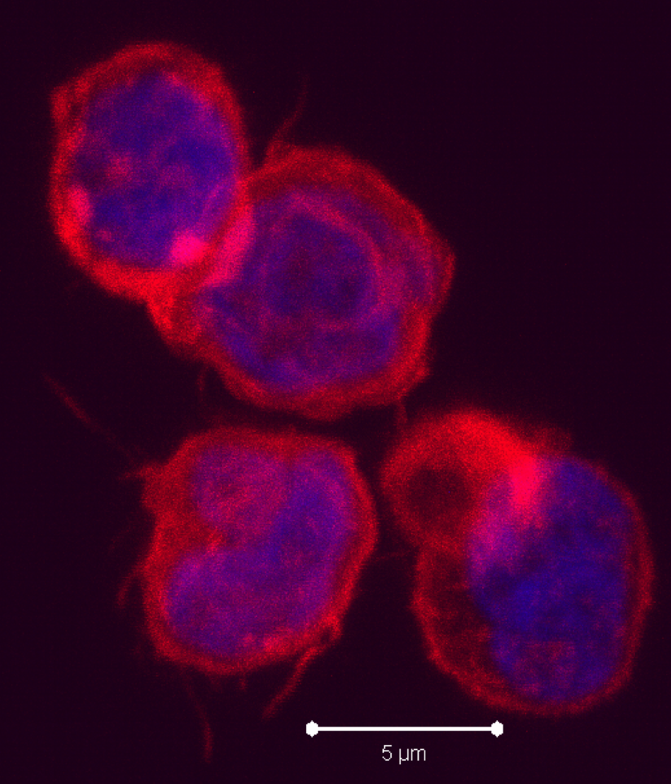
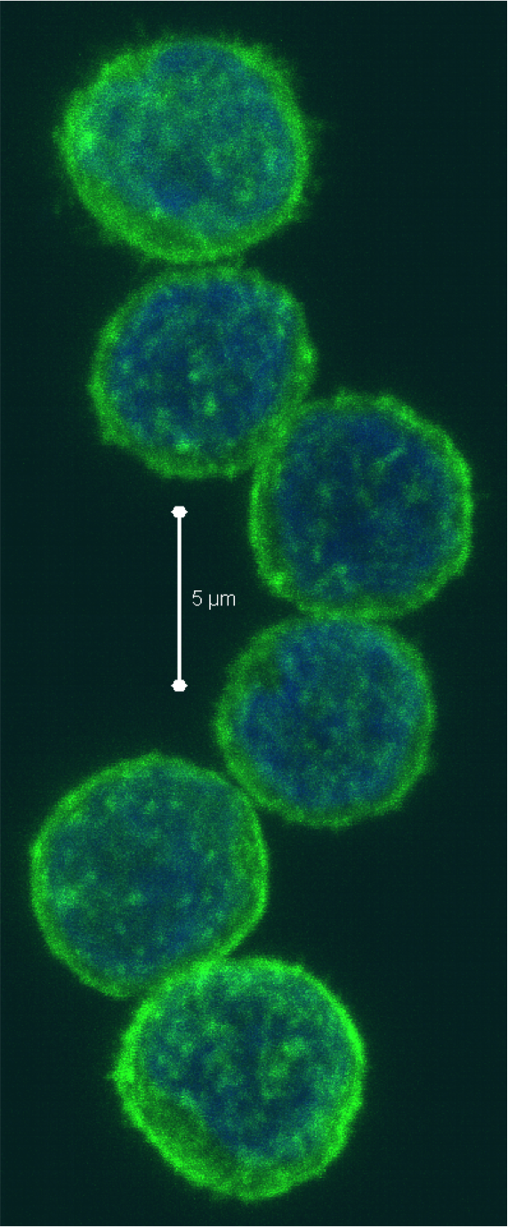
Natural killer (NK) cells represent up to 15% of human peripheral blood mononuclear cells (PBMC), and range from 5-20% of peripheral blood lymphocytes.  NK cells generally fall into three subtypes: CD56dim CD16+, CD56brightCD16+/- and CD56 CD16+ NK cells, the prevalence and functions of which I have previously discussed.  NK cells are considered to be a promising avenue in cell-based anti-tumor immunotherapeutics.  However, the relatively low numbers of these cell types in PBMC have constituted a technical challenge in these efforts and in other studies needing large numbers of NK cells.  In the April 2013 issue of Clinical & Experimental Immunology, Wang et. al, describe an in vitro method for the preferential expansion of human NK cells from PBMC.

NK cell expansion in vitro systems requires multiple signals for survival, proliferation, and activation.  In a previous study, Fujisaki et. al (200describe the image9) demonstrated that highly cytotoxic CD56+ NK cells could be highly preferentially expanded when cultured with a version of the chronic myeloid leukemia K562 cell line, which was genetically altered to express a membrane-bound form of IL-15 and the 41BB ligand (CD137L).  Under this protocol, NK cells expanded an average of 21.6-fold after 7 days and 277-fold after 21 days in culture, and at 21 days reached a purity of 98.6%.  CD3+ T cells on the other hand fell to an average of 3.1% of the cells remaining after 21 days.  Importantly however, is not only the expansion of NK cells, but the functionality of the expanded cell product.  The NK cells generated by this method had enhanced killing potential in vitro.  In xenograft models of acute myeloid leukemia (AML) in immune deficient NOD/scid-IL2RGnull mice, these NK cells were able to elicit potent anti-leukemic activity.  Thus, this method generates large numbers of highly functional human NK cells.

In the current study by Wang et. al, a similar method was utilized in which the K562 cell line was engineered to express a membrane-bound form of IL-21 along with CD137L.  On average under these conditions, NK cells expanded from less than 30% of PBMC to over 85% after 7 days and 95% after 3 weeks, while CD3+ T cells went from 60% initially to 6% at seven days and 1% at three weeks.  Proliferation of NK cells was continual over eight weeks in culture, and by 3 weeks reached over 100-fold, although the exact numbers and ranges were not explicitly stated in the paper.  Thus, NK cells are highly selectively expanded using this method, similarly to the method used by Fujisaki et. al.

In answer to the functionality of NK cells generated under these conditions, Wang et. al demonstrated enhanced expression of activating and inhibitory NK receptors.  Significantly enhanced cytotoxic killing potential after culture was shown, being maximal after one and three weeks in culture whereafter it decreased but still remained higher than resting NK cells.  Thus, these expanded NK cells are also highly functional.

It would be interesting to see a direct comparison of the extent and quality of NK cell expansion from human PBMC by CD137L combined with the membrane-bound form of IL-15 as was done by Fujisaki et. al versus the membrane-bound form of IL-21 developed by Wang et. al.  IL-21 is a strong and preferential activator of STAT3.  Wang et al did establish a role for STAT3 in the induction of these cells.  IL-15 is a strong activator of STAT5 and activates STAT3 to a lesser extent.  However, IL-15 has been shown to strongly induce expression of the STAT3-activating cytokine IL-10.  Thus, for optimal clinical applications of expanded NK cells, it is important to determine how the different cytokine-STAT signals contribute to NK cell proliferation, survival, and activation.

Further Reading:

Membrane-bound interleukin-21 and CD137 ligand induce functional human natural killer cells from peripheral blood mononuclear cells through STAT-3 activation.  Wang X, Lee DA, Wang Y, Wang L, Yao Y, Lin Z, Cheng J, Zhu S. Clin Exp Immunol. 2013 Apr;172(1):104-12. doi: 10.1111/cei.12034.

Expansion of highly cytotoxic human natural killer cells for cancer cell therapy.  Fujisaki H, Kakuda H, Shimasaki N, Imai C, Ma J, Lockey T, Eldridge P, Leung WH, Campana D. Cancer Res. 2009 May 1;69(9):4010-7. doi: 10.1158/0008-5472.CAN-08-3712. Epub 2009 Apr 21.

Natural Killer Cell subtypes and markers in human PBMC

Types of immune cells present in human PBMC

Prospects for the use of NK cells in immunotherapy of human cancer.  Ljunggren HG, Malmberg KJ. Nat Rev Immunol. 2007 May;7(5):329-39.

Properties of the K562 cell line, derived from a patient with chronic myeloid leukemia.  Klein E, Ben-Bassat H, Neumann H, Ralph P, Zeuthen J, Polliack A, Vánky F. Int J Cancer. 1976 Oct 15;18(4):421-31.

Characterization of cytokine differential induction of STAT complexes in primary human T and NK cells.  Yu CR, Young HA, Ortaldo JR. J Leukoc Biol. 1998 Aug;64(2):245-58.

IL-15-induced IL-10 increases the cytolytic activity of human natural killer cells.  Park JY, Lee SH, Yoon SR, Park YJ, Jung H, Kim TD, Choi I. Mol Cells. 2011 Sep;32(3):265-72. doi: 10.1007/s10059-011-1057-8. Epub 2011 Jul 29.

Are Terminally Differentiated Effector Memory Cells present in those “Naïve” CD4+ T cells you isolated from human PBMC?

Immunologists study many aspects regarding differentiation of T cells and function of T cell lineages.  The results and interpretations from these studies always rely on the robustness of the experimental setup.  A question that I posed to myself recently, when testing protocols for differentiation of naïve CD4+ T cells into various functional lineages (TH1, TH2, TH17, TREG), was whether or not CD4+ terminally differentiated effector memory (TEMRA) cells are still present in the population of “naïve” CD4+ T cells obtained following isolation from peripheral blood mononuclear cells (PBMC).

Following antigen exposure, CD4+ and CD8+ T cells undergo differentiation thorough various stages.  While the exact path of differentiation remains under exploration, a current mainstream hypothesis is that naïve cells (TN) progress through central memory (TCM), then effector memory (TEM), then finally terminally differentiated effector memory (TEMRA) states.  Expression of surface markers have been used to identify human T cells in these various states, including CD45RA, CD45RO, CCR7, CD62L, CD27, and CD28.  After antigen exposure, naïve T cells, which are CD45RA+CD45ROCCR7+CD62L+CD27+CD28+ lose expression of CD45RA and gain expression of CD45RO.  As memory T cells progress from TCM to TEM cells, they additionally lose expression of CCR7, CD45RA+, CD27, and CD28.  Finally, TEMRA cells regain expression of CD45RA, but remain identifiable from naïve T cells by their lack of CCR7, CD62L, CD27, and CD28 expression.

The function of CD4+ TEMRA cells parallels that of CD8+ TEMRA cells.  These cells are cytolytic and express IFN-gamma after activation through their TCR or stimulation with PMA/ionomycin. CD4+ TEMRA cells also have shorter telomeres than naïve, TCM, and TEM populations, and lower homeostatic proliferation capacity.

While TEMRA cells are well described for CD8+ T cells, they often are ignored as part of the CD4+ compartment.  Despite the lack of attention that  CD4+ TEMRA cells are given in the literature, I observe them quite frequently in human PBMC from healthy donors, on average being 4% of CD4+ T cells (range 0-15%) and 11% of CD4+CD45RA+ cells (range 0-40%).  Additionally, the percentage of CD4+ TEMRA cells that I observe has a strong correlation with IFN-gamma production by CD4+CD45RA+ T cells from the same donor.

CD4 naive TEMRA cells PBMC

Figure: A. Expression of CD45RA vs. CD62L in human CD4+ PBMCs from four donors.  CD4+ TEMRA cells are CD45RA+CD62L (lower right quadrant). B. Expression of CD45RA vs. IFN-gamma in human CD4+ PBMCs from the same four donors stimulated with PMA/ionomycin. CD45RA+IFN-gamma+ cells are likely CD4+ TEMRA cells.

Considering CD4+ TEMRA cells are not only commonly present but highly functional, I wondered if they would be present in the population of “naïve” CD4+ T cells obtained following isolation from PBMC.  If fluorescence-activated cell sorting (FACS) is used for cell isolation, then this is an easier issue to avoid as all of the necessary markers used to differentiate naïve CD4+ T cells from other cell subsets can be included in the marker staining panel.  However, many researchers use commercially available magnetic bead-based kits or other similar methodologies to obtain a “naïve” CD4+ T cell population.  Because there is no single marker that would isolate a naïve CD4 T cell from PBMC, negative selection kits for untouched isolation of naïve CD4+ T cells are commercially available as “one-step” kits.  These are available from companies including Miltenyi Biotec, Stem Cell Technologies, and R&D Systems.

Analysis of the antigens negatively selected for by these kits revealed the following lists: Miltenyi Biotec: CD45RO, CD8, CD14, CD15, CD16, CD19, CD25, CD34, CD36, CD56, CD123, TCRγ/δ, HLA-DR, and CD235a (glycophorin A).

Stem Cell Technologies: CD45RO, CD8, CD14, CD16, CD19, CD20, CD36, CD56, CD66b, CD123, TCRγ/δ, and CD235a (glycophorin A).

Unfortunately, nothing in the literature indicated that any of these markers targeted removal of TEMRA cells, and the manufacturer’s data sheets only show that the final product obtained by using these kits are CD4+CD45RA+ cells.  Technical support from both Miltenyi Biotec and Stem Cell Technologies came to the same conclusion: the CD4+ TEMRA cells are not removed.

The conclusion:  Isolation of naïve CD4 cells without TEMRA cells may still be possible if this is necessary for your assays.  Following usage of one of the above mentioned kits, positive selection for CCR7, CD62L, CD27, or CD28 can be tested.

The final question is whether these cells can affect the experimental results from for instance, studies on T-helper (TH) subset differentiation.  While it is unknown if these cells themselves could differentiate into TH subtypes, they certainly can produce IFN-gamma in culture which inhibits TH subset differentiation along non-TH1 lineages, underscoring the necessity for the inclusion of anti-IFN-gamma antibodies when differentiating TH subtypes other than TH1.

Further reading:

eBiosciences Human CD & Other Cellular Antigens Chart

**Phenotypic heterogeneity of antigen-specific CD4 T cells under different conditions of antigen persistence and antigen load.  Harari A, Vallelian F, Pantaleo G. Eur J Immunol. 2004 Dec;34(12):3525-33.

Phenotype and function of human T lymphocyte subsets: consensus and issues.  Appay V, van Lier RA, Sallusto F, Roederer M. Cytometry A. 2008 Nov;73(11):975-83. doi: 10.1002/cyto.a.20643.

Phenotypic and functional profiling of CD4 T cell compartment in distinct populations of healthy adults with different antigenic exposure.  Roetynck S, Olotu A, Simam J, Marsh K, Stockinger B, Urban B, Langhorne J. PLoS One. 2013;8(1):e55195. doi: 10.1371/journal.pone.0055195. Epub 2013 Jan 28.

Sensitive gene expression profiling of human T cell subsets reveals parallel post-thymic differentiation for CD4+ and CD8+ lineages. Appay V, Bosio A, Lokan S, Wiencek Y, Biervert C, Küsters D, Devevre E, Speiser D, Romero P, Rufer N, Leyvraz S. J Immunol. 2007 Dec 1;179(11):7406-14.

Characterization of CD4(+) CTLs ex vivo.  Appay V, Zaunders JJ, Papagno L, Sutton J, Jaramillo A, Waters A, Easterbrook P, Grey P, Smith D, McMichael AJ, Cooper DA, Rowland-Jones SL, Kelleher AD. J Immunol. 2002 Jun 1;168(11):5954-8.

Altered proportions of naïve, central memory and terminally differentiated central memory subsets among CD4+ and CD8 + T cells expressing CD26 in patients with type 1 diabetes.  Matteucci E, Ghimenti M, Di Beo S, Giampietro O. J Clin Immunol. 2011 Dec;31(6):977-84. doi: 10.1007/s10875-011-9573-z. Epub 2011 Sep 2.

A New Subset of Negative Regulatory CD8 T Cells in Human PBMC


T cells
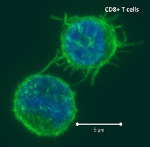
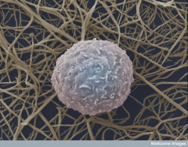
Negative regulatory CD4 T cells
are well characterized and highly studied.  However their CD8 counterparts are not well defined, particularly in humans.  Regulatory CD8 T cells suppress activated CD4 T cells and have proposed roles in various human diseases including multiple sclerosis, ovarian carcinoma and infection with HIV, and many subsets have been described using various markers.  In a recent issue of PLoS One, Hu et. al, describe a population of CD3+CD8+CD161CD56+ T cells within human peripheral blood mononuclear cells (PBMC) that exhibit a cytolytic negative regulatory function.

This group previously published a study where they isolated CD8 T cell clones that were able to lyse autologous T cell receptor (TCR) activated CD4 T cells (Hu et al., 2011).  Surface marker characterization of these regulatory CD8 T cell clones by flow cytometry found that they expressed CD56, CD62L and CD95 but not CD16, CD161, CXCR4 and CCR7.

Because CD161 and CD56 are generally co-expressed markers in NK and NKT cells but are not expressed on conventional CD8 T cells, the authors reasoned that these markers (CD161CD56+) in addition to CD3 and CD8 may provide a robust way to distinguish this population of regulatory CD8 T cells from conventional CD8 T cells, NK cells, and NKT cells by flow cytometry.  Thus in the PLoS One study, the author’s objectives included identification and characterization of this subset of regulatory CD8 T cells in normal human PBMC.

A population of CD3+CD8+CD161CD56+ regulatory CD8 T cells were identified in PBMC and compared with conventional CD8 T cells (CD3+CD8+CD161CD56) and NKT cells (CD3+CD8+CD161+CD56+).  On average, regulatory CD8 T cells occurred at a frequency of 3.2% of total CD8 T cells.  Regulatory CD8 T cells resembled terminally differentiated effector CD8 cells by expressing CD45RA, but not CD45RO or CCR7, and had lower levels of CD62L and CD27.  NKT cells in contrast expressed CD45RO.  For a further discussion of expression of CD45RA, CD45RO, CCR7, CD62L, and CD27 by naïve, central memory, effector memory, and terminally differentiated effector T cell populations, I refer you to a previous post.

Expression of these and numerous other markers were examined in resting and activated regulatory CD8 T cells including CD127, CD25, CD28, CD69, CD94, NKG2a, CD8β, and TCRVα24, and the details can be found in the paper.  Additionally, morphological examination of these cells revealed a larger cytoplasm with some granules, and an irregular nucleus, characteristic of activated T cells and NK cells, but not resting conventional CD8 T cells.

Finally the authors demonstrated that activated CD56+ but notCD56, CD8+CD161 T cells could lyse autologous and allogeneic activated CD4 T cell targets, similarly to the regulatory CD8 T cell clones previously described.

Thus, this study describes the identification of a CD161CD56+ CD8 T cell subset capable of negative regulatory function: cytolysis of activated CD4 T cells.  Many questions remain for further exploration of this interesting population of cells.  Multiple other negative regulatory CD8 T cell subsets have been described including FoxP3+ CD8 T cells.  Determining the differences between various regulatory CD8 T cell subsets regarding marker expression and function should be addressed.  Additionally, the CD8 T cell clones previously described by this group expressed IFN-gamma following activation.  As these negative regulatory CD8 T cells also phenotypically resemble terminally differentiated effector CD8 cells, these populations should be directly functionally compared in future studies.

Identification of Cytolytic CD161(-)CD56(+) Regulatory CD8 T Cells in Human Peripheral Blood.  Hu D, Weiner HL, Ritz J. PLoS One. 2013;8(3):e59545. doi: 10.1371/journal.pone.0059545. Epub 2013 Mar 19.

A clonal model for human CD8+ regulatory T cells: unrestricted contact-dependent killing of activated CD4+ T cellsHu D, Liu X, Zeng W, Weiner HL, Ritz J.  Eur J Immunol. 2012 Jan;42(1):69-79. doi: 10.1002/eji.201141618. Epub 2011 Nov 28.

Basic markers of T cell populations in human PBMC

Time of Flight Mass Cytometry (Cytof): Flying way beyond Fluorescent flow

flow cytometry
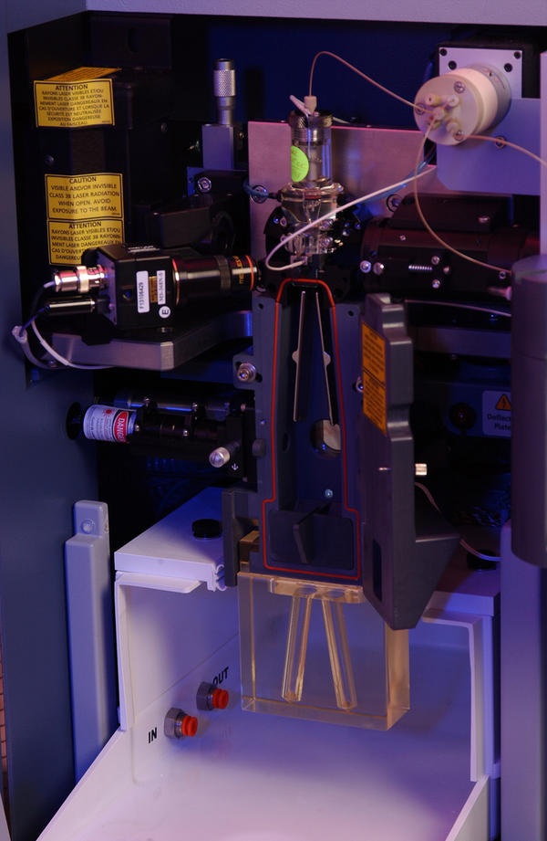
Flow cytometry has been around since the 1950s when Wallace Coulter developed the first flow cytometry device and fluorescence-based flow cytometry was introduced in 1968 by Wolfgang Göhde.  Since then, fluorescence-based flow cytometry and fluorescence-activated cell sorting (FACS) have blown up to become a mainstay of analytical scientific approaches in every field of cell biology, especially immunology.  However, the dominance of fluorescence-based flow cytometry for analytical cellular biology may change with the recent introduction of a new technology: Time of Flight Mass Cytometry (CyTOF).

In fluorescence-based flow cytometry, cells or particles labeled with fluorescent dye-conjugated antibodies or other fluorescent proteins flow in a single file stream past a series of lasers that emit light at specific wavelengths, causing the fluorescent dyes to become excited and emit light caught by detectors.  Thus, a quantitative measure of intensity for each fluorescent parameter, pertaining to the expression level of the antibody-targeted antigen of interest, is obtained for every cell.  The BD Biosciences Influx cell sorter is currently a top of the line fluorescence-based flow cytometer, and supports up to 10 lasers and detection of up to 24 parameters.  However, even with a thorough understanding of flow cytometry, the actual number of utilizable parameters will be typically be far less due to limitations including spectral overlap of fluorescent dyes.

CyTOF utilizes an entirely different technique to quantify protein expression levels on a single cell level: the use of transition element isotopes to label antibodies.  The quantities of isotopes bound to each cell are then analyzed by a time-of-flight mass spectrometer.  While compensation issues due to spectral overlap between fluorophores limits the effective number of parameters assessable by fluorescence-based flow cytometry to far below the theoretical maximums, CyTOF does not suffer from these limitations as there is no requirement for compensation.  In addition, as the metal isotopes used are rare, there is no autofluorescence of cells, another limitation of fluorescence-based flow cytometry. Proof of principle studies have been published by Gary Nolan and colleagues at Stanford University, and have demonstrated the simultaneous use of 34 cell surface and intracellular parameters.  The CyTOF instrument can theoretically detect up to 100 isotopes, thus far extending the ability of researchers to simultaneously assess the expression of many more proteins per cell.

The CyTOF instrument is commercially available from DVS Sciences.  DVS Sciences also offers an expanding list of pre-conjugated metal isotope-labeled antibody reagents and additionally a MAXPAR® labeling kit for conjugation of other antibodies to 33 different metals, allowing researchers to select many additional antigens of interest for analysis.

I have previously stressed the importance of studying cell biology on the single cell level in order to understand the relationships that occur between expression of proteins and signaling states in unique cell populations and on the single cell level.  The addition of CyTOF to the reseracher’s arsenal will allow these types of questions to be addressed on an even more complex level.


Additional Reading:

The history and future of the fluorescence activated cell sorter and flow cytometry: a view from StanfordHerzenberg LA, Parks D, Sahaf B, Perez O, Roederer M, Herzenberg LA. Clin Chem. 2002 Oct;48(10):1819-27.

Single-cell mass cytometry of differential immune and drug responses across a human hematopoietic continuum.  Bendall SC, Simonds EF, Qiu P, Amir el-AD, Krutzik PO, Finck R, Bruggner RV, Melamed R, Trejo A, Ornatsky OI, Balderas RS, Plevritis SK, Sachs K, Pe’er D, Tanner SD, Nolan GP. Science. 2011 May 6;332(6030):687-96. doi: 10.1126/science.1198704.

Mass cytometry: technique for real time single cell multitarget immunoassay based on inductively coupled plasma time-of-flight mass spectrometry.  Bandura DR, Baranov VI, Ornatsky OI, Antonov A, Kinach R, Lou X, Pavlov S, Vorobiev S, Dick JE, Tanner SD. Anal Chem. 2009 Aug 15;81(16):6813-22. doi: 10.1021/ac901049w.

Cytometry by time-of-flight shows combinatorial cytokine expression and virus-specific cell niches within a continuum of CD8+ T cell phenotypes. Immunity. 2012 Jan 27;36(1):142-52. doi: 10.1016/j.immuni.2012.01.002.

Tricks for analyzing PBMC populations by flow cytometry

photo credit: PNNL – Pacific Northwest National Laboratory via photopincc

Cool recent studies utilizing human PBMC

CD4_t_cellThe availability of human peripheral blood mononuclear cells (PBMC) from healthy individuals and from patients with various diseases allows for many studies on normal and abnormal functions of human immune cells.  Because human and murine immune biology differs in many ways, it is important that various methodologies for studying human immunology are established.  The two reports highlighted below demonstrate the usage of human PBMCs for mechanistic and pre-clinical human immune cell studies.

A recent report in The Journal of Immunology by Edwards et. al, demonstrated the usage of healthy human PBMC to elucidate the mechanisms involved in modulation of TH2 T cell responses by Toll-Like Receptor-7 (TLR7) agonists.  Because TLR7 stimulation perturbs TH2 responses, the potential use of rapidly metabolized 8-oxoadenine TLR7 antedrugs for treatment of allergic diseases was explored in this study in collaboration between AstraZeneca and Dainippon Sumitomo.  The TLR7 agonistic antedrug AZ12441970 was found to inhibit TH2 responses via at least two different mechanisms: TLR7-induced production of type I interferons (IFN) and induction of Notch-ligand expression on TLR7-responsive antigen presenting cells.  These led to inhibition of IL-5 production by T cells via the IFN and Notch signaling pathways, respectively.  TLR7-induction of IFNα was found to be intact in PBMCs from asthmatic patients when compared with healthy volunteers, and thus the authors proposed that this therapeutic strategy may be effective in allergic disease patients.

This study provides a demonstration of the usage of human PBMCs for elucidating signaling and immune-cell crosstalk mechanisms as well as determining the potential for the effectiveness of candidate drugs in patients with different disease states.

TLR7 Stimulation of APCs Results in Inhibition of IL-5 through Type I IFN and Notch Signaling Pathways in Human Peripheral Blood Mononuclear Cells. Edwards S, Jones C, Leishman AJ, Young BW, Matsui H, Tomizawa H, Murray CM, Biffen M. J Immunol. 2013 Mar 15;190(6):2585-92. doi: 10.4049/jimmunol.1200780.

The role of the cytokine IL-17 in cancer immunity continues to be controversial.  IL-17 and the IL-17-producing TH17 T cell subsets have been shown to have both pro-tumor as well as anti-tumor immune modulating functions in different cancer contexts.  Monocytes isolated from human PBMC can be differentiated into various myeloid cell types including dendritic cells (DC), providing a tool for studies on these human immune cell types.  In a recent Plos One article, Olsson Åkefeldt et. al, explore the role of IL-17 in survival of human monocyte-derived DCs in vitro, and the relevance of this during chemotherapy.

IL-17 was found to significantly prolong DC survival in vitro, however the cells took on expression of macrophage markers (CD14/CD68) and exhibited a pre-M2 macrophage phenotype.  The prolonged survival was associated with upregulated expression of pro-survival gene BCL-2A1.  Interestingly, IL-17 plus IFNγ treatment in vitro rendered these M2 macrophage-like DCs resistant to cell death induced by 11 of 17 tested chemotherapeutic agents.  Thus, to determine if IL-17 treatment would benefit patients by allowing DC survival during therapy, future studies should address whether this chemoresistance of IL-17 treated DCs occurs in patients undergoing chemotherapy, and to determine how IL-17 affects the anti- versus pro-tumor function of these DCs in various types of cancer.

Chemoresistance of Human Monocyte-Derived Dendritic Cells Is Regulated by IL-17A. Olsson Åkefeldt S, Maisse C, Belot A, Mazzorana M, Salvatore G, Bissay N, Jurdic P, Aricò M, Rabourdin-Combe C, Henter JI, Delprat C. PLoS One. 2013;8(2):e56865. doi: 10.1371/journal.pone.0056865. Epub 2013 Feb 18.

Studies like these are examples of the utility of using human PBMCs to elucidate mechanisms of human immune cell biology under normal and diseased conditions.

Can shutting down the IRF3 kinases be the magic pill to treat obesity?

amlexanoxCan amlexanox, a small molecule drug that has been approved for asthma, allergic rhinitis and aphthous ulcers be an effective treatment for obesity? Many are excited about this prospective presented in a recent article in Nature Medicine by Reilly et. al. as simply Googling “amlexanox” and “obesity” resulted in a plethora of news articles on this report.  Interestingly, the two highly related proteins allegedly inhibited by amlexanox that led to this result in mice are the IRF3-kinases, TBK1 (TANK-binding Kinase-1, T2K, NAK) and IKK-ε (IκB kinase-epsilon, IKK-i), whose major known functions are during pathogen infections: the activation of IRF-3 (interferon regulatory kinase-3), the major transcription factor regulating expression of interferon-β (IFNβ).

Inflammation is considered a key link between obesity and insulin resistance.  The canonical NF-ĸB signaling pathway has been shown to playing a major role in this linkage.  IKK-ε and TBK1 are two IKK-β-related kinases with an unclear role in NF-ĸB activation, and thus their role in obesity and insulin resistance was explored in this and a previous study by the same group (Chiang et. al).

In this study, the authors show that expression and kinase activity of IKK-ε and TBK1 were increased in mice fed a high-fat diet and in response to TNFα in an in vitro adipocyte inflammation model. In the previous study, IKK-ε deficient mice were partially resistant to development of obesity and insulin resistance when fed a high fat diet.  Thus, the authors sought to determine if inhibiting these kinases would have a therapeutic effect on obesity.  A screen for small molecule inhibitors to block IKK-ε and TBK1 identified amlexanox, a drug currently used to treat asthma, allergic rhinitis and aphthous ulcers.  Amlexanox treatment inhibited the in vitro kinase activity of both IKK-ε and TBK1 at a much lower concentration than it did the related canonical NF-ĸB pathway kinase IKK-β.

To determine the effect of in vivo inhibition of these kinases on obesity indexes, mice fed a high fat diet were treated with amlexanox.  Amlexanox-treated mice gained significantly less weight than non-treated mice, and treatment of mice with pre-established diet-induced obesity led to a significant, but reversible weight loss accompanied by a decrease in adipose tissue, without a lower food intake.  Additionally, ob/ob mice which are genetically disposed to overt obesity due to deficient leptin expression, also lost adipose tissue mass when treated with amlexanox.

Interestingly, compared with mice fed a normal diet, mice on a high fat diet have a decreased core body temperature. Amlexanox-treated mice had body temperatures raised to normal levels along with increased oxygen consumption, indicating that an increase in energy expenditure of these mice may contribute to their weight loss.  Other indexes of obesity and glucose intolerance also returned to normal levels following amlexanox treatments.

Despite the promising results achieved with this drug in decreasing obesity and insulin resistance, the mechanisms by which inhibition of these kinases lead to this effect remains unclear.  Thus, future studies need to clarify the effects of amlexanox on IKK-ε and TBK1 regulation of the NF-ĸB pathway and obesity-associated inflammation, as well as address effects on the IRF3-interferon pathway.  Finally, it will be important to determine if other molecules are targeted by this drug.


Further Reading:

An inhibitor of the protein kinases TBK1 and IKK-ɛ improves obesity-related metabolic dysfunctions in mice.  Reilly SM, Chiang SH, Decker SJ, Chang L, Uhm M, Larsen MJ, Rubin JR, Mowers J, White NM, Hochberg I, Downes M, Yu RT, Liddle C, Evans RM, Oh D, Li P, Olefsky JM, Saltiel AR. Nat Med. 2013 Mar;19(3):313-21.

The protein kinase IKKepsilon regulates energy balance in obese mice.  Chiang SH, Bazuine M, Lumeng CN, Geletka LM, Mowers J, White NM, Ma JT, Zhou J, Qi N, Westcott D, Delproposto JB, Blackwell TS, Yull FE, Saltiel AR. Cell. 2009 Sep 4;138(5):961-75.

Turning off the inflammatory, but not the metabolic, flames.  Calay ES, Hotamisligil GS. Nat Med. 2013 Mar 6;19(3):265-7.

Deficiency of T2K leads to apoptotic liver degeneration and impaired NF-kappaB-dependent gene transcription.  Bonnard M, Mirtsos C, Suzuki S, Graham K, Huang J, Ng M, Itié A, Wakeham A, Shahinian A, Henzel WJ, Elia AJ, Shillinglaw W, Mak TW, Cao Z, Yeh WC. EMBO J. 2000 Sep 15;19(18):4976-85.

Heterozygous TBK1 mutations impair TLR3 immunity and underlie herpes simplex encephalitis of childhood.  Herman M, Ciancanelli M, Ou YH, Lorenzo L, Klaudel-Dreszler M, Pauwels E, Sancho-Shimizu V, Pérez de Diego R, Abhyankar A, Israelsson E, Guo Y, Cardon A, Rozenberg F, Lebon P, Tardieu M, Heropolitanska-Pliszka E, Chaussabel D, White MA, Abel L, Zhang SY, Casanova JL. J Exp Med. 2012 Aug 27;209(9):1567-82.

TANK-binding kinase 1 (TBK1) controls cell survival through PAI-2/serpinB2 and transglutaminase 2.  Delhase M, Kim SY, Lee H, Naiki-Ito A, Chen Y, Ahn ER, Murata K, Kim SJ, Lautsch N, Kobayashi KS, Shirai T, Karin M, Nakanishi M. Proc Natl Acad Sci U S A. 2012 Jan 24;109(4):E177-86.

*Image courtesy of RXlist.com*

Markers and functions of human CD4+ follicular helper T cells

human white blood cell
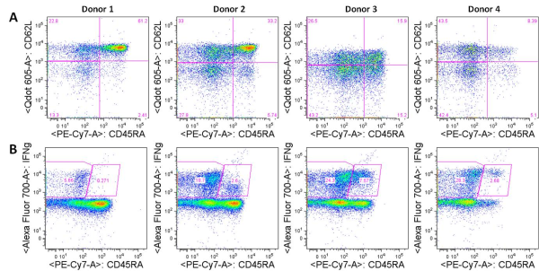
CXCR5 is a chemokine receptor expressed by and used to identity human CD4+ follicular helper T cells (TFH).  TFH cells, as their name implies, promote the differentiation and survival of memory and plasma B cells in the B cell follicular and germinal center regions of secondary lymphoid organs. CXCR5+ TFH-like central memory CD4+ T cells (CD4+ TCM) also circulate in peripheral blood and can be detected among human peripheral blood mononuclear cells (PBMC).  CXCR5+ cells comprise 20-25% of CD4+ TCM cells in human PBMC.  However, what are the identifying markers and functional differences of CXCR5+ vs. CXCR5 CD4+ T cells from human PBMC and the prototypical CXCR5+ TFH cells in secondary lymphoid organs?

I have previously discussed markers that can be used for identification of human CD4+ TH1, TH2, and TH17 T cell helper subsets as well as CD4+ FoxP3+ regulatory T cells.  CXCR5 can be upregulated transiently on activated T cells, however a subset of PBMC T cells constitutively express CXCR5, indicating this is a uniquely functioning subset identifiable by this marker using flow cytometry or other methods.  PBMC CXCR5+ CD4+ TCM cells exhibit many but not all features of TFH cells present in secondary lymphoid organs, and thus may be the circulating memory counterpart of TFH cells.

CXCL13, the chemokine ligand for CXCR5, is highly expressed in B cell follicles and likely plays an important role in recruitment of CXCR5+ TFH cells to B cell zones.  Expression of ICOS by TFH cells has been demonstrated to be essential for their function in B cell help.  Additionally, TFH cells are higher expressers of CXCL13, as well as IL-21, IL-10, Bcl-6, and PD-1 than other helper T cell subsets.

Studies by Chevalier et al, and Morita et. al. compared the functional properties of CXCR5 and CXCR5+ CD4+ TCM cells from human PBMC.  PBMC CXCR5+ CD4+ cells are resting central memory cells in phenotype, being CD45RA, CCR7+ and CD62L+, but not expressing activated TFH markers such as ICOS and CD69.  Upon stimulation, CXCR5+ CD4+ TCM promote significantly higher B cell plasmablast differentiation and Ig secretion than CXCR5 CD4+ TCM cells, attributable to enhanced expression of ICOS and IL-10.  However, Bcl-6 expression was not found to be different between these PBMC subsets, and conclusions for expression levels and role of IL-21 were contradictory between these studies.

The question of whether PBMC CXCR5+ CD4+ TCM are distinct from TH1, TH2, and TH17 T cells was addressed by these studies as well. While Chevalier et al. found that CXCR5+ TCM cells were more non-polarized and secreted comparatively lower levels of cytokines associated with TH1, TH2, and TH17 T cells, Morita et. al, identified TH1, TH2, and TH17 T cells within CXCR5+ compartment, albeit at somewhat different frequencies than CXCR5 cells. Thus PBMC CXCR5+ CD4+ TCM are a heterogenous subset with features of both TFH cells and the various TH1, TH2, and TH17 subsets.  Further interrogation of the functions of these populations are needed.

PBMC CXCR5+ CD4+ cells have been identified as a highly relevant population to study in the context of vaccination and human disease.  Patients with systemic lupus erythematosus (SLE) have higher percentages of circulating CD4+CXCR5+ ICOS+ cells.  Patients with autoimmune juvenile dermatomyositis (JDM) were found to have an altered CXCR5+ compartment where the overall frequency of CXCR5+ cells was not different, but the ratio of TH2 and TH17 to TH1 cells within the CXCR5+ population was enhanced and associated with disease activity.

In the vaccine setting, emergence of a population of circulating ICOS+CXCR3+CXCR5+CD4+ T cells was found in individuals 7 days after influenza vaccination, and correlated with increased antibody titers and B cell plasmablasts. These cells could also induce plasma cell differentiation in vitro and  thus are important in the development of vaccine elicited protective antibody responses.

In conclusion, PBMC CXCR5+ CD4+ T cells are an important cellular subset to study in the context of human disease.  These cells are likely the circulating memory component of TFH cells, and alterations in frequencies and functions are associated with various human diseases and protective antibody responses following vaccination.


Further Reading:

Human blood CXCR5(+)CD4(+) T cells are counterparts of T follicular cells and contain specific subsets that differentially support antibody secretion.  Morita R, Schmitt N, Bentebibel SE, Ranganathan R, Bourdery L, Zurawski G, Foucat E, Dullaers M, Oh S, Sabzghabaei N, Lavecchio EM, Punaro M, Pascual V, Banchereau J, Ueno H. Immunity. 2011 Jan 28;34(1):108-21.

CXCR5 expressing human central memory CD4 T cells and their relevance for humoral immune responses.  Chevalier N, Jarrossay D, Ho E, Avery DT, Ma CS, Yu D, Sallusto F, Tangye SG, Mackay CR. J Immunol. 2011 May 15;186(10):5556-68.

Expansion of circulating T cells resembling follicular helper T cells is a fixed phenotype that identifies a subset of severe systemic lupus erythematosus. N. Simpson, P.A. Gatenby, A. Wilson, S. Malik, D.A. Fulcher, S.G. Tangye, H. Manku, T.J. Vyse, G. Roncador, G.A. Huttley et al.  Arthritis Rheum., 62 (2010), pp. 234–244.

Induction of ICOS+CXCR3+CXCR5+ TH Cells Correlates with Antibody Responses to Influenza Vaccination.  Bentebibel S. E. et al.  Sci Transl Med. 13 March 2013: Vol. 5, Issue 176, p. 176ra32.

Photo credit: wellcome images / Foter.com / CC BY-NC-ND

Natural Killer Cell subtypes and markers in human PBMC

Natural killer cells
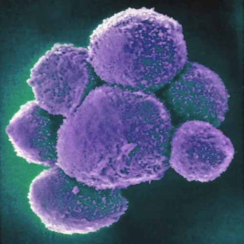
Natural Killer (NK) cells are a cytotoxic innate immune lymphocyte cell type.  In humans, NK cells comprise up to 15% of peripheral blood mononuclear cells (PBMC), and 5-20% of the PBMC lymphocyte population.  Several subtypes of NK cells exist in humans.  In this post, I will discuss phenotypic properties and markers of NK subtypes present in human PBMC.

Three subtypes of NK cells are recognized: CD56dim CD16+, CD56brightCD16+/- and CD56 CD16+ NK cells. The CD56dim CD16+ and CD56brightCD16+/- subsets are best studied and are phenotypically classified as a more cytotoxic and a more cytokine producing subset of NK cells, respectively.  NK cell activation is mediated by the balance between engagement of activating receptors including NKp46, NKp30, NKp44, NKG2D, CD16, 2B4, NKp80, and DNAM-1, and HLA-I binding inhibitory receptors including killer immunoglobulin-like receptors (KIRs), LIR1/ILT2 and NKG2A/CD94.  NK cells can also be activated in response to cytokines such as IL-2, IL-12, IL-15, and IL-18.

CD56dim CD16+ NK cells:  This subtype comprises the majority, up to 90%, of PBMC NK cells and is considered the most cytotoxic subset.  CD16 is the FCγ receptor III, and can thus bind the FC portion of IgG antibodies and mediate antibody dependant cell-mediated cytotoxicity (ADCC) of antibody-bound target cells.  Expression of inhibitory receptors differs among NK subsets, and this subset exhibits lower expression of KIRs and ILT2 but higher expression of NKG2A/CD94 compared with CD56bright NK cells. Expression of granzyme B and perforin is also high in this subset compared with CD56bright NK cells.  A recent report by De Maria et. al, demonstrated that this subset does in fact robustly produce cytokines including IFNγ early after activation.

CD56brightCD16+/- NK cells: This subtype comprises up to 10% of NK cells in PBMC, but is the major NK subtype in tissues and secondary lymphoid organs.  This subset is conventionally known as the cytokine producing subset of NK cells, and rapidly produces cytokines and chemokines including IFNγ, TNFα, GM-CSF, and RANTES after activation.

Interestingly, in HIV-viremic individuals, a third CD56 CD16+ NK population is significantly expanded in PBMC comprising between 20-55% of NK cells.  This population in healthy individuals and aviremic HIV-infected individuals is rare, under 10% of total NK cells.  Compared with CD56+ NK cells, the CD56 CD16+ NK cells from HIV-viremic patients exhibited lower expression of activating receptors NKp46, NKp30, and NKp44, lower cytotoxic activity, higher expression levels of inhibitory receptors, and lower expression levels of cytokines including IFNγ, TNFα, and GM-CSF.  This subset is also expanded in individuals with chronic HCV infection.  Thus, the expansion of this poorly functional NK subset is likely clinically relevant in chronic viral disease.

In summary, these NK populations can be differentiated by expression of CD16 and CD56.  Of note, NKT (natural killer-like T) cells can also express these markers along with CD3.  Thus, to differentiate these cells from NKT cells, the inclusion of CD3 as a cell identification marker is critical in analysis of these cells by flow cytometry or other methods.


Further Reading:

CD56 negative NK cells: origin, function, and role in chronic viral disease.  Björkström NK, Ljunggren HG, Sandberg JK. Trends Immunol. 2010 Nov;31(11):401-6.

The biology of human natural killer-cell subsets. Cooper MA, Fehniger TA, Caligiuri MA. (2001) Trends Immunol 22: 633–640.

Natural killer cell distribution and trafficking in human tissues.  Carrega P, Ferlazzo G. Front Immunol. 2012;3:347.

Revisiting human natural killer cell subset function revealed cytolytic CD56(dim)CD16+ NK cells as rapid producers of abundant IFN-gamma on activation.  De Maria A, Bozzano F, Cantoni C, Moretta L. Proc Natl Acad Sci U S A. 2011 Jan 11;108(2):728-32.

Natural killer cells in HIV-1 infection: dichotomous effects of viremia on inhibitory and activating receptors and their functional correlates.  Mavilio D, Benjamin J, Daucher M, Lombardo G, Kottilil S, Planta MA, Marcenaro E, Bottino C, Moretta L, Moretta A, Fauci AS. Proc Natl Acad Sci U S A. 2003 Dec 9;100(25):15011-6.

Characterization of CD56−/CD16+ natural killer (NK) cells: a highly dysfunctional NK subset expanded in HIV-infected viremic individuals. Mavilio D, Lombardo G, Benjamin J, Kim D, Follman D, et al.. (2005) Proc Natl Acad Sci U S A. 102: 2886–2891.

The Immunoscore: bringing immunological parameters to the clinic for cancer patient prognosis

Cancer stages
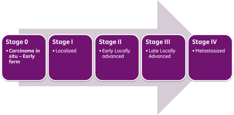
Classical prognosis of cancer patients utilizes the AJCC/UICC (American Joint Committee on Cancer / International Union Against Cancer) “TNM” classification system, in which T (Tumor) is indicative of primary tumor size and invasion properties, N (Nodes) indicates the extent of tumor invasion into draining and regional lymph nodes, and M (Metastasis), describes the presence and extent of metastatic lesions at diagnosis.  The combinations of these parameters are then used to assess a patient’s stage at diagnosis and predict patient outcome.  The exact parameter definitions vary for each cancer type.

However, it has long been known that while the TNM system provides a fairly good estimate for patient populations overall, there is still significant heterogeneity within each stage as to tumor recurrence and ultimate outcome.  This is unsurprising given this staging system evaluates only tumor characteristics and fails to account for other patient parameters. In particular, the integrity of the patient’s immune system, our inherent natural protection against tumor development, has been shown to have a significant impact on disease progression and patient outcome.

In colorectal cancer as well as other cancer types, much progress has been made in ascertaining the prognostic significance of cytotoxic CD8+ T cell infiltration into the tumor microenvironment.  The densities of CD8+ T cell presence in the invasive margin (IM) and the center of the tumor (CT) have been shown to have significant prognostic value.  In a study by Pages et. al. (J Clin Oncol. 2009), assessment of CD8+ T cells and CD45RO+ memory T cell densities in CT/IM tumor regions in stage I and II colorectal cancer patients significantly predicted recurrence and overall survival, showing that application of this system is particularly relevant in early stage patients to better direct treatment strategies.  Multiple cytotoxic CD8+ T cell and TH1 phenotyping markers have shown prognostic significance in human cancer patients, including CD8, CD3, CD45RO, and Granzyme B expression.  However, as discussed by Dr. Jerome Galon in the Oct 3, 2012 J Transl Med. article, CD8 and CD3 represent the most robust markers for adoption into routine clinical practice as CD45RO, and Granzyme B expression are intensity-dependant evaluations and thus much more technically difficult to standardize.

Thus, the proposed immunoscore relies on immunohistochemistry staining for CD8+ and CD3+ T cells in CT/IM tumor regions using standardized antibodies and protocols.  Quantitative assessment of their densities is then determined and scored on whole tissue slides using specified slide scanning and staining analysis software.

In 2012, an international task force, led by Dr. Galon was established to promote the routine usage of this classification system in clinical diagnosis of cancer patients.  The goals of this taskforce include feasibility and standardization of the quantitative immunohistochemistry protocol used to derive the score, worldwide validation of the immunoscore for colorectal cancer patient prognosis, as well as the application of this classification system for other cancer types.  Thus, the immunoscore may soon become a standard clinical practice and aid in better prognostic stratification of patients and therapeutic guidance.


Further Reading:

http://www.immunescore.org/

Website: American Joint Committee on Cancer Staging

Cancer classification using the Immunoscore: a worldwide task forceGalon J, Pagès F, Marincola FM, Angell HK, Thurin M, Lugli A, Zlobec I, Berger A, Bifulco C, Botti G, Tatangelo F, Britten CM, Kreiter S, Chouchane L, Delrio P, Arndt H, Asslaber M, Maio M, Masucci GV, Mihm M, Vidal-Vanaclocha F, Allison JP, Gnjatic S, Hakansson L, Huber C, Singh-Jasuja H, Ottensmeier C, Zwierzina H, Laghi L, Grizzi F, Ohashi PS, Shaw PA, Clarke BA, Wouters BG, Kawakami Y, Hazama S, Okuno K, Wang E, O’Donnell-Tormey J, Lagorce C, Pawelec G, Nishimura MI, Hawkins R, Lapointe R, Lundqvist A, Khleif SN, Ogino S, Gibbs P, Waring P, Sato N, Torigoe T, Itoh K, Patel PS, Shukla SN, Palmqvist R, Nagtegaal ID, Wang Y, D’Arrigo C, Kopetz S, Sinicrope FA, Trinchieri G, Gajewski TF, Ascierto PA, Fox BA.  J Transl Med. 2012 Oct 3;10:205.

The immune score as a new possible approach for the classification of cancer.  Galon J, Pagès F, Marincola FM, Thurin M, Trinchieri G, Fox BA, Gajewski TF, Ascierto PA. J Transl Med. 2012 Jan 3;10:1.

The immune contexture in human tumours: impact on clinical outcome. Fridman WH, Pages F, Sautes-Fridman C, Galon J.  Nat Rev Cancer 2012, 12:298-306.

In situ cytotoxic and memory T cells predict outcome in patients with early-stage colorectal cancerPagès F, Kirilovsky A, Mlecnik B, Asslaber M, Tosolini M, Bindea G, Lagorce C, Wind P, Marliot F, Bruneval P, Zatloukal K, Trajanoski Z, Berger A, Fridman WH, Galon J. J Clin Oncol. 2009 Dec 10;27(35):5944-51.

Image courtesy of simplyanon on Wikipedia

Generation of CD4+ Th1 cells from human PBMC

CD4+ T helper type 1 (TH1) cells are the effector T cell population that governs cell mediated immune responses against intracellular pathogens including viruses and intracellular bacteria.  TH1 cells mediate their effect by secreting cytokines such as interferon-gamma (IFNγ) and IL-2, and express cell surface markers including CXCR3 and CCR5 and the characteristic TH1 master transcription factor T-bet (TBX21) which can also be used for detection of TH1cells by flow cytometry, as discussed in a previous blog post.

Differentiation of naïve human CD4+ T cells down the TH1 pathway involves cytokines such as IL-12 which activates STAT4, and induces expression of IFNγ and T-bet.  As such, in vitro protocols differentiating peripheral blood mononuclear cells (PBMC)-derived naïve CD4+ T cells into TH1 cells involves incubation with IL-12 in the context of T cell activation through the T cell receptor (TCR) complex.

In my experience, TH1 cells are by far the easiest CD4+ helper T cell population to generate in vitro.  In order to generate TH1 cells from human PBMC, naïve CD4+ T cells must first be isolated.  Multiple methods of naïve CD4+ T cell isolation can be utilized, and magnetic bead- based methods are common and easy methods.  Companies such as Miltenyi Biotech and Stem Cell Technologies offer kits for isolation of untouched naïve CD4+ T cells from PBMC by negative isolation methodologies.

Following isolation, naïve CD4+ T cells are activated through the TCR complex.  Tissue culture plates can be coated with anti-CD3 (OKT1) and anti-CD28 antibodies in PBS prior to culture.  Alternatively, naïve CD4+ T cells can be cultured with Dynal CD3/CD28 T Cell Expander Dynabeads (Life Technologies) at a 1 bead per cell ratio.  A third alternative involves coating tissue culture plates with anti-CD3 alone and obtaining CD28 co-stimulation by the addition of autologous monocytes isolated from PBMCs into the culture.

To generate TH1 cells, recombinant human IL-12 is added alone, or at a lower dose in combination with anti-IL-4 blocking antibodies to inhibit the counteractive effects of IL-4 and TH2 pathways on TH1 cell polarization.  Finally recombinant human IL-2 is added to promote T cell proliferation.  Media and cytokines/blocking antibodies are refreshed every two to three days depending on the cell density, and as the cells expand the time to refresh the media shortens.

Lymphocyte activationTH1 cells can be generated and assayed for functions including IFNγ expression in as few as three days.  If long term or clonal T cells assays are of interest, cells can be expanded in the presence of IL-2 for 2-3 weeks following single cell cloning.  As previously discussed, TH1 cells can be identified by IFNγ expression following a 4-6 hour incubation with TCR activation by plate bound anti-CD3 plus anti-CD28, CD3/CD28 Dynabeads, or PMA/ionomycin in the presence of brefeldin-A.  Cells are then fixed, permeabilized, and stained for cell surface markers and intracellular IFNγ.

Finally, as a comparison, tandem experiments can be run in which naïve CD4+ T cells are maintained under non-polarizing (TH0) conditions.  For this, often no cytokines aside from IL-2 are added.  However the addition of anti-IL-12 and anti-IL-4 may be necessary to inhibit any cells from differentiating down TH1 or TH2 pathways by production of these cytokines by the T cells themselves.

In conclusion, generation of CD4+ TH1cells from human PBMC is a relatively simple and straightforward protocol, and very high percentages of TH1cells can be obtained through optimized protocols.


Further Reading:

Differentiation of effector CD4 T cell populations (*).  Zhu J, Yamane H, Paul WE.  Annu Rev Immunol. 2010;28:445-89.

Memory and flexibility of cytokine gene expression as separable properties of human T(H)1 and T(H)2 lymphocytes.  Messi M, Giacchetto I, Nagata K, Lanzavecchia A, Natoli G, Sallusto F.  Nat Immunol. 2003 Jan;4(1):78-86.

A critical function for transforming growth factor-beta, interleukin 23 and proinflammatory cytokines in driving and modulating human T(H)-17 responses.  Volpe E, Servant N, Zollinger R, Bogiatzi SI, Hupé P, Barillot E, Soumelis V. Nat Immunol. 2008 Jun;9(6):650-7.